Multi-Omic Single-Cell Landscape of Lung Cancer Progression Reveals Dynamic Microenvironmental Rewiring
This comprehensive single-cell analysis of human lung tissue across normal, early, and advanced tumor stages unveils profound shifts in cellular populations, gene expression, and cell-cell interactions within the tumor microenvironment. Key findings include the robust identification of aneuploid lung epithelial cells as the primary malignant population, widespread genomic instability with recurrent oncogene amplifications (e.g., EGFR, EIF3E), and a hyper-proliferative state driven by cell cycle dysregulation. The immune landscape progressively shifts towards immunosuppression, characterized by reduced anti-tumor T cells and NK cells, increased regulatory T cells (Tregs), and a polarization of macrophages towards pro-tumorigenic M2-like phenotypes. Stromal cells, particularly fibroblasts, are reprogrammed into cancer-associated fibroblasts (CAFs) that actively remodel the extracellular matrix and support tumor growth. These changes are orchestrated through complex cell-cell communication networks involving critical pathways like EGFR, TGF-beta, PGE2, and integrin signaling, offering critical insights into lung cancer pathogenesis and potential therapeutic targets.
Contents
- Dataset overview
- UMAP Visualization of Lung Single-Cell RNA-seq Data
- UMAP Visualization of Major Cell Type Scores, Ploidy Status, and Cell Type Annotations
- Overall Celltype_subset Marker Expression Validation
- Copy Number Variation Analysis in Tumor-Origin and Unassigned Lung Cells
- CNV-based UMAP of Single-Cell Data Reveals Distinct Malignant and Non-Malignant Cell Populations
- Minor Cell Type Population Analysis Across Lung Cancer Stages
- Lung Cancer Microenvironment: T Cell and Innate Lymphoid Cell Subset Shifts
- Changes in T Cell Subset Proportions Across Lung Tumor Progression
- Macrophage Subset Population Shifts in Lung Cancer Progression
- Macrophage Subset Population Shifts Across Lung Cancer Progression
- Ploidy Population Analysis of Tumor-Origin and Unassigned Cells Across Lung Conditions
- Condition-Specific Cell-Cell Interaction Patterns in Lung Tissue
- Condition-Specific Cell-Cell Interaction Analysis in Lung Tissue
- Cell-Cell Interaction Analysis of Immune Checkpoint and Cell Cycle Related Genes in Lung Cancer Progression
- Condition-Specific Cell-Cell Interaction Patterns in Lung Tissue
- Lung Epithelial Cell Condition-Specific Surfaceome Markers Analysis
- Macrophage Condition-Specific Surfaceome Markers in Lung Tissue
- Fibroblast Condition-Specific Surfaceome Marker Analysis in Lung Tissue
- Condition-Specific Surfaceome Markers in CD4+ T cells Across Lung Conditions
- Differential Expression of Cell Cycle Genes in Lung Epithelial Cells Across Tumor Stages
- 폐 상피세포의 플로이드 상태 및 질병 단계별 유전자 온톨로지(GSA) 분석 결과
- Gene Set Enrichment Analysis Reveals Distinct Pathway Deregulation Across Lung Cancer Cell Types
- Discussion
- Query List
0. Dataset overview
Dataset Summary
- This dataset contains single-cell RNA-seq data from human lung tissue, comprising 84,300 cells and 23,489 genes.
- It includes metadata such as 'Tumor', 'Sample', 'Cell_type', 'Patient id', 'Tissue origins', 'Histology', 'Gender', 'Age', 'Smoking', 'Pathology', 'EGFR_mutation', and 'Stage'.
- Key categorical annotations include conditions ('Tumor(adv)', 'Tumor(early)', 'Normal'), and hierarchical cell type classifications ('celltype_major', 'celltype_minor', 'celltype_subset').
- Ploidy information is available through 'ploidy_dec' (Aneuploid, Diploid), with 'Lung Epithelial cell' identified as the tumor origin cell type.
- The dataset is pre-processed and contains computed results for Cell-Cell Interaction (CCI), Differential Gene Expression (DEG), Gene Set Enrichment Analysis (GSEA), and Gene Ontology (GO/GSA).
- Reference condition 'Normal' is used for 'DEG_vs_ref', 'GSEA_vs_ref', and 'GSA_vs_ref_up' analyses.
- Precomputed results include CCI (per condition and per sample), DEG (comparing one condition vs rest/reference), GSEA (comparing one condition vs rest/reference), GSA_up (comparing one condition vs rest/reference), ploidy inference labels, and CNV estimates ('X_cnv').
- Various observation (obs) and variable (var) columns provide extensive cell and gene metadata.
1. UMAP Visualization of Lung Single-Cell RNA-seq Data
[Analysis Visualization Results]...
Analysis Overview
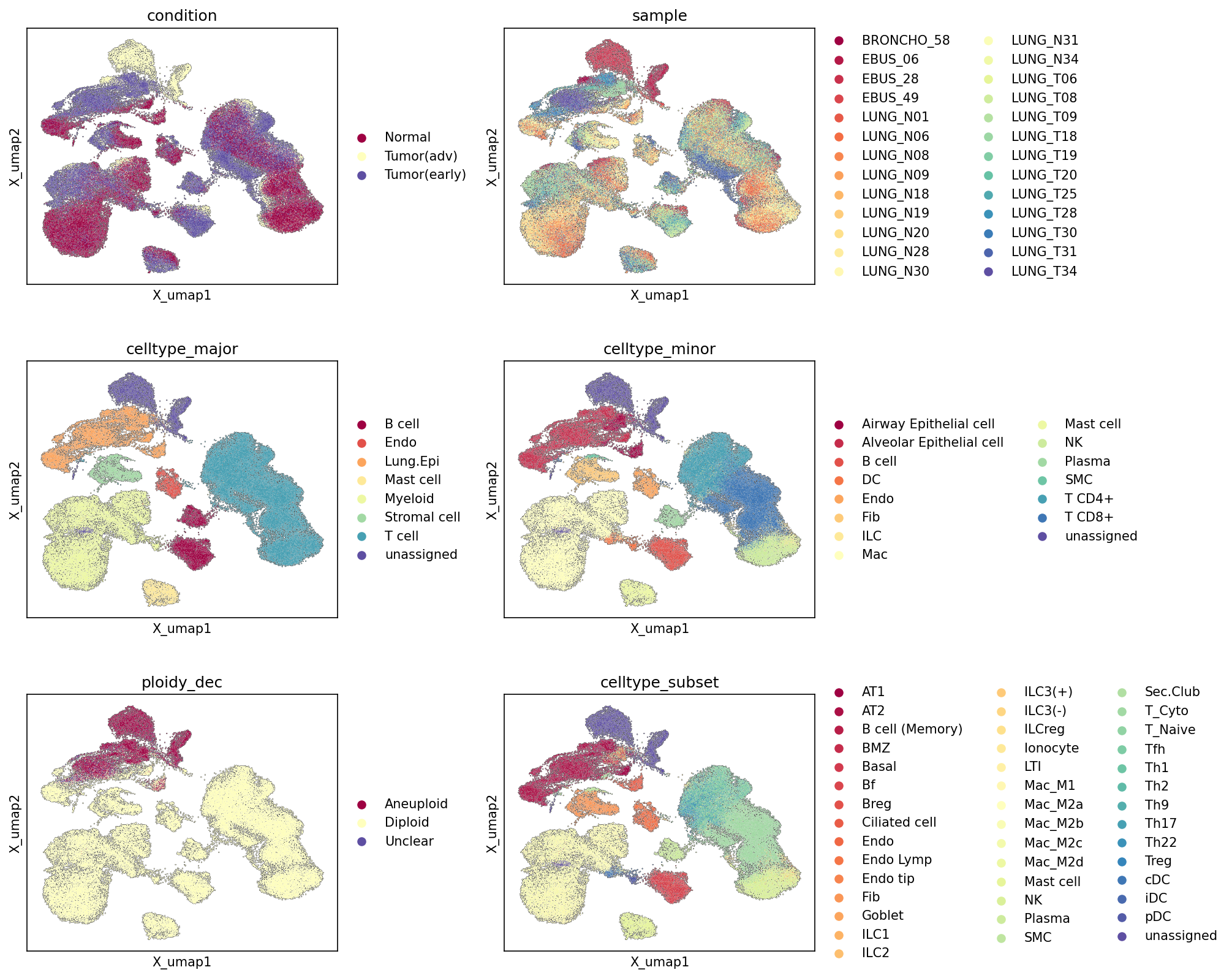
This analysis presents a series of Uniform Manifold Approximation and Projection (UMAP) plots, providing an overview of the single-cell RNA-sequencing dataset. Cells are visualized in a 2-dimensional embedding space and colored by various metadata attributes including condition, sample, celltype_major, celltype_minor, ploidy_dec, and celltype_subset. These visualizations are crucial for assessing the overall structure of the dataset, evaluating the quality of cell type annotation, identifying sample-specific biases (batch effects), and exploring the distribution of key biological features like ploidy across the cellular landscape.
Visual Summary
Condition
The UMAP colored by condition reveals a clear separation of cells from Normal tissue (red) from those in Tumor(adv) (yellow) and Tumor(early) (purple) conditions. The Normal cells tend to occupy a distinct region, primarily in the upper-left and lower-right parts of the embedding. Tumor(adv) and Tumor(early) conditions show significant overlap, particularly in the central and upper-central regions, suggesting shared cellular compositions and transcriptional states in different tumor stages. However, some clusters show enrichment for either Tumor(adv) or Tumor(early) specifically, indicating condition-specific cellular populations or states.
Sample
The sample colored UMAP indicates a generally good mixing of cells from different samples across the major cell clusters. While some smaller sub-clusters might show enrichment for specific samples, the overall distribution suggests that the primary biological variance (e.g., cell type differences) rather than sample-specific batch effects largely drives the clustering structure. This is important for ensuring the generalizability of findings across patients.
Celltype_major
This UMAP shows that major cell types (e.g., T cell, Myeloid cell, Lung Epithelial cell) form well-defined, distinct clusters. For instance, T cell (light blue) forms a large, distinct cluster on the right, Myeloid cell (light green) occupies the lower-central region, and Lung Epithelial cell (orange) is prominent in the upper-left. B cell (dark red), Endothelial cell (red), Mast cell (yellow), and Stromal cell (dark green) also form characteristic, separate groups. The clear separation of these major populations validates the robustness of the cell type annotation at this level.
Celltype_minor
Further refinement of cell types is evident in the celltype_minor UMAP. Subtypes like T cell CD8+ and T cell CD4+ are clearly delineated within the broader T cell cluster. Similarly, Macrophage and Dendritic cell populations are distinct within the Myeloid cell major group, and Alveolar Epithelial cell and Airway Epithelial cell within Lung Epithelial cells. This level of detail confirms that the embedding successfully resolves functionally distinct cell populations within the major cell lineages.
Ploidy_dec
The ploidy_dec UMAP highlights the distribution of aneuploid, diploid, and unclear cells. A significant cluster of Aneuploid cells (dark red) is concentrated in the upper-left region of the UMAP. This region strongly overlaps with areas enriched for Lung Epithelial cell and Tumor(adv)/Tumor(early) conditions. Diploid cells (yellow) are broadly distributed across the entire UMAP, as expected for non-malignant cells, but are also present within the tumor-associated regions, likely representing immune, stromal, and some normal epithelial cells. The clear localization of Aneuploid cells to specific tumor-associated epithelial clusters is a key observation.
Celltype_subset
The celltype_subset UMAP provides the most granular view of cell type annotations, showing detailed subpopulations within celltype_minor groups. Examples include Alveolar type 1 (AT1) and Alveolar type 2 (AT2) within Alveolar Epithelial cells, and various macrophage subtypes (e.g., Macrophage (M1), Macrophage (M2A)) within the macrophage population. The visual separation of these fine-grained subsets indicates a high resolution of cell identities within the dataset and supports the quality of the annotation at this detailed level.
Biological Interpretation
The UMAP visualizations provide compelling biological insights into the cellular landscape of lung tissue across normal, early-stage, and advanced-stage tumor conditions.
- Tumor Microenvironment Heterogeneity: The distinct clustering by condition underscores the significant transcriptional and cellular differences between normal lung tissue and the tumor microenvironment. The partial overlap between Tumor(early) and Tumor(adv) conditions suggests some shared malignant and immune cell features, while also indicating the emergence of unique cellular states or populations as the tumor progresses.
- Malignant Cell Identification: The strong co-localization of Aneuploid cells with Lung Epithelial cell clusters, predominantly within the Tumor(adv) and Tumor(early) conditions, provides robust evidence for the identification of malignant epithelial cells. Given that Lung Epithelial cells are identified as the Tumor origin celltype, this finding is highly consistent with the expected biology of lung cancer, where chromosomal instability and aneuploidy are hallmarks of malignancy [PubMed Search].
- Immune and Stromal Cell Infiltration: The presence of diverse T cell, Myeloid cell, B cell, Stromal cell, and Endothelial cell populations across both tumor conditions and normal tissue, but often in distinct regions or states, reflects the complex interplay within the tumor microenvironment. For instance, specific immune cell subsets might be recruited or activated differently in early vs. advanced tumors compared to normal tissue.
- Robust Cell Type Annotation: The consistent and hierarchical clustering observed from celltype_major down to celltype_subset confirms the high quality and resolution of the cell type annotations. This robust annotation is critical for downstream analyses such as differential gene expression or cell-cell interaction studies, ensuring that comparisons are made between truly distinct cellular populations.
Annotation Notes
The UMAP plots collectively demonstrate a high quality of data integration, embedding, and cell type annotation for this single-cell RNA-seq dataset.
- Cell Type Resolution: The progressive refinement of cell types from major groups to minor and subset levels, each forming coherent and distinct clusters, indicates excellent resolution and confidence in the cell identity assignments.
- Batch Effect Management: The adequate mixing of samples across the UMAP suggests that sample-specific batch effects are not a dominant factor driving the global clustering, reinforcing the reliability of condition and cell type-specific observations.
- Ploidy Corroboration: The distinct clustering of aneuploid cells, largely confined to the tumor epithelial compartment, serves as a strong internal validation for the identification of malignant cells within the tumor samples.
- "Unassigned" Cells: A small proportion of cells remain "unassigned" across the celltype_major, celltype_minor, and celltype_subset annotations. While these are relatively few, further investigation into these populations might reveal novel or rare cell types, or indicate cells with ambiguous transcriptional profiles that require more refined annotation strategies. However, their limited presence does not undermine the overall quality of the current annotations.
2. UMAP Visualization of Major Cell Type Scores, Ploidy Status, and Cell Type Annotations
[Analysis Visualization Results]...
Analysis Overview
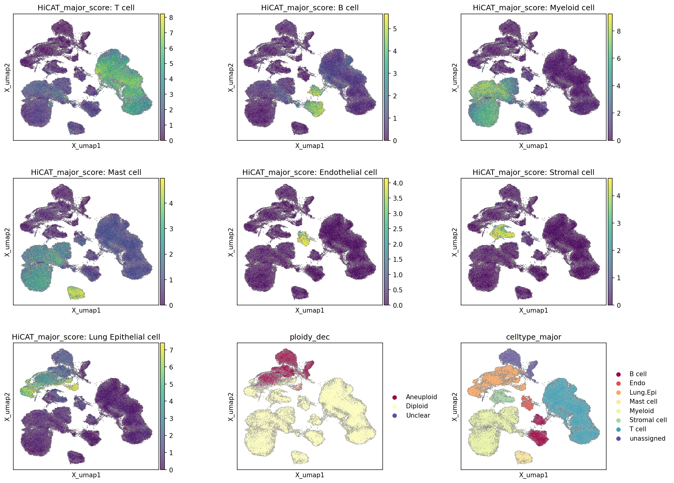
This analysis utilizes UMAP to visualize the distribution of major cell types based on their calculated expression scores (HiCAT_major_score), the inferred ploidy status (Diploid/Aneuploid), and the pre-assigned major cell type annotations (celltype_major) across 84,300 single cells from human lung tissue. The purpose is to assess the consistency of cell type identification, understand the spatial relationships between different cell populations, and identify potentially malignant cells based on their ploidy state within the embedding.
Visual Summary
The UMAP plots effectively delineate distinct cellular populations within the single-cell RNA-seq dataset.
- Cell Type Score Plots (HiCAT_major_score): Each of the seven plots (T cell, B cell, Myeloid cell, Mast cell, Endothelial cell, Stromal cell, Lung Epithelial cell) shows regions of high score (yellow/green) corresponding to specific clusters on the UMAP. These high-scoring regions for each cell type generally form well-separated clusters, indicating distinct gene expression signatures for each major cell type. For example, T cells show a large, well-defined cluster with high scores in the upper-right region. Lung Epithelial cells also show a prominent cluster in the upper-left, characterized by high scores.
- Ploidy Status Plot (ploidy_dec): This plot reveals a clear separation of Aneuploid (red) and Diploid (light yellow) cells. A significant population of Aneuploid cells primarily occupies the upper-left region of the UMAP, forming one or more distinct clusters. The majority of the remaining cells are identified as Diploid.
- Major Cell Type Annotation Plot (celltype_major): This plot displays the final assigned major cell types using a distinct color for each. It visually confirms the clustering patterns observed in the score plots, with T cells (teal) forming a large cluster in the upper-right, Myeloid cells (light green) and B cells (maroon) in the lower-right, and Lung Epithelial cells (light orange) prominently in the upper-left. Stromal cells (yellow-green) and Endothelial cells (red-orange) occupy central regions.
Biological Interpretation
- Robust Cell Type Identification: The strong concordance between the HiCAT_major_score plots and the celltype_major annotation plot demonstrates robust and consistent identification of major cell types. High scores for a specific cell type (e.g., T cell) consistently map to the same UMAP region that is ultimately labeled as that cell type in the celltype_major annotation. This provides confidence in the accuracy of the cell type assignments based on characteristic gene expression profiles.
- Malignant Cell Identification: A critical observation is the clear clustering of Aneuploid cells, which predominantly co-localize with the Lung Epithelial cell cluster in the upper-left region of the UMAP. Given that "Lung Epithelial cell" is specified as the "Tumor origin celltype" in the data context, this strongly suggests that these Aneuploid Lung Epithelial cells represent the malignant epithelial cell population within the tumor samples. Aneuploidy, or an abnormal number of chromosomes, is a well-established hallmark of cancer cells and is a strong indicator of malignancy PMID: 24209995.
- Immune and Stromal Microenvironment: The remaining Diploid cells comprise various immune (T cells, B cells, Myeloid cells, Mast cells) and stromal populations (Endothelial cells, Stromal cells). Their distribution on the UMAP indicates a diverse tumor microenvironment (TME) or lung tissue architecture. T cells form a large, distinct cluster, suggesting a significant immune infiltrate. Myeloid cells and B cells also form notable clusters, indicating their presence in the tissue.
- Spatial Segregation of Cell States: The UMAP embedding visually separates cells not only by their primary identity but also by physiological state, with aneuploidy creating a distinct separation for potential tumor cells from the host diploid cells.
Clinical or Translational Implications
- Identification of Tumor Cells: The clear delineation of Aneuploid Lung Epithelial cells provides a robust method for identifying and isolating malignant cells from the tumor microenvironment in this dataset. This is crucial for downstream analyses focused on tumor biology, resistance mechanisms, or novel therapeutic targets.
- Characterization of the Tumor Microenvironment: The successful identification and clustering of various immune and stromal cell types allow for detailed investigation of their roles in tumor progression, immune evasion, and response to therapy in lung cancer. Understanding the cellular composition and interactions within the TME is fundamental for developing effective immunotherapies and targeted treatments.
- Quality Control for Annotation: The consistency observed between HiCAT_major_score and celltype_major validates the quality of the cell type annotation pipeline, ensuring that subsequent functional analyses (e.g., DEG, GSEA) are performed on accurately identified cell populations.
3. Overall Celltype_subset Marker Expression Validation
[Analysis Visualization Results]...
Analysis Overview
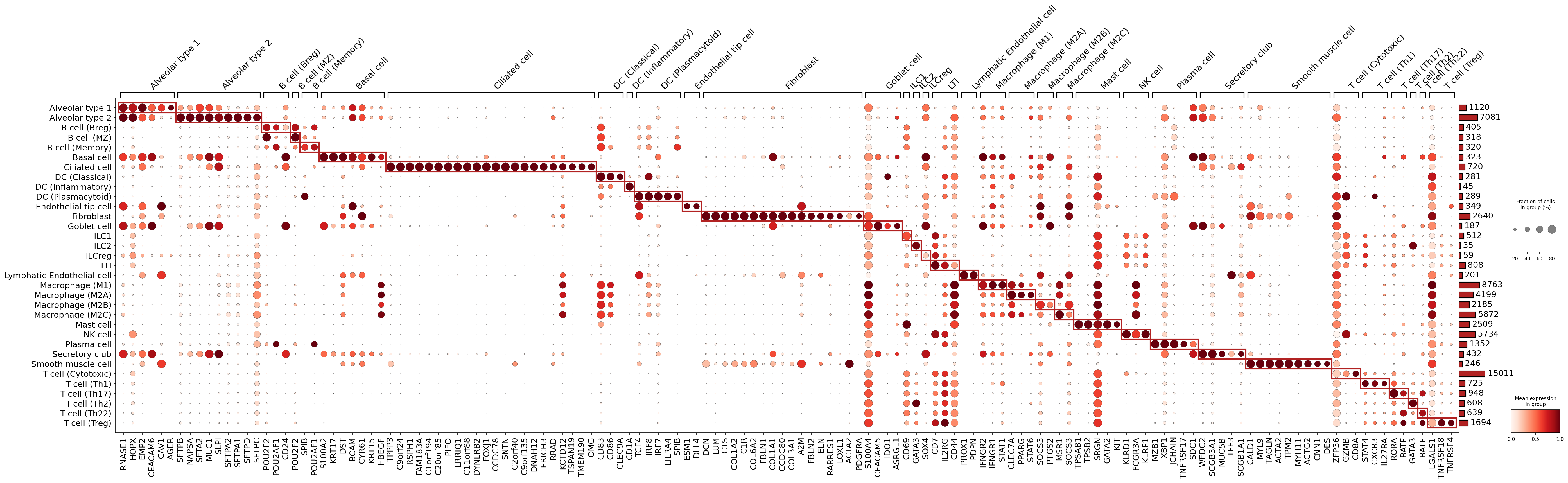
This analysis generates a dot plot visualizing the expression of marker genes across all identified celltype_subset populations from human lung single-cell RNA-seq data. The primary goal is to assess the specificity and enrichment of these markers to validate the quality and distinctness of the cell type annotations. Marker genes were selected based on their differential expression and surfaceome localization.
Visual Summary
The dot plot effectively illustrates the expression patterns of 140 marker genes across 39 distinct celltype_subset groups.
- Specificity and Enrichment: A prominent diagonal pattern of dark red, large circles, often enclosed by red boxes, indicates that numerous marker genes are highly and specifically expressed within their assigned cell type subsets. This pattern signifies strong segregation and distinct transcriptional identities for most cell populations.
- Expression Level and Prevalence: The color intensity of each dot corresponds to the mean expression level of the gene, while the dot size represents the fraction of cells within that group expressing the gene. High intensity (dark red) and large size (large circle) denote robust and widespread expression of a marker within a specific cell type.
- Cell Type Abundance: The bar chart on the right side of the plot shows the number of cells belonging to each celltype_subset, providing context for the robustness of marker detection and the relative representation of each cell type in the dataset.
- Shared Markers: While most markers show high specificity, some genes exhibit expression in closely related cell types (e.g., different macrophage or T cell subtypes), reflecting shared lineage or functional programs.
Biological Interpretation
The observed marker gene expression profiles largely align with established biological knowledge for human lung cell types, strongly supporting the accuracy and resolution of the celltype_subset annotations.
Lung Epithelial Cells
- Alveolar type 1 (AT1) cells are distinctly marked by *AGER* and *HOPX*, consistent with their role in gas exchange [GeneCards].
- Alveolar type 2 (AT2) cells show high expression of surfactant proteins such as *SFTPC* and *SFTPB*, critical for lung function [GeneCards].
- Basal cells are characterized by keratins like *KRT5* and *KRT15*, indicative of their progenitor function in airway repair [GeneCards].
- Ciliated cells express *FOXJ1* and *DNAH12*, key genes for cilia formation and function [GeneCards].
- Secretory club cells are identified by *SCGB1A1* and *SCGB3A1*, reflecting their role in airway protection [GeneCards].
Immune Cells
- Macrophage subtypes (M1, M2A, M2B, M2C) display canonical markers such as *CD68* (general macrophage marker) and distinct polarization markers like *CD80*/*CD86* for M1-like macrophages and *CD163* for M2-like macrophages [GeneCards], [GeneCards]. *SPP1* (Osteopontin) is notably expressed in Macrophage (M2B), consistent with its role in tumor microenvironment remodeling.
- Dendritic cells (DCs), including Classical DCs (marked by *CLEC9A*) and Plasmacytoid DCs (marked by *LILRA4*), exhibit distinct profiles, confirming their specialized immune functions [GeneCards].
- Plasma cells are clearly identified by markers like *JCHAIN*, *SDC1* (CD138), and *XBP1*, reflecting their antibody-secreting function [GeneCards].
- T cell subsets demonstrate specific markers, such as *CD8A* and *GZMB* for Cytotoxic T cells, *IL2RA* for T regulatory cells, and *RORC* for Th17 cells, which align with their diverse immune roles.
- NK cells are characterized by *KLRD1* and *FCGR3A*, consistent with their cytotoxic activity [GeneCards].
- Mast cells show high expression of *KIT* (CD117) and *TPSAB1*, key markers for their identification and function in allergic responses [GeneCards].
- ILC subsets (ILC1, ILC2, ILCreg, ILC3) also show distinguishing markers (e.g., *TBX21* for ILC1, *GATA3* for ILC2), supporting their distinct innate immune functions.
Stromal and Endothelial Cells
- Fibroblasts are marked by extracellular matrix components like *COL1A1*, *DCN*, and *LUM*, alongside *PDGFRA*, consistent with their structural and regulatory roles [GeneCards], [GeneCards].
- Smooth muscle cells express *ACTA2* and *TAGLN*, indicative of their contractile function.
- Endothelial cells (general and specialized subtypes like Lymphatic Endothelial cells) are identified by canonical markers such as *PECAM1* (CD31), *CDH5* (VE-cadherin), and specific markers like *PROX1* and *PDPN* for lymphatic endothelium [GeneCards].
Annotation Notes
The comprehensive marker expression dot plot serves as a strong validation of the celltype_subset annotations. The clear definition of most cell types by unique and biologically relevant marker genes provides high confidence in the cell assignments within this dataset. The find_cfg parameter surfaceome_only: True successfully identified external-facing markers that are particularly useful for distinguishing cell types and potentially relevant for cell-cell interaction studies or therapeutic targeting. While a few rare cell types, such as Ionocytes, show fewer highly specific surfaceome markers in this visualization, the overall clarity and specificity of the vast majority of cell type annotations are excellent. This robust annotation forms a solid foundation for subsequent analyses, including investigation of condition-specific changes or cell-cell interactions.
4. Copy Number Variation Analysis in Tumor-Origin and Unassigned Lung Cells
[Analysis Visualization Results]...
Analysis Overview
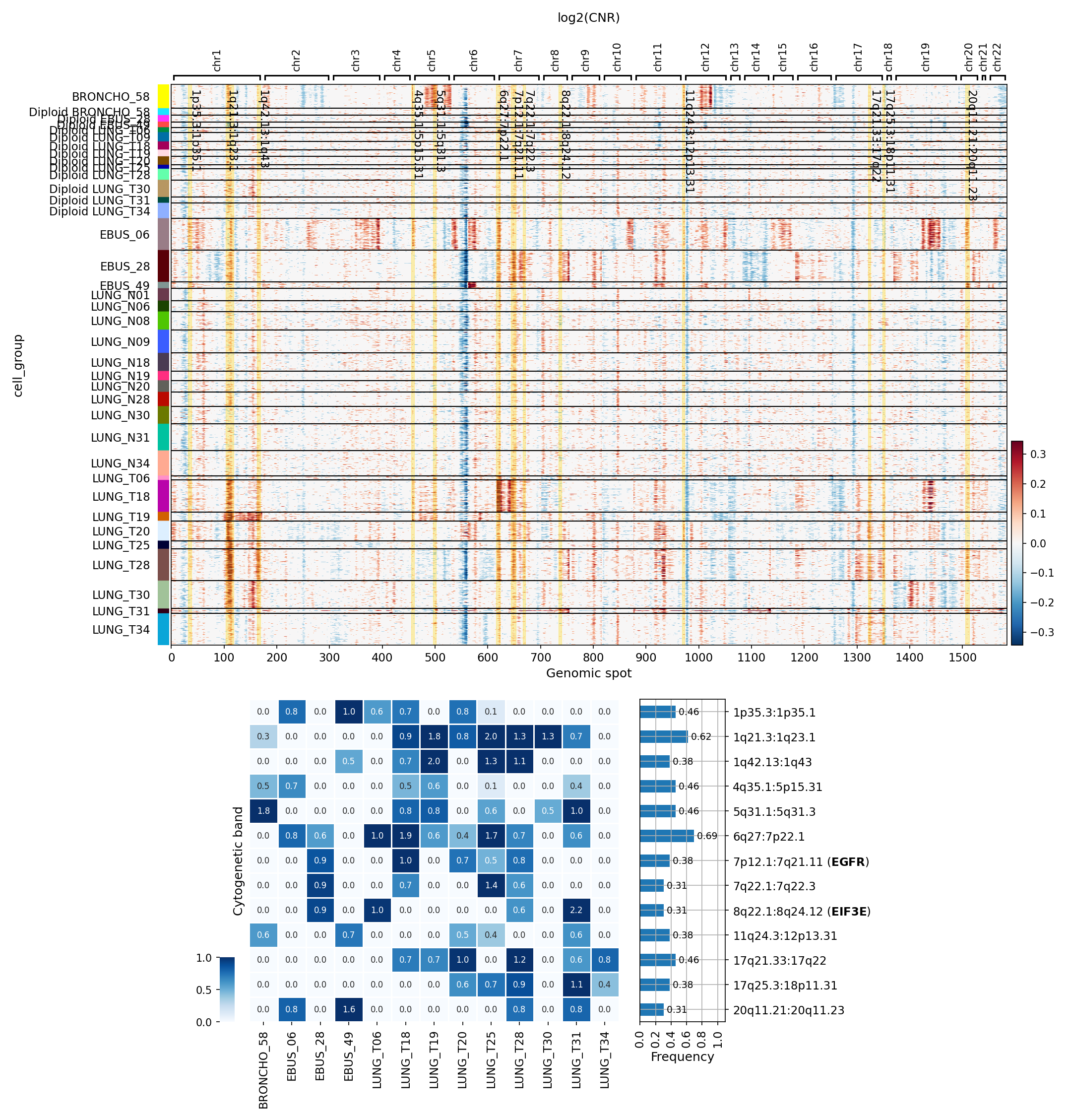
This analysis investigates copy number variations (CNVs) in tumor-origin cells (Lung Epithelial cells) and unassigned cells across various lung tissue samples, grouped by sample. The goal is to identify genomic aberrations characteristic of tumor cells and assess genomic instability. The visualization includes a heatmap displaying log2(CNR) (log2 Copy Number Ratio) values across genomic spots for each sample group, along with a summary heatmap highlighting the frequency of significantly amplified cytogenetic bands across samples.
Visual Summary
CNV Heatmap (log2(CNR))
The heatmap displays log2(CNR) values, where red indicates amplifications and blue indicates deletions. Genomic spots are ordered along the x-axis, representing chromosomes 1 through 22. Samples are grouped on the y-axis, with an explicit distinction made for samples predominantly classified as "Diploid" versus "Aneuploid" based on their ploidy inference.
- Diploid Samples: Samples prefixed with "Diploid" (e.g., BRONCHO_58, LUNG_06, LUNG_18, LUNG_T31, LUNG_T34) show minimal to no significant genomic aberrations, as expected for diploid cells. Their log2(CNR) values largely cluster around zero, indicating stable copy numbers.
- Aneuploid Samples: The majority of samples, particularly those not prefixed with "Diploid" (e.g., EBUS_06, LUNG_N01, LUNG_T06, LUNG_T19, LUNG_T28), display extensive and often recurrent CNVs. These samples exhibit widespread regions of both amplification (red) and deletion (blue).
- Recurrent Amplifications: Notable regions of amplification frequently observed across multiple aneuploid samples include segments on chromosome 1q, 7p, 8q, 17q, and 20q.
- Recurrent Deletions: Some deletions are also present, although less uniformly recurrent or pronounced in this heatmap (e.g., scattered blue regions on chr3p, chr9p).
- Inter-sample Heterogeneity: While common patterns exist, there is clear heterogeneity in the specific CNV profiles among different aneuploid samples, reflecting the unique genomic landscapes of individual tumors.
CNA Summary Heatmap (Amplification Frequency)
The summary heatmap quantifies the frequency of significant amplifications within specific cytogenetic bands across the aneuploid samples. Darker blue indicates higher frequency.
- Highly Frequent Amplifications: Several cytogenetic bands show high frequencies of amplification across the profiled samples. The most prominent include:
1q regions: 1p35.3:1p35.1, 1q21.3:1q23.1, 1q42:1q43
5q region: 5q31.1:5q31.3
- 7p/q regions: 7p12.1:7q21.11 (notably encompassing the *EGFR* gene) and 7q22.1:7q22.3
- 8q region: 8q22.1:8q24.12 (notably encompassing the *EIF3E* gene)
17q regions: 17q21.33:17q22 and 17q25.3:18p11.31
20q region: 20q11.21:20q11.23
- Key Driver Genes Highlighted: The summary explicitly highlights the amplification of *EGFR* on chromosome 7p12.1:7q21.11 and *EIF3E* on chromosome 8q22.1:8q24.12, indicating their frequent involvement in these tumor samples.
- Sample-specific enrichment: Some samples, such as LUNG_T18, LUNG_T19, LUNG_T20, LUNG_T28, and LUNG_T30, consistently show high frequencies of amplifications across multiple cancer-associated cytogenetic bands.
Biological Interpretation
The analysis focused on "Lung Epithelial cell," which is designated as the tumor-origin cell type in this dataset, along with "unassigned" cells. The observed CNVs primarily reflect genomic instability characteristic of these tumor-associated populations.
- Genomic Instability in Tumor Cells: The clear distinction between diploid and aneuploid samples confirms that a significant portion of the lung tissue samples harbor substantial genomic aberrations within the Lung Epithelial cell population. This genomic instability, often leading to aneuploidy and CNVs, is a hallmark of cancer and drives tumor evolution and heterogeneity.
- Recurrent Oncogenic Amplifications: The identification of recurrent amplifications in specific chromosomal regions points to the selection pressure for genes located within these regions, which often include oncogenes or genes that promote cell survival and proliferation.
- EGFR Amplification: The frequent amplification of the region containing *EGFR* (Epidermal Growth Factor Receptor) is particularly significant in lung cancer. *EGFR* is a well-established oncogene and driver mutation in non-small cell lung cancer (NSCLC), particularly in adenocarcinoma, which aligns with the "Lung" tissue context and "Tumor" conditions. Amplification of *EGFR* often leads to its overexpression and constitutive activation, promoting cell growth and survival. GeneCards: EGFR
- EIF3E Amplification: Amplification of the *EIF3E* (Eukaryotic Translation Initiation Factor 3 Subunit E) gene on 8q22.1:8q24.12 is also recurrent. *EIF3E* plays a crucial role in the initiation of protein synthesis and has been implicated in the pathogenesis and progression of various cancers, including lung cancer, often by promoting cell proliferation and inhibiting apoptosis. GeneCards: EIF3E
- Other Recurrent Regions: Amplifications in 1q, 5q, 17q, and 20q are also commonly observed in lung cancers and may harbor other oncogenes or regulatory elements contributing to tumor development and progression. For example, 1q is frequently amplified in various cancers and can contain genes involved in cell cycle regulation and proliferation.
- Ploidy and Tumor Progression: The ploidy status (ploidy_dec in obs) accurately reflects the observed CNV patterns. "Aneuploid" samples exhibit extensive CNVs, suggesting these are likely advanced or genetically unstable tumors, whereas "Diploid" samples show stable genomes. This validates the ploidy inference and categorizes samples based on their genomic landscape.
- Implications for "unassigned" cells: If "unassigned" cells within these samples contribute significantly to the aneuploid CNV signal, it suggests they may represent tumor cells that were not confidently assigned to a specific epithelial subtype, or other cell types that have acquired tumor-like genomic instability in the tumor microenvironment. Given the dominant signal from Lung Epithelial cells as the tumor origin, it's more probable that "unassigned" cells showing CNVs are indeed of cancerous origin.
Clinical or Translational Implications
- Targeted Therapy Stratification: The recurrent amplification of *EGFR* strongly suggests that a subset of these lung cancer patients could be candidates for EGFR tyrosine kinase inhibitor (TKI) therapies. Detecting such amplifications, along with mutations, is a standard practice in lung cancer diagnostics. PubMed Search: EGFR TKI Lung Cancer
- Prognostic Marker: The presence and extent of CNVs, especially those affecting known oncogenes, can serve as prognostic indicators, potentially correlating with tumor aggressiveness, recurrence risk, or overall survival in lung cancer patients.
- Identification of Novel Targets: The recurrent amplification of *EIF3E* and other regions warrants further investigation into their functional roles in lung cancer and their potential as novel therapeutic targets. Understanding the mechanisms by which these genes contribute to tumor biology could lead to the development of new drugs.
- Understanding Tumor Heterogeneity: The observed sample-to-sample variability in CNV patterns underscores the genetic heterogeneity of lung cancer. This heterogeneity can contribute to differential treatment responses and the development of resistance. Monitoring CNV profiles could aid in personalized treatment strategies.
- Validation of Ploidy Inference: The CNV heatmap visually confirms the accuracy of the ploidy_dec annotation, providing a robust classification of samples based on their genomic stability.
5. CNV-based UMAP of Single-Cell Data Reveals Distinct Malignant and Non-Malignant Cell Populations
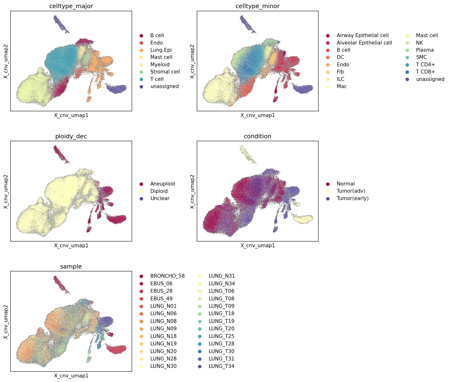
[Analysis Visualization Results]...
Analysis Overview
This analysis visualizes single-cell RNA-seq data projected onto a Uniform Manifold Approximation and Projection (UMAP) embedding, where the embedding space (X_cnv_umap) is primarily driven by estimated Copy Number Variation (CNV) patterns. Cells are colored according to their major cell type, minor cell type, ploidy status, disease condition, and sample origin. The aim is to understand how cellular identities, malignancy status (aneuploidy), and disease conditions manifest in the CNV landscape.
Visual Summary
The UMAP visualization, based on CNV estimates, reveals a clear segregation of cell populations into distinct clusters driven by their chromosomal integrity.
Cell Type Distribution (celltype_major, celltype_minor):
- A large, relatively dense cluster on the left side of the UMAP is predominantly composed of immune cells (T cells, B cells, Myeloid cells) and stromal cells (Fibroblasts, Endothelial cells).
- Conversely, several more dispersed but distinct clusters, particularly in the mid-right and top regions of the UMAP, are largely populated by Lung Epithelial cells (specifically Alveolar Epithelial cells and Airway Epithelial cells).
Ploidy Status (ploidy_dec):
- The large cluster on the left, identified as immune and stromal cells, is overwhelmingly classified as 'Diploid' (pale yellow). This aligns with the expected ploidy of non-malignant cells.
- In stark contrast, the Lung Epithelial cell clusters on the right and top of the UMAP are almost exclusively labeled as 'Aneuploid' (dark red). A small number of 'Unclear' cells are scattered throughout.
Condition Association (condition):
- The 'Diploid' cluster (left) shows a strong enrichment for 'Normal' condition cells (dark red), as expected for healthy tissue components.
- The 'Aneuploid' clusters (right/top), corresponding to Lung Epithelial cells, are heavily dominated by cells from 'Tumor(early)' (pale yellow) and 'Tumor(adv)' (dark blue) conditions. There is considerable overlap between early and advanced tumor cells within the aneuploid space.
Sample Origin (sample):
- The 'Diploid' cluster contains cells from a wide variety of samples, including both normal (LUNG_N series) and tumor (LUNG_T series, BRONCHO, EBUS) samples, representing the common presence of non-malignant cells across all biopsy types.
- The 'Aneuploid' clusters display clear sample-specific contributions, with certain tumor samples (e.g., BRONCHO_58, EBUS_06, LUNG_T19, LUNG_T20, LUNG_T25, LUNG_T28, LUNG_T30, LUNG_T31, LUNG_T34) being highly enriched within these regions. This indicates distinct, patient-specific CNV profiles among the malignant cell populations.
Biological Interpretation
The CNV-driven UMAP provides compelling biological insights into the cellular landscape of lung tissue under normal and cancerous conditions.
- Discrimination of Malignant vs. Non-Malignant Cells: The most striking finding is the robust separation of cells based on their CNV profiles, which effectively segregates non-malignant cells (immune, stromal) from malignant ones. Non-malignant cells maintain a diploid genomic state, clustering together due to their relatively stable copy number profiles. In contrast, tumor-derived Lung Epithelial cells exhibit widespread aneuploidy, forming distinct clusters reflecting their unique and often complex chromosomal alterations. This confirms that CNV is a powerful feature for distinguishing tumor cells, especially the 'Lung Epithelial cell' population which is defined as the tumor origin cell type in the data context.
- Evidence of Tumor Cell Aneuploidy: The strong co-localization of 'Lung Epithelial cell' clusters with 'Aneuploid' labels and cells from 'Tumor(early)' and 'Tumor(adv)' conditions provides strong evidence that these specific epithelial cells are the neoplastic cells driving the lung cancer. Aneuploidy is a hallmark of cancer, contributing to tumor heterogeneity and adaptation.
- Reference: Hanahan, D., & Weinberg, R. A. (2011). Hallmarks of cancer: the next generation. *Cell*, 144(5), 646–674. PubMed Search: "Hallmarks of cancer aneuploidy"
- Tumor Heterogeneity: The observation that multiple distinct aneuploid Lung Epithelial cell clusters exist, and that different tumor samples contribute uniquely to these regions, underscores the genomic heterogeneity inherent in lung cancer. This heterogeneity can exist both between patients (inter-patient) and potentially within a single tumor (intra-tumor), influencing disease progression and therapeutic responses.
- Identification of Tumor-Associated Microenvironment: The presence of diploid immune and stromal cells within samples from tumor conditions highlights the significant infiltration of the tumor microenvironment (TME) by non-malignant cells. These cells, despite being from tumor biopsies, retain their normal diploid state and distinct CNV profiles from the malignant cells.
Clinical or Translational Implications
- Improved Tumor Cell Identification: This CNV-based analysis offers a powerful method to confidently identify and isolate true tumor cells from single-cell transcriptomics data, even in highly heterogeneous tumor samples. This is critical for downstream analyses aimed at understanding tumor-specific biology, identifying therapeutic targets, or developing diagnostic biomarkers without contamination from non-malignant cells.
- Insights into Tumor Evolution and Progression: The distinct aneuploid patterns observed across different tumor samples and conditions (early vs. advanced) suggest that CNVs could serve as markers for tumor progression or predictors of clinical outcome. Analyzing the specific CNV patterns within these clusters could uncover common or unique genomic alterations associated with different stages or subtypes of lung cancer.
- Personalized Medicine Approaches: The high degree of inter-patient CNV heterogeneity observed suggests that therapeutic strategies might need to be tailored to the specific genomic landscape of each patient's tumor. This supports the rationale for precision oncology, where individual tumor CNV profiles could guide treatment selection.
6. Minor Cell Type Population Analysis Across Lung Cancer Stages
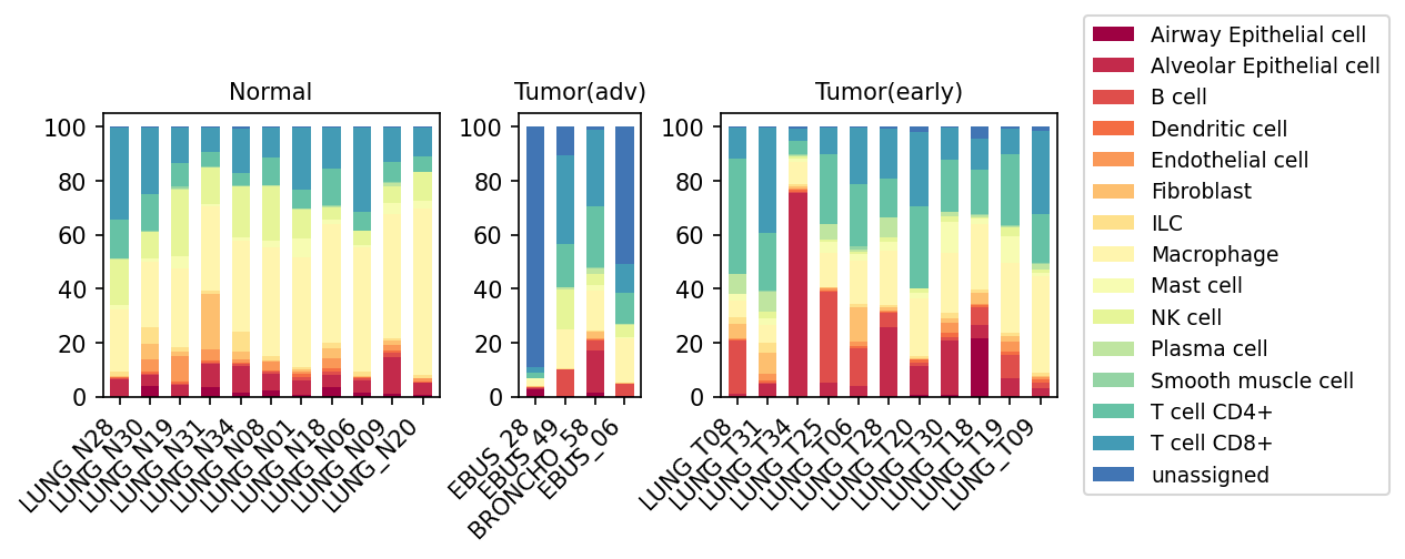
[Analysis Visualization Results]...
Analysis Overview
This analysis presents the proportional distribution of minor cell types within individual samples, grouped by condition: Normal, Tumor (early), and Tumor (advanced). The stacked bar plots allow for a visual comparison of cellular composition shifts in the lung tissue microenvironment as it progresses from a normal state to early and advanced tumor stages. Each bar represents a single sample, and the segments within the bar show the relative abundance of different minor cell types.
Visual Summary
- Normal Lung Tissue Composition: Normal lung samples exhibit a diverse cellular landscape, predominantly characterized by T cells (both CD4+ and CD8+), Macrophages, and ILCs. Alveolar and Airway Epithelial cells, along with Stromal cells (Fibroblasts, Endothelial cells, Smooth muscle cells), are consistently present in smaller proportions.
- Early Tumor Stage (Tumor(early)): A notable characteristic in early tumor samples is the significant increase in Alveolar Epithelial cell proportions in several samples (e.g., LUNG_T25, LUNG_T06, LUNG_T28, LUNG_T18, LUNG_T19). Concurrently, there appears to be a relative reduction in the overall proportion of T cells compared to normal tissue. Macrophages maintain a substantial presence in this stage.
- Advanced Tumor Stage (Tumor(adv)): In advanced tumor samples, the proportion of "unassigned" cells shows a pronounced increase in some samples (e.g., EBRUS_28, EBRUS_49), which could indicate highly aberrant or dedifferentiated cell populations that are difficult to classify precisely. T cell proportions, particularly CD8+ T cells, appear to be further diminished or remain low. Macrophages continue to be a significant component of the microenvironment. The epithelial cell fraction, while still present, does not consistently dominate as starkly as in some early tumor samples.
- Immune Cell Trends: Across the progression from normal to advanced tumor, there is a general trend of decreasing T cell (CD4+ and CD8+) proportions, especially CD8+ T cells. Conversely, Macrophages consistently represent a substantial fraction of the cellular landscape in both early and advanced tumor conditions.
Biological Interpretation
The observed shifts in cell type proportions provide crucial insights into the evolving tumor microenvironment (TME) during lung cancer progression.
- Epithelial Neoplasia: The dramatic increase in Alveolar Epithelial cells in the "Tumor(early)" stage directly reflects the neoplastic proliferation originating from Lung Epithelial cells, which are identified as the "Tumor origin celltype." This expansion of malignant epithelial cells is a hallmark of early tumor development.
- Immune Evasion and Suppression: The reduction in both CD4+ and CD8+ T cell populations, particularly the cytotoxic CD8+ T cells, from normal to tumor conditions (and potentially further in advanced stages), suggests the establishment of an immunosuppressive TME. This T cell exclusion or anergy is a common mechanism by which tumors evade anti-tumor immunity.
- Macrophages in Tumor Progression: The sustained or potentially increased presence of Macrophages throughout tumor development (both early and advanced stages) highlights their critical and often pro-tumorigenic role. Macrophages are highly plastic and can be polarized towards M2-like phenotypes, which contribute to tumor growth, angiogenesis, immune suppression, and metastasis.
- Stromal Remodeling: The consistent presence of Fibroblasts and Endothelial cells in tumor samples underscores the dynamic interplay between tumor cells and the surrounding stroma. Fibroblasts can differentiate into Cancer-Associated Fibroblasts (CAFs) that contribute to extracellular matrix remodeling and create a supportive, pro-tumorigenic niche. Endothelial cells are essential for angiogenesis, supplying nutrients and oxygen to the growing tumor.
- Unassigned Cell Population in Advanced Cancer: The emergence of a significant "unassigned" cell population in advanced tumors may indicate cellular states that deviate significantly from known reference cell types. These could represent highly dedifferentiated tumor cells, stem-like cancer cells, or novel, uncharacterized cell types that arise in aggressive disease. This also points to a potential challenge in precisely annotating highly heterogeneous or aberrant cellular states in advanced disease.
Clinical or Translational Implications
The findings from this cell population analysis hold several clinical and translational implications for lung cancer:
- Biomarker Identification and Prognosis: The relative abundance of specific cell types, such as the high proportion of Alveolar Epithelial cells in early tumors or the reduced T cell infiltrates, could serve as potential diagnostic or prognostic biomarkers. Changes in the T cell-to-Macrophage ratio could also be prognostic indicators.
- Immunotherapy Strategies: The observed reduction in T cell populations in tumor stages suggests that strategies aimed at restoring or enhancing anti-tumor T cell immunity (e.g., immune checkpoint inhibitors, adoptive cell therapies) are critical. Furthermore, targeting tumor-associated macrophages (TAMs) to reprogram their pro-tumorigenic functions could be a complementary therapeutic approach.
- Targeting the Tumor Microenvironment: The persistent presence of Fibroblasts and Endothelial cells in the TME suggests opportunities for developing therapies that target stromal components, such as anti-angiogenic agents or CAF-modulating drugs, to impede tumor growth and metastasis.
- Understanding Disease Heterogeneity: The variability in cell type proportions among individual samples, even within the same condition, highlights the inherent heterogeneity of lung cancer, which needs to be considered for personalized treatment approaches.
- Refining Cell Annotation: The presence of a substantial "unassigned" population in advanced tumors indicates a need for deeper characterization of these cell states, which could reveal novel therapeutic targets or mechanisms of resistance specific to advanced disease.
7. Lung Cancer Microenvironment: T Cell and Innate Lymphoid Cell Subset Shifts
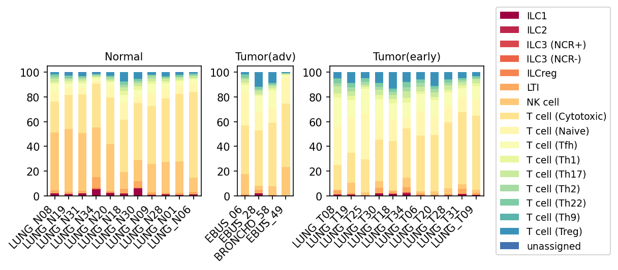
[Analysis Visualization Results]...
Analysis Overview
This analysis investigates the proportional distribution of various T cell subsets, Natural Killer (NK) cells, and Innate Lymphoid Cells (ILCs) within the broader "T cell" major cell type category across normal lung tissue and lung tumors at early and advanced stages. The stacked bar plot visualizes how the composition of these immune cell populations changes across different samples grouped by their condition, providing insights into immune remodeling during lung cancer progression.
Visual Summary
The stacked bar plot effectively illustrates the relative proportions of 18 distinct lymphoid cell subsets, including various T cell types, NK cells, and ILCs, within each sample. Samples are grouped into three conditions: Normal, Tumor (adv), and Tumor (early).
- Normal Samples: These samples generally show a high proportion of NK cells (light orange) and T cell (Cytotoxic) (light yellow), often collectively accounting for over 70% of the depicted lymphoid cells. T cell (Naive) also contributes a notable fraction. Other T helper subsets (Th1, Th17, Th2, Th22, Th9, Tfh) and ILCs (red/maroon shades) are present in smaller, more consistent proportions.
- Tumor (adv) Samples: A marked shift in composition is observed in advanced tumor samples. There is a noticeable decrease in the proportion of NK cells and, in several samples (e.g., EBUS_06, EBUS_28), a reduction in T cell (Cytotoxic) cells compared to normal tissue. Concurrently, a relative increase in T cell (Naive) is often apparent. T cell (Treg) (dark blue) proportions, while still minor, show a subtle increase in some advanced tumor samples. Significant inter-sample heterogeneity is also evident, with some samples (e.g., EBUS_58) retaining higher proportions of cytotoxic cells.
- Tumor (early) Samples: Early tumor samples exhibit an intermediate phenotype. The proportions of NK cells and T cell (Cytotoxic) are generally reduced compared to normal, but this reduction appears less pronounced than in many advanced tumor samples. T cell (Naive) populations often show a relative increase. Similar to advanced tumors, there's a trend for a slight increase in T cell (Treg) cells, although they remain a small fraction of the total lymphoid cells. Sample-to-sample variability is also present in this group.
Biological Interpretation
The observed shifts in lymphoid cell populations highlight dynamic changes in the lung tumor immune microenvironment, impacting both innate and adaptive immunity.
- Compromised Anti-Tumor Immunity: The consistent reduction in NK cells and T cell (Cytotoxic) proportions from normal to early and advanced tumor stages suggests a progressive weakening of the host's anti-tumor immune response. NK cells are critical for innate tumor surveillance and direct killing of cancer cells, while cytotoxic T cells are the primary effectors of adaptive anti-tumor immunity. Their depletion or reduced representation can contribute to immune evasion and tumor progression. [PubMed search: NK cell dysfunction lung cancer, Cytotoxic T cell exhaustion cancer]
- Emergence of Immunosuppression: The relative increase in T cell (Treg) populations in tumor conditions, albeit subtle, is a critical biological finding. Tregs are potent immunosuppressive cells that can inhibit the activity of cytotoxic T cells and other anti-tumor immune cells, fostering a tolerogenic environment that promotes tumor growth. [GeneCards: FOXP3]
- Recruitment of Naive T Cells: The increased proportion of T cell (Naive) in tumor samples could indicate continuous recruitment of undifferentiated T cells into the tumor microenvironment without adequate activation or differentiation into effector cells. This might reflect a failure of effective anti-tumor immune priming within the tumor-draining lymph nodes or an inability to overcome immunosuppressive signals locally.
- Disease Progression Signature: The gradual shift in immune cell composition from normal to early to advanced tumor stages suggests a progressive immune remodeling process. Early-stage tumors may initiate changes that become more pronounced and detrimental to anti-tumor immunity in advanced disease.
- Limited Role of Other Subsets: Most other T helper subsets and ILC populations maintain relatively low proportions across all conditions, suggesting that while they have specific immune roles, their proportional contribution to the overall lymphoid cell changes depicted here is less dominant compared to NK cells, cytotoxic T cells, naive T cells, and Tregs.
Clinical or Translational Implications
The findings from this analysis carry several important clinical and translational implications for lung cancer:
- Biomarker for Disease Progression: The ratios and absolute numbers of NK cells, cytotoxic T cells, and Tregs could serve as valuable biomarkers for monitoring lung cancer progression or predicting patient prognosis. A lower NK/cytotoxic T cell ratio or a higher Treg proportion might correlate with worse outcomes.
- Immunotherapeutic Strategies: The identified changes suggest potential targets for immunotherapy. Strategies aimed at restoring NK cell activity, enhancing cytotoxic T cell function (e.g., through checkpoint blockade or adoptive cell therapies), or depleting/inhibiting Treg cells could be explored to re-invigorate anti-tumor immunity in lung cancer patients. [PubMed search: Immunotherapy lung cancer NK cells, Treg targeted therapy cancer]
- Personalized Treatment Approaches: The observed inter-patient heterogeneity, particularly in the advanced tumor group (e.g., sample EBUS_58 showing a more "normal-like" cytotoxic profile), underscores the need for personalized medicine. Immune profiling of individual tumors could help stratify patients who might respond differently to various immunotherapeutic interventions.
- Stage-Specific Intervention Design: Given the progressive nature of immune remodeling, immunotherapeutic interventions might be optimized for specific disease stages. Early interventions could aim to prevent immune evasion, while advanced stages might require more robust immune activation strategies.
8. Changes in T Cell Subset Proportions Across Lung Tumor Progression
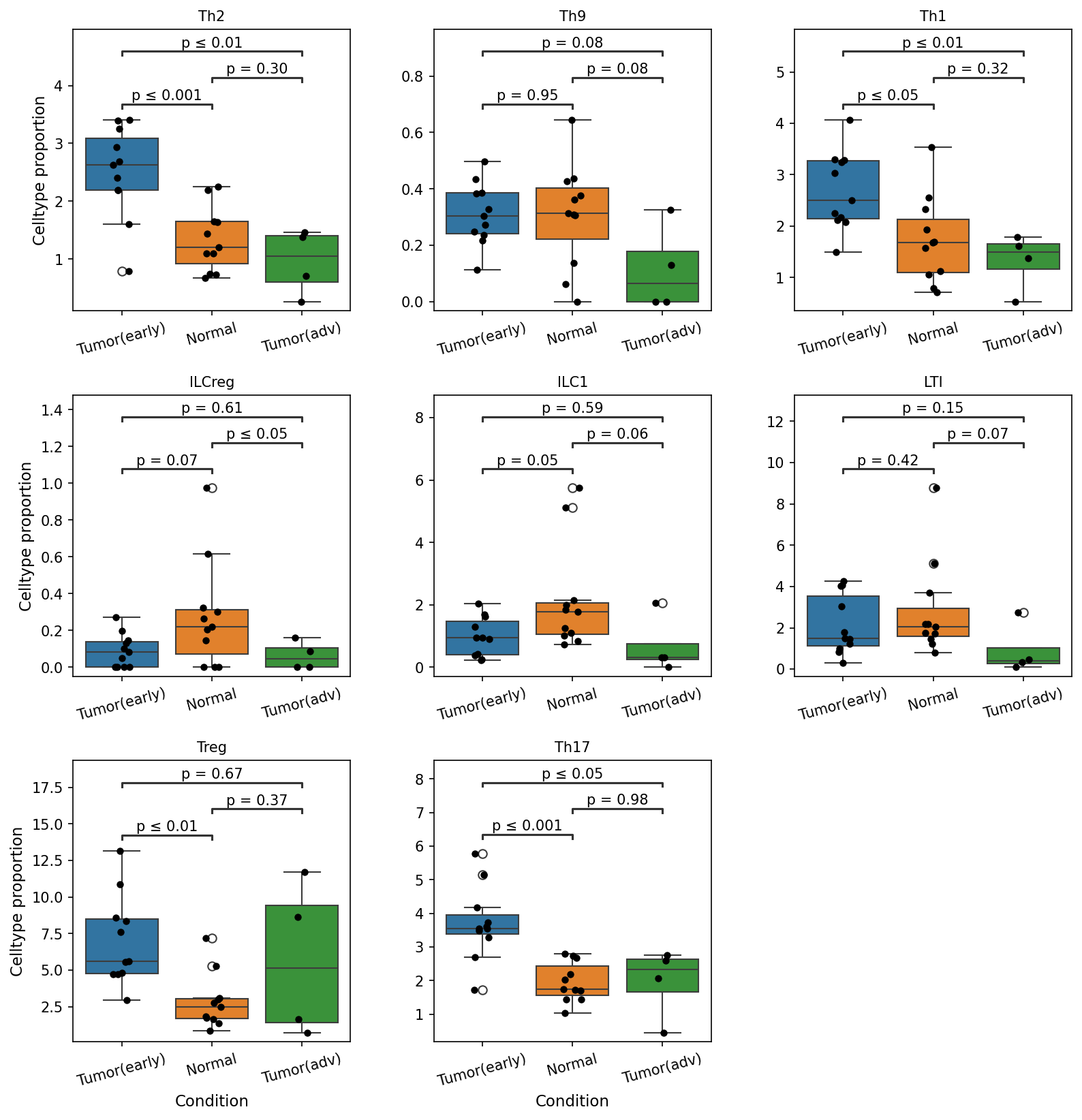
[Analysis Visualization Results]...
Analysis Overview
This analysis investigates the proportions of various T cell and Innate Lymphoid Cell (ILC) subsets within the overall cell population across different conditions: "Tumor(early)", "Normal", and "Tumor(adv)" (advanced tumor). The goal is to identify statistically significant shifts in the immune landscape during lung tumor development and progression. Box plots visualize the cell type proportion for each condition, with p-values indicating significant differences between group comparisons (p-value cutoff: 0.1).
Visual Summary
The box plots display the proportion of several T cell and ILC subsets across three conditions: Tumor(early), Normal, and Tumor(adv). Key observations regarding statistically significant differences (p ≤ 0.1) are:
- Th2 cells: Significantly elevated in "Tumor(early)" compared to "Normal" (p ≤ 0.001) and "Tumor(adv)" (p ≤ 0.01). Proportions are comparable between "Normal" and "Tumor(adv)" (p = 0.30).
- Th1 cells: Significantly higher in "Tumor(early)" compared to "Normal" (p ≤ 0.05) and "Tumor(adv)" (p ≤ 0.01). No significant difference between "Normal" and "Tumor(adv)" (p = 0.32).
- Treg cells: Show a significantly higher proportion in "Tumor(early)" compared to "Normal" (p ≤ 0.01). No significant differences were observed between other comparisons.
- Th17 cells: Significantly increased in "Tumor(early)" when compared to "Normal" (p ≤ 0.001) and "Tumor(adv)" (p ≤ 0.05). "Normal" and "Tumor(adv)" show similar proportions (p = 0.98).
- ILCreg cells: The proportion is significantly lower in "Tumor(adv)" compared to "Normal" (p ≤ 0.05). A trend of lower proportion is also observed in "Tumor(early)" compared to "Normal" (p = 0.07).
- ILC1 cells: Proportions are significantly higher in "Tumor(early)" than in "Tumor(adv)" (p = 0.05). Similarly, "Normal" tissues exhibit higher ILC1 proportions compared to "Tumor(adv)" (p = 0.06).
- LTI cells: The proportion of LTI cells is significantly lower in "Tumor(adv)" compared to "Normal" (p = 0.07).
- Th9 cells: No statistically significant differences in proportions were observed between any of the conditions (p > 0.1).
Biological Interpretation
The analysis reveals dynamic shifts in the T cell and ILC landscape during lung cancer progression.
- Immune Activation and Regulation in Early Tumor: The elevated proportions of Th1, Th2, Th17, and Treg cells in early-stage tumors suggest a robust, albeit potentially dysregulated, immune response to the nascent tumor.
- Th1 cells are typically associated with anti-tumor immunity through IFN-$\gamma$ production, promoting cytotoxic T cell responses PubMed Search: Th1 cells cancer immunity. Their increase in early tumor might indicate an initial host defense.
- Th2 cells are often linked to allergic responses and can sometimes promote tumor growth or immune evasion through mechanisms like M2 macrophage polarization PubMed Search: Th2 cells tumor progression.
- Th17 cells are pro-inflammatory and their role in cancer is complex, being either pro- or anti-tumorigenic depending on the tumor microenvironment and cytokine milieu GeneCards: RORC (Th17 marker). Their increase suggests an inflammatory environment.
- Treg cells are immunosuppressive, crucial for maintaining immune tolerance and preventing autoimmunity. Their elevated presence in early tumors could reflect an early mechanism by the tumor to establish an immunosuppressive environment, counteracting effector T cell responses UniProt: FOXP3 (Treg marker).
- Immune Suppression and Dysregulation in Advanced Tumor: Conversely, advanced tumors ("Tumor(adv)") show significantly reduced proportions of several critical immune subsets when compared to early tumors or normal tissue:
- The significant decrease in Th1, Th2, Th17, ILC1, ILCreg, and LTI cells in advanced tumors suggests a profound shift towards an immunosuppressed or exhausted state in the tumor microenvironment as the disease progresses.
- ILC1 cells are important producers of IFN-$\gamma$ and contribute to anti-tumor immunity PubMed Search: ILC1 cancer. Their reduction in advanced tumors implies a weakened innate immune surveillance.
- ILCreg cells and LTI cells are involved in immune regulation and lymphoid tissue organization, respectively PubMed Search: ILCreg immunity, PubMed Search: LTI cells lymphoid organ development. Their decrease in advanced disease could contribute to a disorganized or impaired immune architecture that favors tumor escape.
Clinical or Translational Implications
The observed shifts in T cell and ILC subset proportions between early-stage, normal, and advanced lung tumors provide valuable insights with potential clinical and translational relevance:
- Biomarkers for Disease Progression: The distinct immune profiles, particularly the reduction of various T cell and ILC subsets from early to advanced tumor stages, could serve as prognostic biomarkers to stratify patients based on their disease stage and immune status.
- Immunotherapy Strategies: These findings highlight the dynamic nature of the anti-tumor immune response.
- In early-stage disease, where both effector (Th1, Th17) and regulatory (Treg, Th2) populations are elevated, therapeutic interventions might focus on tipping the balance towards effective anti-tumor immunity by enhancing effector function or dampening regulatory mechanisms.
- In advanced disease, the diminished presence of key immune subsets like Th1, ILC1, ILCreg, and LTI suggests a more immunosuppressed microenvironment. Immunotherapies in this setting might need to focus on reactivating exhausted immune cells, recruiting new effector cells, or restoring immune architecture.
- Understanding Immune Evasion: The reduction of anti-tumor immune cells (e.g., Th1, ILC1) and regulatory cells (ILCreg, LTI) in advanced tumors implies that the tumor may have successfully evaded or suppressed host immunity. Further investigation into the mechanisms driving these population changes could uncover novel targets for intervention.
9. Macrophage Subset Population Shifts in Lung Cancer Progression
[Analysis Visualization Results]...
Analysis Overview
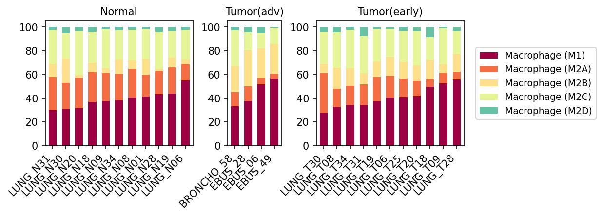
This analysis investigates the relative proportions of different macrophage subsets (M1, M2A, M2B, M2C, M2D) within the total macrophage population across various lung tissue samples, categorized into Normal, Tumor (early), and Tumor (advanced) conditions. The stacked bar plot visualizes the distribution of these macrophage phenotypes for each individual sample, providing insight into the changes in macrophage polarization profiles during lung cancer progression.
Visual Summary
The stacked bar plot presents the relative contribution of five distinct macrophage subsets (M1, M2A, M2B, M2C, M2D) to the total macrophage population in each sample, grouped by clinical condition: Normal, Tumor (advanced), and Tumor (early).
- Normal Samples: In normal lung tissue samples, Macrophage (M1) (maroon) and Macrophage (M2A) (orange) represent the most substantial proportions of the macrophage population. M1 macrophages consistently constitute a large fraction, often ranging from 30% to over 50% in individual normal samples. M2B (light yellow), M2C (light green/yellow), and M2D (teal/light blue) subsets are present but typically in smaller, less variable proportions.
- Tumor (early) Samples: In early-stage tumor samples, there is a general trend of a slight decrease in the M1 macrophage proportion compared to some normal samples, while the M2A (orange) and M2B (light yellow) populations appear to be relatively increased. The overall shift towards M2-like phenotypes seems to initiate at this stage, though M1 macrophages still maintain a significant presence in many samples.
- Tumor (advanced) Samples: Samples from advanced tumors show a more pronounced shift in macrophage polarization. The proportion of Macrophage (M1) (maroon) generally decreases, becoming less dominant compared to normal and early-stage tumor tissues, often falling below 40%. Concomitantly, the proportions of Macrophage (M2A) (orange) and Macrophage (M2B) (light yellow) notably increase, often becoming the major or co-dominant macrophage subsets. The M2C and M2D populations remain relatively minor across all conditions but show some variability.
In summary, there is a clear trend towards a reduced proportion of M1 macrophages and an increased prevalence of M2A and M2B macrophages as lung cancer progresses from normal to early and then advanced stages.
Biological Interpretation
Macrophages are highly plastic immune cells that can differentiate into various functional subsets, broadly categorized as M1 (pro-inflammatory, anti-tumor) and M2 (anti-inflammatory, pro-tumor, tissue repair) phenotypes. This analysis reveals a significant shift in macrophage polarization dynamics within the lung tumor microenvironment (TME).
- M1 Macrophages (Maroon): These macrophages are typically activated by pro-inflammatory signals (e.g., IFN-γ, LPS) and are characterized by their ability to produce pro-inflammatory cytokines, present antigens effectively, and exert direct anti-tumor effects. The observed decrease in M1 macrophage proportions in tumor conditions, especially in advanced stages, suggests a suppression of anti-tumor immunity in the TME.
- M2 Macrophages (Orange, Light Yellow, Light Green/Yellow, Teal/Light Blue): M2 macrophages are a heterogeneous group involved in tissue remodeling, angiogenesis, immune suppression, and promoting tumor growth and metastasis.
- M2A (Orange) and M2B (Light Yellow): The increasing proportions of M2A and M2B macrophages in tumor conditions, particularly in advanced tumors, are notable. M2A macrophages are typically associated with wound healing and allergic responses, pathways that can be hijacked by tumors to promote growth and fibrosis. M2B macrophages are known for their immunoregulatory functions. Their enrichment suggests a microenvironment that supports tumor progression by fostering immune suppression and tissue restructuring.
- M2C and M2D: While less abundant, these subsets are also generally associated with immune suppression (M2C) and pro-angiogenic/tumor-promoting functions (M2D, often referred to as Tumor-Associated Macrophages or TAMs, especially in the context of TLR activation). Their presence, even at low levels, contributes to the overall immune-suppressive TME.
The observed shift from a relatively balanced M1/M2A profile in normal lung tissue to a dominance of M2A and M2B macrophages in advanced lung tumors indicates a significant re-programming of the macrophage population. This polarization towards M2-like phenotypes contributes to immune evasion by suppressing anti-tumor T cell responses, promoting angiogenesis, and facilitating tumor invasion and metastasis, which are hallmarks of cancer progression.
Clinical or Translational Implications
The distinct shifts in macrophage subset populations observed in lung cancer have several important clinical and translational implications:
- Biomarker Potential: The ratio of M1 to M2 macrophage subsets, or the specific abundance of M2A and M2B, could serve as a prognostic biomarker for lung cancer progression or response to therapy. Patients with a higher M2A/M2B prevalence might have a worse prognosis or be less responsive to immunotherapies that rely on effective anti-tumor immune responses.
- Therapeutic Targets: The observed M2 polarization presents an attractive therapeutic target. Strategies aimed at:
- Re-polarizing M2-like macrophages to M1 phenotypes: This could enhance anti-tumor immunity and potentially synergize with existing immunotherapies like checkpoint inhibitors.
- Inhibiting M2 macrophage functions: Targeting specific signaling pathways or molecules crucial for M2 polarization or their pro-tumor functions (e.g., angiogenesis, immune suppression) could impede tumor growth and metastasis.
- Depleting M2 macrophages: Selective depletion of M2-like macrophages could be another approach, though careful consideration of off-target effects would be necessary.
- Disease Monitoring: Monitoring macrophage polarization profiles in tumor biopsies or liquid biopsies could provide insights into disease progression and aid in selecting appropriate treatment strategies for individual patients with lung cancer.
10. Macrophage Subset Population Shifts Across Lung Cancer Progression
[Analysis Visualization Results]...
Analysis Overview
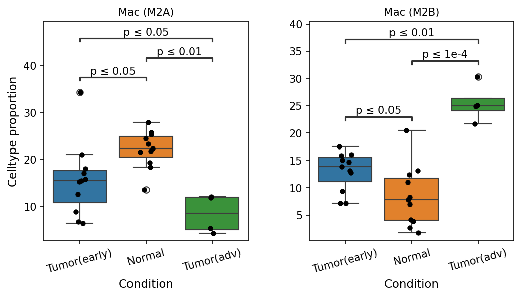
This analysis presents box plots illustrating the statistically significant differences in the proportion of specific macrophage subsets, Mac (M2A) and Mac (M2B), across different conditions: "Tumor(early)", "Normal", and "Tumor(adv)". The objective is to identify how the abundance of these macrophage populations changes during lung cancer development and progression, using "Normal" tissue as a reference.
Visual Summary
The box plots show the celltype proportion on the y-axis for each macrophage subset, grouped by condition on the x-axis, with individual sample data points overlaid. Statistical significance is indicated by p-values.
Mac (M2A) Population Dynamics:
- In Normal lung tissue, Mac (M2A) cells exhibit the highest median proportion, around 23-24%.
- Their proportion significantly decreases in Tumor(early) stage (p ≤ 0.05) compared to Normal tissue.
- The decrease becomes even more pronounced and statistically significant in Tumor(adv) stage, where their proportion is significantly lower than both Normal (p ≤ 0.01) and Tumor(early) (p ≤ 0.05) conditions. This indicates a progressive decline of Mac (M2A) macrophages with advancing tumor stage.
Mac (M2B) Population Dynamics:
- Conversely, Mac (M2B) cells show the lowest median proportion in Normal lung tissue, around 8%.
- Their proportion significantly increases in Tumor(early) stage (p ≤ 0.05) compared to Normal tissue.
- This increase is further amplified and becomes highly significant in Tumor(adv) stage. Mac (M2B) proportions in Tumor(adv) are significantly higher than both Normal (p ≤ 0.01) and Tumor(early) (p ≤ 1e-4) conditions, suggesting a substantial expansion of Mac (M2B) macrophages during tumor progression.
Biological Interpretation
Macrophages are highly plastic immune cells that can adopt various functional states, broadly categorized into M1 (pro-inflammatory, anti-tumor) and M2 (anti-inflammatory, pro-tumor, tissue repair) phenotypes. The M2 category itself is heterogeneous, encompassing subtypes like M2a, M2b, M2c, and M2d, each with distinct activation signals and functions.
The observed shifts in Mac (M2A) and Mac (M2B) populations suggest a critical re-polarization of the tumor-associated macrophage (TAM) landscape during lung cancer progression:
- Decline of Mac (M2A) Macrophages: M2A macrophages are typically activated by IL-4 and IL-13, and are involved in allergic responses and anti-parasitic immunity. They also play roles in wound healing and tissue repair. In some contexts, M2A macrophages have been implicated in anti-tumor responses or in maintaining tissue homeostasis. Their significant reduction from normal to early, and further to advanced tumor stages, suggests a loss of these specific macrophage functions, which could contribute to a less controlled tumor microenvironment or a diminished capacity for tissue repair/homeostasis https://www.ncbi.nlm.nih.gov/pmc/articles/PMC7966779/.
- Expansion of Mac (M2B) Macrophages: M2B macrophages are activated by immune complexes (e.g., via FcγR) in combination with TLR agonists (like LPS) or IL-1R ligands. They are characterized by the production of both pro-inflammatory cytokines (ee.g., TNFα, IL-1β, IL-6) and anti-inflammatory mediators (e.g., IL-10), making their role complex and context-dependent. In the context of tumor progression, their significant increase from normal to early, and dramatically to advanced tumor stages, suggests a potentially prominent pro-tumorigenic role. This expansion may contribute to chronic inflammation, immune evasion, angiogenesis, and metastasis, all of which are hallmarks of cancer progression https://www.ncbi.nlm.nih.gov/pmc/articles/PMC8909890/ https://www.ncbi.nlm.nih.gov/pmc/articles/PMC6678665/.
The inverse relationship between Mac (M2A) and Mac (M2B) populations strongly points towards a dynamic shift in macrophage polarization within the lung tumor microenvironment, favoring phenotypes that likely support tumor growth and progression as the disease advances. This "re-education" of macrophages towards pro-tumorigenic states is a well-established mechanism in various cancers, including lung cancer.
Clinical or Translational Implications
The observed shifts in macrophage subsets have significant clinical and translational implications for lung cancer:
- Biomarker Potential: The proportions of Mac (M2A) and Mac (M2B) could serve as potential biomarkers for lung cancer diagnosis, staging, or prognosis. A higher Mac (M2B) to Mac (M2A) ratio, particularly in advanced stages, might indicate a more aggressive disease state or poorer patient outcome.
- Therapeutic Targeting: The predominant increase of Mac (M2B) macrophages in advanced lung tumors suggests that targeting these specific pro-tumorigenic macrophage subsets could be a viable therapeutic strategy. This could involve approaches to:
- Inhibit the recruitment or survival of M2B macrophages.
- Reprogram M2B macrophages towards anti-tumor M1-like phenotypes.
- Block their pro-tumorigenic functions (e.g., cytokine production, immune suppression).
- Immunotherapy Enhancement: Understanding the precise roles of different macrophage subsets can help in designing more effective immunotherapies. For instance, modulating TAM polarization could potentially enhance the efficacy of checkpoint inhibitors or other immune-modulating drugs in lung cancer patients https://www.ncbi.nlm.nih.gov/pmc/articles/PMC7000806/.
- Disease Monitoring: Monitoring the proportions of these macrophage subsets over time in patients could provide insights into disease progression or response to treatment.
11. Ploidy Population Analysis of Tumor-Origin and Unassigned Cells Across Lung Conditions
[Analysis Visualization Results]...
Analysis Overview
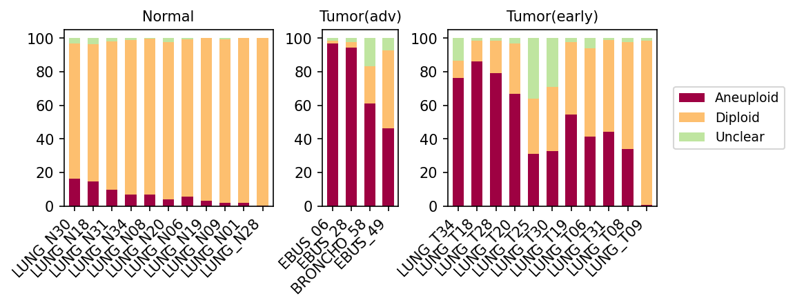
This analysis investigates the ploidy status (Aneuploid, Diploid, or Unclear) within lung epithelial cells and unassigned cell populations, comparing normal lung tissue, early-stage lung tumors, and advanced-stage lung tumors. The plot_celltype_population tool was used to visualize the proportional distribution of these ploidy states across individual samples within each condition. This provides insight into chromosomal aberrations, a key characteristic of cancer, in the presumptive tumor-initiating cells and other unclassified cells.
Visual Summary
The bar plot displays the percentage of Aneuploid (burgundy), Diploid (orange), and Unclear (light green) cells for each sample, grouped by condition: Normal, Tumor(adv), and Tumor(early).
- Normal Samples: In normal lung tissue samples, the vast majority of "Lung Epithelial cells" and "unassigned" cells are Diploid (orange bars dominating, typically >85%). Aneuploid cells (burgundy) constitute a very minor fraction, generally less than 15%, and "Unclear" cells are negligible. This is the expected baseline for healthy tissue.
- Tumor(adv) Samples: Advanced tumor samples show a striking shift towards Aneuploidy. Most samples (e.g., EBUS_06, EBUS_28) exhibit a very high proportion of Aneuploid cells, often exceeding 90%. While some samples (BRONCHO_58, EBUS_49) retain a noticeable diploid population (around 20-40%), Aneuploidy remains the dominant ploidy state in all advanced tumor samples.
- Tumor(early) Samples: Early-stage tumor samples present a more heterogeneous picture compared to advanced tumors but consistently show a higher prevalence of Aneuploid cells than normal tissue. Several early tumor samples display a very high proportion of Aneuploid cells (>70-80%, e.g., LUNG_T34, LUNG_T18, LUNG_T28), similar to observations in advanced tumors. However, other early tumor samples show a more balanced distribution between Aneuploid and Diploid cells, with Diploid cells still forming a substantial fraction (e.g., LUNG_T25, LUNG_T30, LUNG_T19, LUNG_T06, LUNG_T31, LUNG_T08, LUNG_T09). The "Unclear" category also appears slightly more prominent in some early tumor samples compared to normal or advanced.
Biological Interpretation
The observed ploidy patterns strongly corroborate the known association between aneuploidy and cancer progression, particularly in cells of tumor origin.
- Aneuploidy as a Hallmark of Cancer: The substantial increase in aneuploid cells in tumor samples, especially in advanced stages, underscores aneuploidy as a critical hallmark of malignancy. Aneuploidy, the presence of an abnormal number of chromosomes, drives genomic instability and contributes to tumor heterogeneity and evolution [1].
- Tumor-Origin Cell Transformation: The analysis specifically targets "Lung Epithelial cell," which is identified as the "Tumor origin celltype" in the data context. The shift from predominantly diploid cells in normal lung epithelial populations to highly aneuploid populations in tumor conditions directly reflects the malignant transformation and proliferation of these cells.
- Progression-Associated Aneuploidy: There is a clear trend of increasing aneuploidy from normal tissue to early-stage tumors, and further to advanced-stage tumors. This suggests that the accumulation of chromosomal aberrations is an ongoing process during tumor development and progression, potentially contributing to more aggressive phenotypes in advanced disease.
- Heterogeneity in Early Tumors: The variability in aneuploidy levels among early-stage tumor samples could reflect the intrinsic genomic heterogeneity of early tumors. Some tumors may acquire significant chromosomal abnormalities early on, while others might develop them more gradually, influencing their biological behavior and potential response to treatment.
- Role of Unassigned Cells: The inclusion of "unassigned" cells in this analysis is insightful. If these "unassigned" cells also display high levels of aneuploidy in tumor samples, it could indicate that they are likely highly aberrant tumor cells that were difficult to classify precisely using standard markers, or other transformed cells within the tumor microenvironment. This highlights the challenge of cell type annotation in complex tumor ecosystems.
Clinical or Translational Implications
The findings have several potential clinical implications:
- Diagnostic and Prognostic Biomarker: The prevalence of aneuploidy in lung epithelial cells could serve as a robust diagnostic marker for detecting malignancy, particularly when assessing lung biopsies. Furthermore, the degree of aneuploidy, especially in tumor-origin cells, may have prognostic value, with higher levels potentially indicating more aggressive disease or advanced staging [2].
- Monitoring Disease Progression: Tracking the ploidy status of tumor-origin cells, perhaps through liquid biopsies or serial tissue sampling, could offer a method for monitoring disease progression and evaluating treatment efficacy. A decrease in aneuploid cell populations following therapy might indicate a positive response.
- Therapeutic Targeting: Understanding the mechanisms that drive aneuploidy and genomic instability in lung cancer could pave the way for developing targeted therapies that exploit these vulnerabilities. Therapeutic strategies aimed at restoring euploidy or inhibiting aneuploidy-driven cancer cell fitness are areas of active research [3].
---
References:
- Aneuploidy as a Hallmark of Cancer:
- Aneuploidy as a Prognostic Marker:
- Targeting Aneuploidy in Cancer Therapy:
12. Condition-Specific Cell-Cell Interaction Patterns in Lung Tissue
[Analysis Visualization Results]...
Analysis Overview
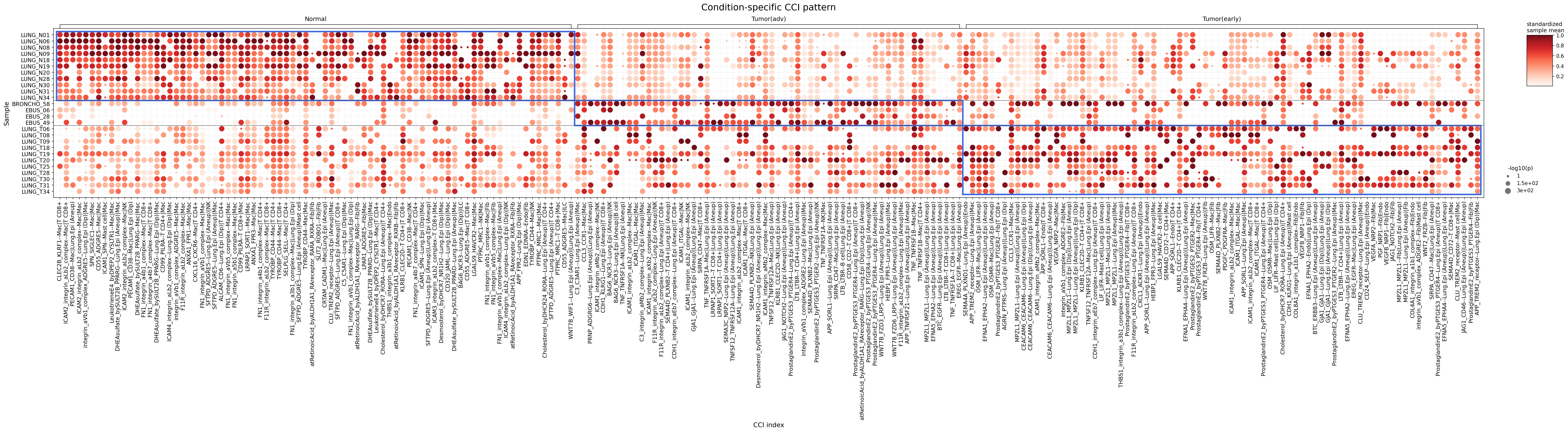
This analysis investigates condition-specific cell-cell interaction (CCI) patterns involving Lung Epithelial cells, Fibroblasts, Macrophages, and T cells (CD4+ and CD8+) across Normal, Tumor(early), and Tumor(adv) conditions in human lung single-cell RNA-seq data. The dot plot visualizes the strength (color intensity) and statistical significance (dot size) of up to 80 prominent cell-cell interactions per condition, allowing for a comparison of the tumor microenvironment's communicative landscape at different disease stages. Notably, the analysis distinguishes interactions based on the inferred ploidy status (Aneuploid or Diploid) of Lung Epithelial cells, Fibroblasts, and Macrophages, offering deeper insight into the role of malignant or reprogrammed cells.
Visual Summary
The visualization clearly delineates distinct CCI profiles across the Normal, Tumor(early), and Tumor(adv) conditions.
- Normal Condition: Displays a specific set of strong and significant interactions, predominantly involving diploid (Dip) cell populations such as Mac(Dip), Fib(Dip), and Lung.Epi(Dip) interacting with each other and with T cells. Key interactions observed include those mediated by Integrin (e.g., ICAM2, FN1) and PECAM1, suggesting a role in maintaining tissue homeostasis and cell adhesion.
- Tumor(early) Condition: Shows a shift from the normal pattern. Some normal interactions appear attenuated, while new, distinct interactions emerge. Notably, interactions involving aneuploid (AneuP) cell populations, such as Lung.Epi(AneuP) and Mac(AneuP), become more prominent. Ligand-receptor pairs like WNT7B-FZD6_LRP5 between Lung.Epi(AneuP) and Fibroblast, and certain Integrin-mediated interactions with Lung.Epi(AneuP), begin to gain strength.
- Tumor(adv) Condition: Exhibits a profoundly altered and often intensified CCI landscape compared to early tumor and normal conditions. A very large number of highly significant and strong interactions are observed. Interactions predominantly involve aneuploid cells (Lung.Epi(AneuP), Mac(AneuP), Fib(AneuP)). A striking feature is the widespread and strong activity of ProstaglandinE2 signaling (via PTGES3-PTGER4/PTGER2/PTGER3) involving Lung Epithelial cells and Macrophages (both Aneuploid and Diploid), and Fibroblasts. Other significant interactions include TNF-TNFRSF1A and various Integrin-mediated pairs involving Lung.Epi(AneuP) with T cells or Macrophages, and interactions between Lung.Epi(AneuP) and Endothelial cells.
Biological Interpretation
The observed changes in CCI patterns reflect a dynamic evolution of the lung tumor microenvironment (TME) during cancer progression.
- Normal Tissue Homeostasis: In the normal lung, the strong integrin-mediated interactions (e.g., ICAM2-integrin_aLb2_complex, FN1-integrin complexes) and PECAM1 interactions likely contribute to maintaining tissue integrity, cell-matrix adhesion, and healthy immune surveillance involving macrophages and T cells PubMed search for integrins cell adhesion lung.
- Emergence of Pro-tumorigenic Signaling in Early Stages: The shift towards interactions involving aneuploid cells in Tumor(early) suggests that early malignant transformation instigates specific communication changes. The appearance of WNT signaling (e.g., WNT7B-FZD6_LRP5 between Lung.Epi(AneuP) and Fibroblast) is notable, as WNT pathways are critical for development and frequently dysregulated in cancer, promoting cell proliferation and survival PubMed search for WNT signaling lung cancer.
- Advanced Tumor Microenvironment Remodeling: The advanced tumor stage is characterized by a highly active and distinct CCI network, driven primarily by interactions involving aneuploid tumor-origin Lung Epithelial cells.
- Prostaglandin E2 (PGE2) Signaling: The striking prevalence and strength of ProstaglandinE2 signaling in Tumor(adv), particularly involving Lung.Epi(AneuP) and Mac(AneuP)/Fibroblasts, is a critical observation. PGE2 is a potent lipid mediator known to promote tumor growth, angiogenesis, and immunosuppression within the TME by influencing various immune cells, including macrophages GeneCards: PTGES3 PubMed search for PGE2 immunosuppression cancer. This suggests a strong pro-tumorigenic and immune-evasive environment.
- Inflammatory and Immune Modulation: Increased TNF-TNFRSF1A interactions between Lung.Epi(AneuP) and T cells/Macrophages indicate an intensified inflammatory milieu, which can have dual effects (anti-tumor or pro-tumor) depending on context, but often contributes to chronic inflammation that fuels cancer progression and immune escape in advanced stages GeneCards: TNFRSF1A.
- Stromal Reprogramming: The sustained and evolving interactions between Lung Epithelial cells and Fibroblasts (many of which are likely reprogrammed into Cancer-Associated Fibroblasts, CAFs) through WNT and Integrin pathways highlight the active role of the stroma in supporting tumor growth and invasion.
- Immune Cell Recruitment and Dysfunction: The altered interactions involving Macrophages (especially Aneuploid ones) and T cells suggest recruitment, polarization (e.g., M2-like macrophages), and functional suppression of immune cells within the advanced TME.
Clinical or Translational Implications
The analysis of condition-specific CCI patterns offers valuable clinical and translational insights for lung cancer.
- Biomarkers for Disease Progression: Specific CCI pairs that exhibit significant changes between normal, early, and advanced tumor stages could serve as novel biomarkers for early detection, prognosis, or monitoring disease progression in lung cancer patients.
- Therapeutic Targets in the Tumor Microenvironment: The strong and prevalent ProstaglandinE2 signaling in advanced tumors represents a particularly attractive therapeutic target. Inhibiting this pathway could disrupt critical pro-tumorigenic and immunosuppressive feedback loops within the TME, potentially enhancing anti-tumor immune responses or directly inhibiting tumor growth PubMed search for PGE2 pathway inhibitors cancer therapy. Other pathways, such as WNT and TNF-TNFRSF1A, also warrant investigation as potential targets for modulating the TME.
- Understanding Immune Evasion: The intricate communication involving aneuploid tumor cells, macrophages, and T cells in advanced cancer highlights mechanisms of immune evasion. Targeting specific ligand-receptor interactions could help reprogram the immune microenvironment to be more hostile to the tumor.
- Stratification of Patients: The distinct CCI patterns, particularly those distinguishing early from advanced tumors, could aid in stratifying patients for personalized treatment approaches based on their specific TME characteristics.
13. Condition-Specific Cell-Cell Interaction Analysis in Lung Tissue
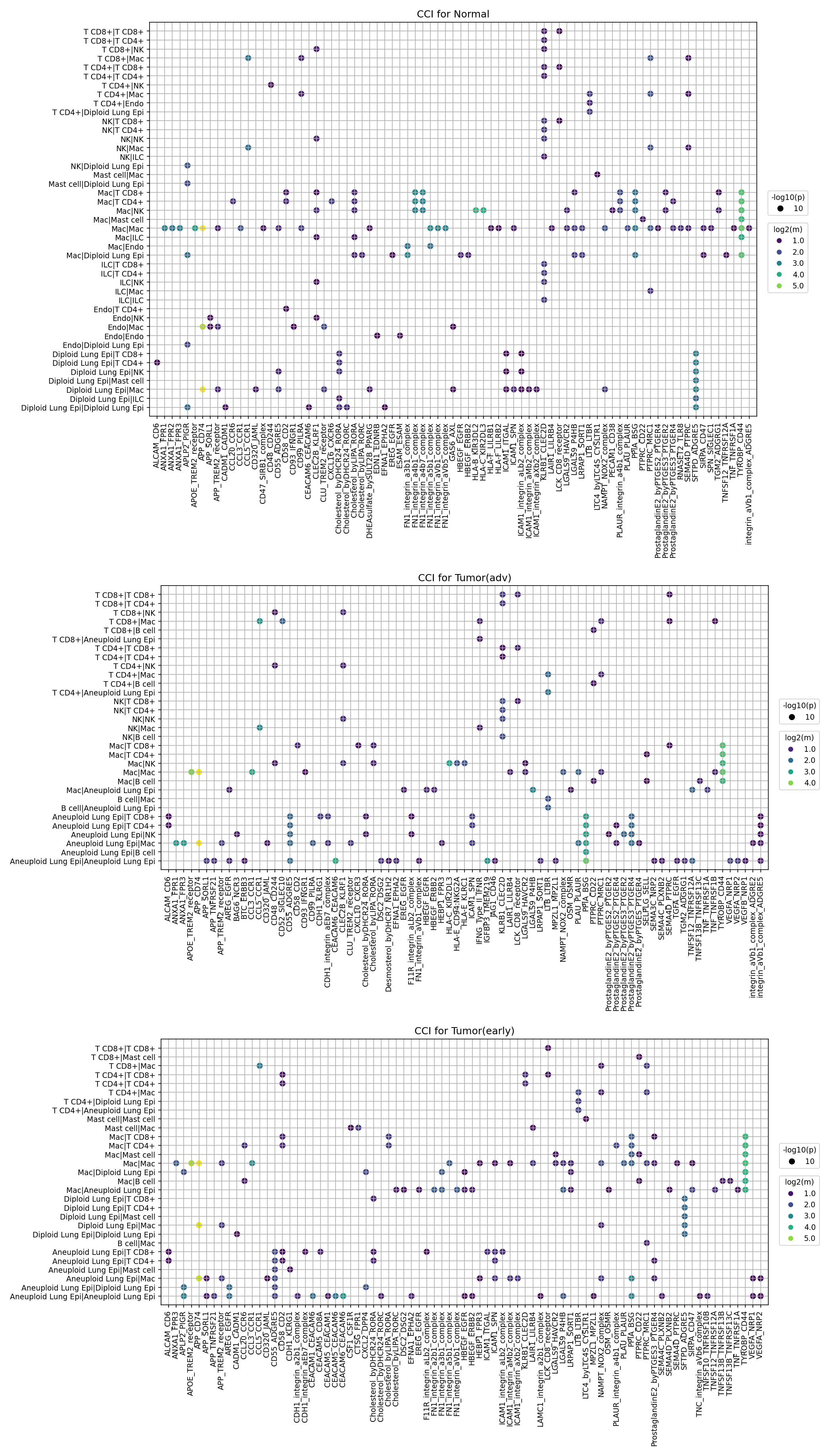
[Analysis Visualization Results]...
Analysis Overview
This analysis investigates cell-cell interactions (CCIs) across different conditions—Normal, Tumor(early), and Tumor(adv)—within lung single-cell RNA sequencing data, utilizing CellPhoneDB. The plot_cci_dots tool visually represents these interactions, where the size of each dot indicates the statistical significance of the interaction (-log10(p-value)), and the color represents the mean expression level (log2(mean)) of the ligand-receptor pair. This approach highlights how cellular communication networks evolve with tumor progression, focusing on ligand-receptor biology relevant to disease mechanisms and potential therapeutic interventions.
Visual Summary
The three dot plots display the top 80 most significant cell-cell interactions for Normal, Tumor(early), and Tumor(adv) conditions, respectively.
- Overall Interaction Pattern: A clear trend emerges from Normal to Tumor(adv) conditions. The Normal tissue exhibits a relatively sparser network of interactions, with fewer highly significant (larger dots) and less intensely expressed (brighter colors) ligand-receptor pairs. In contrast, Tumor(early) shows an increase in both the number and intensity of interactions, a trend that becomes profoundly amplified in Tumor(adv) where a dense network of strong and highly significant interactions is observed.
Cell Type Involvement
- Aneuploid Lung Epithelial Cells: Notably, interactions involving "Aneuploid Lung Epi" cells (which represent the likely tumor-initiating or tumor cells given the "Lung Epithelial cell" tumor origin and ploidy_dec annotation) are significantly more prevalent and robust in both tumor conditions, especially in Tumor(adv). These cells engage in extensive communication with various immune and stromal cell types, as well as with other Aneuploid Lung Epithelial cells.
- Macrophages (Mac): Macrophages are consistently highly interactive across all conditions but show an increased number and strength of interactions with Aneuploid Lung Epithelial cells and T cells in tumor environments.
- T Cells (CD8+, CD4+): T cells also participate in numerous interactions, with shifts in their communication patterns observed in tumor contexts, particularly in their engagement with macrophages and aneuploid epithelial cells.
- Other Stromal/Immune Cells: Endothelial cells, Fibroblasts, and B cells show condition-specific interactions, contributing to the evolving tumor microenvironment.
Key Ligand-Receptor Pairs
- Integrin Complexes: Interactions involving Fibronectin (FN1) and Tenascin C (TNC) with various integrin complexes (e.g., FN1_integrin_a5b1_complex, FN1_integrin_avb1_complex, TNC-integrin_avb1_complex) are markedly upregulated and more significant in Tumor(adv) compared to Normal. These are prominent in interactions between Aneuploid Lung Epithelial cells, Macrophages, and Fibroblasts.
- SPP1-Integrin Axis: The SPP1-integrin interactions (e.g., SPP1-integrin_avb1_complex, SPP1-integrin_avb3_complex) are particularly strong and widespread in Tumor(adv), especially involving Macrophages and Aneuploid Lung Epithelial cells.
- VEGF Axis: VEGFA-NRP2, VEGFA-VEGFR1, and VEGFA-VEGFR2 interactions become more pronounced in tumor conditions, indicating increased angiogenesis.
- TNF Superfamily: Interactions involving various members of the TNF superfamily (e.g., TNFSF10-TNFRSF10B, TNFSF12-TNFRSF12A, TNFSF13-TNFRSF13B) show increased activity in tumor conditions, often involving immune cells and epithelial cells.
- CXCL12-CXCR4: This chemokine axis appears more active in tumor stages, contributing to immune cell trafficking and tumor progression.
Biological Interpretation
The observed shifts in cell-cell interaction patterns from Normal to early and advanced lung tumors provide critical insights into the evolving tumor microenvironment (TME) and mechanisms of tumor progression.
- Tumor Cell-Centric Microenvironment Remodeling: The pronounced increase in interactions involving "Aneuploid Lung Epi" cells in tumor stages underscores their central role in actively shaping the TME. These cells, likely representing malignant epithelial cells, establish intricate communication networks with immune cells (Macrophages, T cells) and stromal cells (Fibroblasts, Endothelial cells), orchestrating processes critical for tumor growth, invasion, and immune evasion. Their self-interactions (Aneuploid Lung Epi|Aneuploid Lung Epi) also become more prominent, potentially indicating enhanced tumor cell cohesion or communication facilitating collective invasion.
- Extracellular Matrix (ECM) Remodeling and Invasion: The dramatic upregulation of integrin-mediated interactions involving Fibronectin (FN1), Tenascin C (TNC), and Secreted Phosphoprotein 1 (SPP1) in advanced tumors highlights extensive ECM remodeling. These interactions are crucial for:
- Cell Adhesion and Migration: Integrins mediate cell-ECM and cell-cell adhesion, influencing cell migration and invasion, key steps in metastasis. [Ref: Integrins in cancer: functional crosstalk with growth factors and cytokines. Annu Rev Pathol. 2018;13:427-463. PubMed ID: 29096956]
- Pro-tumorigenic Signaling: SPP1 (also known as Osteopontin), an ECM protein, interacts with integrins to promote tumor cell survival, proliferation, angiogenesis, and metastasis. Its strong presence in interactions with Macrophages and Aneuploid Lung Epithelial cells suggests a significant role in tumor progression and immune modulation within the TME. [Ref: SPP1 (Osteopontin) in Cancer: Functions, Signaling Pathways, and Therapeutic Opportunities. Cell. 2022 Mar 3;185(5):761-782. PubMed ID: 35245417]
- Angiogenesis and Vascular Remodeling: The increased activity of the VEGFA-VEGFR/NRP2 axis in tumor conditions directly indicates enhanced angiogenesis. Tumor cells and stromal cells secrete VEGFA, stimulating endothelial cells to form new blood vessels that supply oxygen and nutrients essential for tumor growth and metastasis. [Ref: VEGF and its receptors in cancer: therapeutic implications. J Clin Oncol. 2007 Apr 10;25(11):1377-85. PubMed ID: 17409214]
- Immune Evasion and Modulation:
- Macrophage Reprogramming: The extensive interactions between Macrophages and Aneuploid Lung Epithelial cells (and other immune cells) suggest macrophage reprogramming within the TME. Macrophages, particularly tumor-associated macrophages (TAMs), can adopt pro-tumorigenic phenotypes (e.g., M2-like) that promote immune suppression, angiogenesis, and matrix remodeling through various ligand-receptor interactions, including those involving integrins and TNF superfamily members. [Ref: Tumor-Associated Macrophages: From Biological Functions to Clinical Implications. Cancers (Basel). 2019 Jun 14;11(6):826. PubMed ID: 31213794]
- Immune Checkpoint & Inflammation: Interactions involving the TNF superfamily (e.g., TNFSF10/TRAIL, TNFSF12/TWEAK, TNFSF13/APRIL) can mediate cell death, inflammation, and immune cell activation or suppression. Their differential activity highlights evolving immune responses and potential immune evasion strategies in the tumor microenvironment. The CXCL12-CXCR4 axis can also contribute to immune suppression by recruiting regulatory T cells or myeloid-derived suppressor cells. [Ref: The CXCL12/CXCR4 chemokine axis in cancer: from biology to new therapeutic strategies. Cancer Metastasis Rev. 2018 Dec;37(4):681-696. PubMed ID: 30413988]
Clinical or Translational Implications
The identified condition-specific cell-cell interactions offer compelling targets for therapeutic intervention and potential biomarkers in lung cancer.
- Therapeutic Target Prioritization:
- Integrin and SPP1 Inhibition: The highly activated SPP1-integrin and FN1/TNC-integrin axes in advanced tumors present promising therapeutic targets. Inhibitors blocking specific integrin receptors (e.g., αvβ3, αvβ1) or targeting SPP1 could disrupt tumor cell adhesion, migration, and survival, as well as reduce pro-tumorigenic macrophage functions. [Ref: Integrin inhibitors in oncology. Cancer Res. 2010 Sep 15;70(18):7004-12. PubMed ID: 20847113]
- Anti-Angiogenic Therapies: The prominence of VEGFA-VEGFR/NRP2 interactions strongly supports the continued relevance of anti-VEGF therapies (e.g., bevacizumab) or next-generation angiogenesis inhibitors in lung cancer, particularly for advanced stages.
- CXCL12-CXCR4 Axis Blockade: Given its role in tumor growth, metastasis, and immune evasion, targeting the CXCL12-CXCR4 axis could be beneficial in inhibiting tumor progression and potentially enhancing the efficacy of immunotherapies.
- Biomarkers for Prognosis and Response: Specific clusters of highly interactive cell pairs or ligand-receptor activities identified in Tumor(adv) could serve as prognostic biomarkers for disease progression or indicators of resistance to current therapies. For example, high levels of SPP1-integrin interactions might predict aggressive disease or responsiveness to integrin-targeted agents.
- Experimental Validation: The specific cell-cell interaction pairs highlighted in this analysis provide a roadmap for experimental validation.
- In vitro studies: Co-culture experiments involving primary tumor cells (Aneuploid Lung Epi), macrophages, and fibroblasts can be used to functionally validate the identified interactions (e.g., using blocking antibodies, genetic knockouts) and assess their impact on tumor cell proliferation, migration, invasion, and immune cell polarization.
- In vivo models: Genetically engineered mouse models or patient-derived xenografts (PDX) could be employed to test the therapeutic efficacy of targeting these specific ligand-receptor interactions in a more complex TME.
- Spatial Transcriptomics: Further investigation with spatial transcriptomics could confirm the proximity and precise cellular contexts of these interactions within the tumor tissue.
14. Cell-Cell Interaction Analysis of Immune Checkpoint and Cell Cycle Related Genes in Lung Cancer Progression
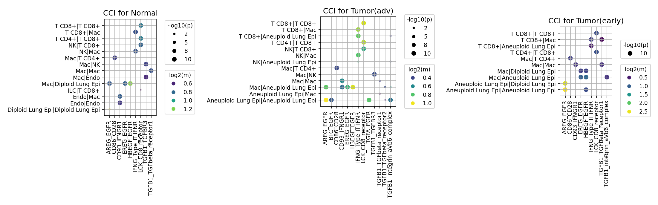
[Analysis Visualization Results]...
Analysis Overview
This analysis investigates cell-cell interactions (CCI) mediated by a curated set of genes associated with immune checkpoint and cell cycle pathways across different conditions of lung tissue: Normal, Tumor (early), and Tumor (advanced). The plot_cci_dots tool was used to visualize significant ligand-receptor interactions between various cell types, including immune cells (T CD8+, T CD4+, NK, Macrophage), endothelial cells, and lung epithelial cells (distinguished by ploidy as Diploid or Aneuploid), focusing on a predefined set of genes. The size of the dot represents the negative log10 p-value of the interaction, indicating statistical significance, while the color intensity reflects the log2 mean expression of the interacting ligand-receptor pair, indicating interaction strength.
Visual Summary
The dot plots reveal distinct patterns of cell-cell communication for selected ligand-receptor pairs across Normal, early tumor, and advanced tumor conditions.
Normal Condition: Displays baseline interactions. Key observations include
- AREG_EGFR, EREG_EGFR, HBEGF_EGFR interactions primarily within Macrophages (Mac|Mac), Endothelial cells (Endo|Endo), and with T cells (T CD8+|Mac, T CD4+|Mac), as well as autocrine Diploid Lung Epi|Diploid Lung Epi.
- CD86-CD28 (T cell co-stimulation) is active within T cells (T CD8+|T CD8+, T CD4+|T CD8+).
- IFNG_Type_II_IFNR interactions are notable in T CD8+|T CD8+ and NK|Mac.
- TGFB1_TGFbeta_receptor1/2 interactions are present across various immune cell pairs (T cells, NK cells, Macrophages).
- Tumor (early) Condition: Shows an expansion and intensification of certain interactions, particularly involving Aneuploid Lung Epithelial cells.
- AREG_EGFR interactions become highly significant and stronger, prominently involving Aneuploid Lung Epithelial cells (Aneuploid Lung Epi|Aneuploid Lung Epi, Aneuploid Lung Epi|Diploid Lung Epi, Aneuploid Lung Epi|Mac). HBEGF_EGFR also appears in Aneuploid Lung Epi|Diploid Lung Epi.
- TGFB1_TGFbeta_receptor1/2 interactions show increased strength and significance across multiple immune cell types and, importantly, between Aneuploid Lung Epi|Mac and Aneuploid Lung Epi|Aneuploid Lung Epi. The TGFB1_integrin_avb6_complex also emerges, particularly for Aneuploid Lung Epi|Aneuploid Lung Epi.
- CD86-CD28 and IFNG_Type_II_IFNR continue to show active interactions within immune cell populations.
- Tumor (advanced) Condition: Characterized by widespread and highly significant interactions, especially those involving Aneuploid Lung Epithelial cells and robust TGFB signaling.
- EGFR Signaling: AREG_EGFR and BTC_EGFR interactions are very strong, particularly between Aneuploid Lung Epi|Mac and Aneuploid Lung Epi|Aneuploid Lung Epi, indicating sustained and enhanced autocrine/paracrine EGFR activation in tumor cells.
- TGFB Signaling: TGFB1_TGFbeta_receptor1/2 interactions are exceptionally strong and pervasive across almost all tested cell-cell pairs, including T CD8+|T CD8+, T CD4+|T CD8+, NK|Mac, Mac|Mac, Mac|Aneuploid Lung Epi, and Aneuploid Lung Epi|Aneuploid Lung Epi. Critically, TGFB1_integrin_avb6_complex displays very strong and significant interactions, especially for Aneuploid Lung Epi|Mac and Aneuploid Lung Epi|Aneuploid Lung Epi.
- Immune Co-stimulation/Inflammation: CD86-CD28 and IFNG_Type_II_IFNR interactions remain active, suggesting ongoing immune responses, although their functional outcome in an advanced tumor microenvironment needs further context.
Biological Interpretation
The analysis highlights a dynamic shift in cell-cell communication patterns during lung tumor progression, with a strong emphasis on EGFR and TGFB signaling pathways.
- EGFR Pathway Activation in Tumor Cells: The increased and prominent interactions involving AREG_EGFR, EREG_EGFR, HBEGF_EGFR, and BTC_EGFR with Aneuploid Lung Epithelial cells (identified as tumor cells based on tumor origin and ploidy status) from early to advanced tumor stages underscore the critical role of EGFR signaling in lung cancer. EGFR is a well-established oncogenic driver, promoting tumor cell proliferation, survival, and migration. The emergence of BTC_EGFR in advanced disease, specifically between Aneuploid Lung Epithelial cells and Macrophages, suggests a complex interplay where tumor-associated macrophages might contribute to sustaining tumor growth via paracrine EGFR ligand secretion. Reference: GeneCards for EGFR
- TGFB Signaling as a Dominant Force in Tumor Progression and Immune Evasion: The most striking observation is the significant and widespread upregulation of TGFB1_TGFbeta_receptor1/2 and TGFB1_integrin_avb6_complex interactions as the tumor progresses. TGFB1 is a pleiotropic cytokine with context-dependent roles in cancer. While it can act as a tumor suppressor in early stages, it becomes a potent pro-tumorigenic and immunosuppressive factor in established tumors. In this context:
- Its increased activity across diverse immune cells (T cells, NK cells, Macrophages) suggests a broad immunosuppressive effect, inhibiting anti-tumor immune responses and fostering an immune-tolerant microenvironment.
- The strong interactions involving Aneuploid Lung Epithelial cells, particularly via the TGFB1_integrin_avb6_complex, indicate that tumor cells are actively engaged in and potentially driving TGFB-mediated processes. This complex is known to activate latent TGFB and is associated with epithelial-mesenchymal transition (EMT), fibrosis, and increased invasiveness in various cancers, including lung cancer. Reference: GeneCards for TGFB1, Reference: PubMed search for "TGFB1 integrin alpha-v beta-6 lung cancer"
- Immune Cell Context in Tumor Microenvironment: While interactions like CD86-CD28 (T cell co-stimulation) and IFNG_Type_II_IFNR (interferon-gamma signaling, indicative of inflammation/anti-tumor response) persist, the concurrent dominance of immunosuppressive TGFB signaling suggests a skewed immune microenvironment. Macrophages, highly interactive with Aneuploid Lung Epithelial cells through both EGFR and TGFB pathways, are likely polarized towards pro-tumorigenic, M2-like phenotypes, contributing to immune suppression and tumor growth rather than anti-tumor immunity.
- Role of Cell Cycle Genes (Indirect): Although direct ligand-receptor pairs for intracellular cell cycle regulators are not observed, the presence of 'Aneuploid Lung Epi' cells directly reflects cell cycle dysregulation and genomic instability characteristic of cancer. The observed EGFR and TGFB signaling pathways are major drivers that promote uncontrolled cell cycle progression and proliferation in these tumor cells.
Clinical or Translational Implications
The differential cell-cell interactions observed across lung cancer progression have significant clinical and translational implications:
- Therapeutic Targeting of EGFR Pathway: The sustained and enhanced EGFR signaling in Aneuploid Lung Epithelial cells reinforces the importance of EGFR as a therapeutic target in lung cancer. EGFR tyrosine kinase inhibitors (TKIs) are standard of care for patients with EGFR-mutated NSCLC. Understanding the specific ligands involved (AREG, BTC, EREG, HBEGF) and the interacting cell types could help refine patient selection for EGFR-targeted therapies or anticipate resistance mechanisms. Reference: PubMed search for "EGFR inhibitors NSCLC clinical trials"
- TGFB Signaling as a Promising Immunotherapy Target: The pronounced upregulation of TGFB1 signaling, particularly the TGFB1_integrin_avb6_complex in advanced tumors, positions TGFB as a critical target for therapeutic intervention. Inhibiting TGFB signaling could help overcome tumor-induced immunosuppression, normalize the tumor microenvironment, and potentially enhance the efficacy of other immunotherapies, such as immune checkpoint blockade. Several TGFB inhibitors are in clinical development. Reference: PubMed search for "TGFB inhibitors cancer therapy"
- Combination Therapy Strategies: Given the co-activation of EGFR and TGFB pathways and their interplay with the immune microenvironment, combination therapies hold great promise. For instance, combining EGFR TKIs with TGFB inhibitors or with immune checkpoint inhibitors could synergistically target tumor proliferation, metastasis, and immune evasion, leading to improved outcomes in lung cancer patients.
- Biomarker Development: The specific and strong cell-cell interactions observed in advanced disease, such as the TGFB1_integrin_avb6_complex involving Aneuploid Lung Epithelial cells, could serve as potential biomarkers for disease progression, therapeutic response, or patient stratification for targeted therapies. Further validation using methods like spatial transcriptomics or multiplexed IHC could confirm these interactions in tissue.
15. Condition-Specific Cell-Cell Interaction Patterns in Lung Tissue
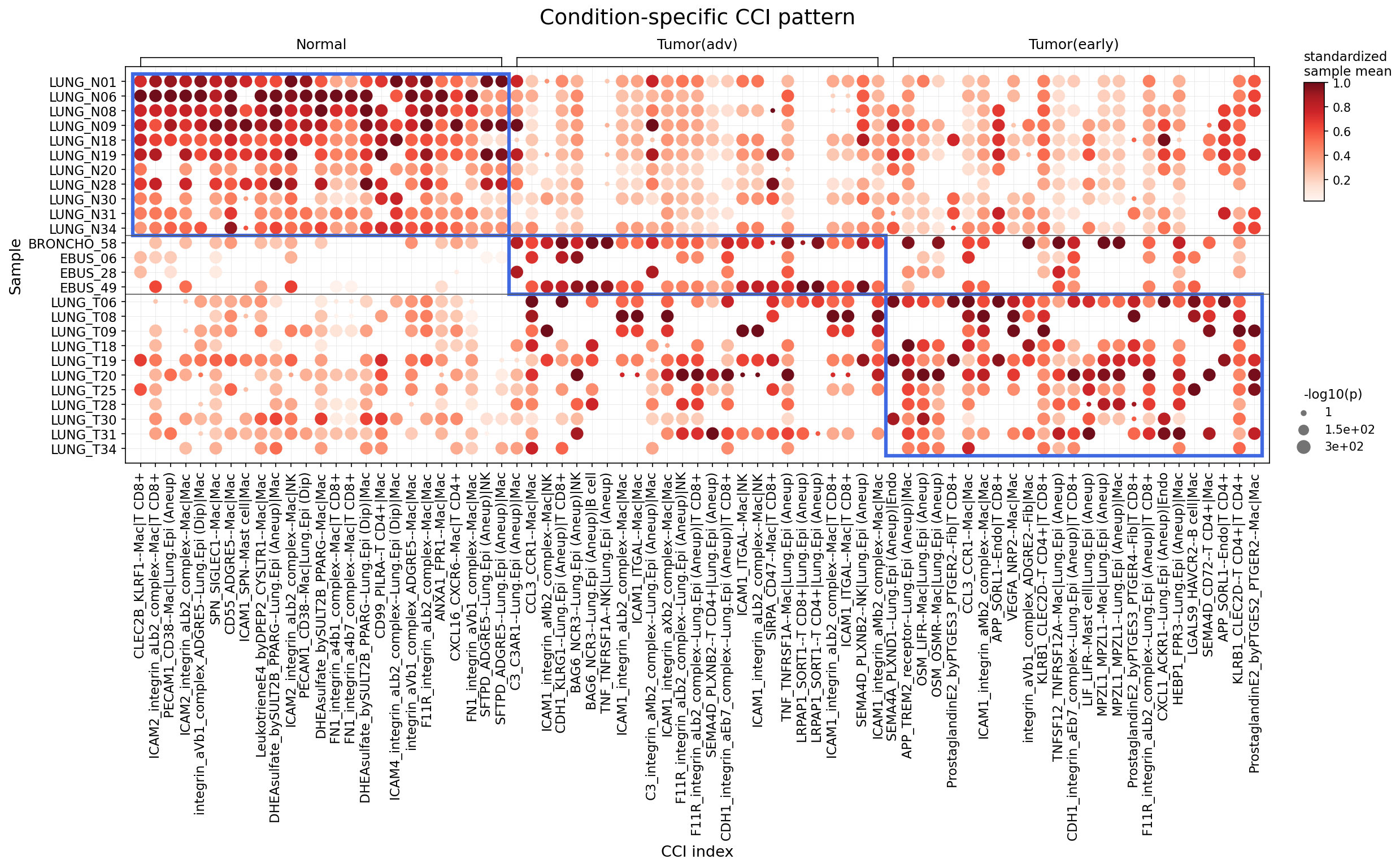
[Analysis Visualization Results]...
Analysis Overview
This analysis identifies statistically significant differences in cell-cell interactions (CCI) across Normal, Tumor(early), and Tumor(adv) conditions within lung tissue, focusing on major immune and stromal cell types (T cell, Myeloid cell, B cell, Mast cell, Stromal cell, Endothelial cell). The provided dot plot visualizes the strength (standardized sample mean, color intensity) and statistical significance (-log10(p-value), dot size) of selected ligand-receptor pairs for each sample, grouped by the condition in which the interactions were found to be significantly enriched. The analysis specifically highlights interactions that are significantly stronger or more prevalent in one condition compared to others.
Visual Summary
The dot plot displays a matrix where rows represent individual samples, and columns represent specific cell-cell interaction pairs (CCI index), grouped by the condition in which they are enriched. Three distinct blue boxes delineate the interaction patterns enriched in Normal, Tumor(adv), and Tumor(early) conditions, respectively.
- Normal Condition (Leftmost block): This section shows a high density of strong (dark red) and significant (large dots) interactions primarily within the Normal lung samples (LUNG_N01 to EBUS_49). These interactions are less pronounced or absent in tumor samples. Key interactions often involve macrophages and lung epithelial cells, as well as T cells.
- Tumor(adv) Condition (Middle block): A different set of interactions emerges as strong and significant in the tumor samples (LUNG_T06 to LUNG_T34) under the 'Tumor(adv)' context. These interactions show distinct patterns compared to Normal, often involving aneuploid lung epithelial cells, which are indicative of malignant cells.
- Tumor(early) Condition (Rightmost block): This block also highlights interactions enriched in the tumor samples (LUNG_T06 to LUNG_T34) but within the 'Tumor(early)' context. While sharing some overlap with 'Tumor(adv)' interactions, this group also exhibits unique or differentially emphasized interaction patterns.
In general, the plot clearly segregates CCI profiles based on disease status, with distinct sets of interactions characterizing normal lung tissue versus early and advanced tumor stages. Aneuploid lung epithelial cells are frequently identified as key players in tumor-associated interactions.
Biological Interpretation
The analysis reveals condition-specific rewiring of cell-cell communication networks in lung cancer, involving critical immune and stromal cell types interacting with both normal and neoplastic epithelial cells.
Normal Lung Environment
In normal lung tissue, the prominent interactions suggest active immune surveillance, tissue homeostasis, and basic cell adhesion.
- Macrophage-centric interactions: Many significant interactions involve Macrophages (Mac) interacting with T cells (T CD8+), Lung Epithelial cells (Lung.Epi), or other Macrophages. Examples include Integrins (integrin_aLb2_complex, integrin_aVb1_complex) and ICAMs (ICAM2_integrin_aLb2_complex), highlighting general cell adhesion and communication crucial for tissue integrity and immune cell trafficking [Ref: PubMed search for "integrin ICAM lung immunity"].
- Innate immunity and complement: Interactions like SFTPD-ADGRE5 (Mac|Mac) and C3-C3AR1 (Lung.Epi (Aneuploid)|Mac) suggest roles for surfactant proteins in innate immunity and complement activation, which are essential defense mechanisms in the lung [Ref: GeneCards SFTPD, C3]. The presence of "Lung.Epi (Aneuploid)" in normal samples for C3-C3AR1 could indicate early cellular changes or a subset of atypical epithelial cells even in morphologically normal tissue.
Tumor Microenvironment: Early and Advanced Stages
Both Tumor(early) and Tumor(adv) conditions exhibit a shift towards interactions that are characteristic of a pro-tumorigenic and immunosuppressive microenvironment, with aneuploid lung epithelial cells (Lung.Epi (Aneuploid)) playing a central role.
Interactions Enriched in Tumor(adv)
- Immune Evasion: The SIRPA-CD47 axis (Mac|Mac, Mac|T CD8+) is highly prominent. CD47 on tumor cells often binds to SIRPA on macrophages and T cells, acting as a "don't eat me" signal that prevents phagocytosis and suppresses anti-tumor immunity. Its strong presence in advanced tumors suggests a key mechanism for immune escape [Ref: PubMed search for "SIRPA CD47 cancer immunotherapy"].
- NK Cell Modulation: Interactions involving NK cells, such as BAG6-NCR3 (Lung.Epi (Aneuploid)|NK), TNF-TNFRSF1A (NK|Lung.Epi (Aneuploid)), and ICAM1-ITGAL (Mac|NK), suggest altered NK cell activity. NCR3 (NKp30) is an activating NK cell receptor, and TNF signaling can have complex effects on NK cells and tumor progression [Ref: UniProt NCR3].
- Pro-tumorigenic Inflammation and Growth: The Oncostatin M (OSM)-LIFR (Mac|Lung.Epi (Aneuploid)) pathway can promote tumor cell proliferation, survival, and metastasis [Ref: PubMed search for "Oncostatin M cancer progression"]. Prostaglandin E2 (PGE2) signaling (PGE2_PTGER2, Fib|Mac) is known to create an immunosuppressive environment and support tumor growth by influencing macrophage polarization and T cell function [Ref: PubMed search for "PGE2 tumor immunosuppression"].
- Angiogenesis and Cell Adhesion: APP-TREM2_receptor (Lung.Epi (Aneuploid)|Endo) and SEMA4D-PLXNB2 (Lung.Epi (Aneuploid)|T CD8+) suggest communication between tumor cells, endothelial cells, and T cells, potentially contributing to angiogenesis and altered immune cell function.
Interactions Enriched in Tumor(early)
While some interactions overlap with Tumor(adv), the 'Tumor(early)' context highlights specific features that might be critical in early disease progression.
- Angiogenesis and Stromal Remodeling: VEGFA-NRP2 (Mac|Mac, Mac|Endo) is a well-established pathway driving angiogenesis, crucial for tumor growth and metastasis. Its prominence underscores the initiation of new blood vessel formation even in early-stage tumors [Ref: GeneCards VEGFA]. Integrin_aVb1_complex_ADGRE2 (Fib|Mac) indicates active cross-talk between fibroblasts and macrophages, important for extracellular matrix remodeling and creating a permissive microenvironment for tumor growth [Ref: PubMed search for "fibroblast macrophage tumor remodeling"].
- Immune Checkpoints and Suppression: GAL9-HAVCR2 (Galectin-9-TIM-3) (B cell|Mac) is often associated with immune exhaustion, particularly of T cells. Its presence here between B cells and macrophages could influence macrophage polarization or B cell anergy, contributing to local immunosuppression early on [Ref: PubMed search for "Galectin-9 TIM-3 immune suppression"].
- T Cell and NK Cell Interactions: KLRB1-CLEC2D (T CD4+|Lung.Epi (Aneuploid)) and CLEC2D-T CD4+ (T CD4+|T CD4+) can modulate T cell and NK cell responses, influencing their activation and cytotoxicity against tumor cells.
- Chemokine Signaling: CCL3-CCR1 (Mac|Mac) indicates active chemokine signaling among macrophages, which can contribute to their recruitment and inflammatory functions within the early tumor microenvironment.
Clinical or Translational Implications
The distinct CCI patterns observed across normal and tumor conditions, especially the enrichment of interactions involving aneuploid lung epithelial cells, offer several potential clinical and translational implications:
- Biomarker Discovery: Specific CCI pairs identified as highly enriched and significant in Tumor(early) or Tumor(adv) could serve as potential diagnostic or prognostic biomarkers for lung cancer. For instance, strong SIRPA-CD47 or VEGFA-NRP2 signals could indicate a more aggressive tumor phenotype or early angiogenic activity, respectively.
- Therapeutic Targets: The highlighted ligand-receptor interactions represent potential therapeutic targets. Inhibiting the SIRPA-CD47 axis, for example, is an active area of cancer immunotherapy aimed at boosting anti-tumor macrophage activity [Ref: PubMed search for "CD47 SIRPA inhibitors cancer"]. Targeting VEGFA-NRP2 could complement existing anti-angiogenic therapies.
- Understanding Disease Progression: The differences between Tumor(early) and Tumor(adv) CCI profiles provide insights into the dynamic evolution of the tumor microenvironment. This understanding can help tailor stage-specific treatment strategies. For example, early-stage interventions might focus more on angiogenesis, while advanced stages might prioritize immune checkpoint blockade or overcoming immune evasion.
- Novel Combination Therapies: Identifying multiple interconnected pathways (e.g., pro-inflammatory PGE2 signaling alongside immune evasive SIRPA-CD47) suggests that combination therapies targeting different nodes of the CCI network might be more effective in disrupting tumor progression and restoring anti-tumor immunity.
16. Lung Epithelial Cell Condition-Specific Surfaceome Markers Analysis
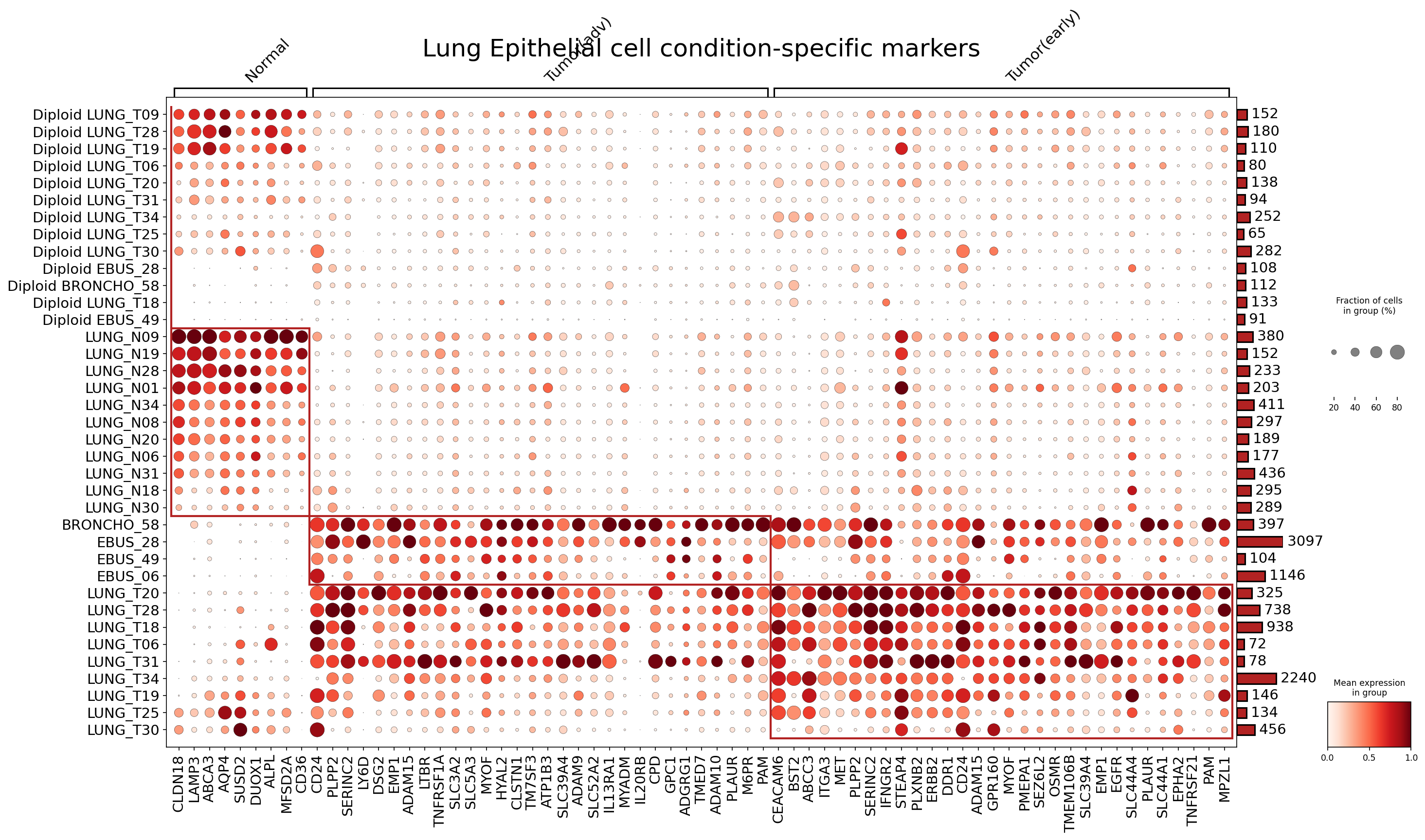
[Analysis Visualization Results]...
Analysis Overview
This analysis identifies condition-specific surfaceome markers in Lung Epithelial cells from single-cell RNA-seq data. The objective was to pinpoint surface-expressed genes that are differentially expressed across Normal, Tumor (advanced), and Tumor (early) conditions. This dot plot visualization highlights up to 50 top differentially expressed surfaceome markers for the Lung Epithelial cell type in each condition, comparing it against all other conditions. The samples are further stratified by their ploidy status where available (e.g., "Diploid" for normal samples). Such markers are crucial for understanding disease progression and identifying potential diagnostic or therapeutic targets.
Visual Summary
The dot plot effectively visualizes the expression patterns of condition-specific surfaceome markers within Lung Epithelial cells across various patient samples grouped by their clinical condition (Normal, Tumor(adv), Tumor(early)).
- Grouping: Samples are organized along the y-axis and grouped under their respective conditions. The "Diploid" prefix for many Normal samples indicates inferred ploidy status, which helps distinguish healthy epithelial cells from potentially aneuploid tumor cells. The absence of "Diploid" for tumor samples aligns with the expectation of aneuploidy in cancer.
- Marker Distribution: A distinct set of surfaceome markers is highly enriched in each condition.
- Normal Samples: Show robust expression of markers such as CLDN18, LAMP3, AQP4, and DUOX1. These markers appear largely absent or lowly expressed in tumor conditions.
- Tumor (advanced) Samples: Exhibit a unique cluster of highly expressed markers including EGFR, SERINC2, LY6D, ADAM15, EMP1, PLAU, PLAUR, CD24, and OSMR.
- Tumor (early) Samples: Display another set of markers, with some overlap with advanced tumor markers, such as CD36, ITGA3, ERBB2, DDR1, and MPZL1. Some markers like CD24, SERINC2, LY6D, and EMP1 are observed in both early and advanced tumor contexts, indicating their role throughout tumor progression.
- Dot Size and Color: The size of each dot correlates with the fraction of cells within a sample group expressing the gene, while the color intensity (from light to dark red) indicates the mean expression level of the gene. Darker, larger dots signify higher expression in a larger proportion of cells, indicating strong, prevalent marker expression.
- Sample Heterogeneity: Within each condition, there is some variability in marker expression across individual samples, suggesting inter-patient heterogeneity in tumor biology, even at similar disease stages.
Biological Interpretation
The differential expression of surfaceome markers in Lung Epithelial cells provides critical insights into the molecular changes occurring during lung tumorigenesis and progression.
- Normal Epithelial Homeostasis: Markers like CLDN18 (Claudin-18), a tight junction protein, are integral to maintaining epithelial barrier integrity and are typically associated with differentiated, healthy lung epithelial cells. LAMP3 (Lysosomal Associated Membrane Protein 3) is involved in antigen presentation and surfactant homeostasis in type II alveolar epithelial cells, suggesting normal lung function. The presence of these markers primarily in normal samples underscores their role in healthy lung tissue. GeneCards: CLDN18 GeneCards: LAMP3
- Early Tumorigenesis Signatures: The early tumor-specific markers, such as CD36 and ERBB2 (HER2), hint at metabolic reprogramming and altered growth signaling pathways even at early stages. CD36 is a fatty acid translocase implicated in lipid metabolism, which is often dysregulated in cancer cells to support rapid proliferation. ERBB2 (HER2) is a well-established oncogene whose overexpression or amplification drives proliferation and survival in various cancers, including a subset of lung cancers. GeneCards: CD36 GeneCards: ERBB2
- Advanced Tumor Progression and Malignancy: The advanced tumor condition is characterized by the prominent upregulation of markers like EGFR, EMP1, and PLAU/PLAUR.
- EGFR (Epidermal Growth Factor Receptor) is a canonical oncogene and a primary driver of non-small cell lung cancer (NSCLC) through its role in cell growth, proliferation, and survival. Its strong expression in advanced tumors is a hallmark of aggressive disease. GeneCards: EGFR
- EMP1 (Epithelial Membrane Protein 1) is associated with increased cell proliferation, invasion, and metastasis in various cancers, including lung cancer.
- PLAU (urokinase-type plasminogen activator) and its receptor PLAUR are critical components of the plasminogen activation system, promoting extracellular matrix degradation, facilitating tumor invasion, and metastasis.
- ADAM15 and ADAM9 (ADAM metallopeptidases) are involved in cell adhesion, migration, and growth factor shedding, contributing to tumor progression.
- CD24 is a glycoprotein often expressed in cancer stem cells and associated with increased invasiveness and poor prognosis. Its presence across tumor stages might indicate its role in maintaining malignant potential. GeneCards: CD24
- Ploidy Status: The distinction of "Diploid" in normal samples confirms that the analysis successfully distinguishes healthy, non-malignant epithelial cells, whereas tumor epithelial cells likely exhibit aneuploidy, a common feature of cancer, which would explain the distinct marker profiles.
Clinical or Translational Implications
The identified condition-specific surfaceome markers in Lung Epithelial cells hold significant promise for clinical applications.
Diagnostic and Prognostic Biomarkers:
- The robust upregulation of markers like EGFR, ERBB2, EMP1, PLAU/PLAUR, ADAMs, CD24, CD36, and ITGA3 in tumor cells compared to normal lung epithelial cells could serve as highly specific diagnostic biomarkers for lung cancer, potentially aiding in early detection and differential diagnosis.
- Differences in marker expression between early and advanced tumor stages could help in disease staging and predicting prognosis. Surface markers are particularly advantageous for diagnostic tests involving biopsies, liquid biopsies (e.g., circulating tumor cells), or imaging, as they are directly accessible on the cell surface.
Therapeutic Targets:
- Several identified markers are already established or emerging therapeutic targets. EGFR and ERBB2 are actionable targets for FDA-approved tyrosine kinase inhibitors and monoclonal antibodies in lung cancer treatment.
- Other highly expressed surface proteins in tumor cells, such as CD24, CD36, EMP1, PLAU/PLAUR, ADAMs, ITGA3, and DDR1, represent potential novel therapeutic targets. These could be exploited for targeted therapies, including antibody-drug conjugates (ADCs), bispecific antibodies, or CAR T-cell therapies, which can selectively eliminate tumor cells while minimizing off-target effects on healthy tissues. PubMed Search: "antibody drug conjugate lung cancer surface markers"
Patient Stratification and Personalized Medicine:
- The observed heterogeneity in marker expression across individual tumor samples suggests that these markers could be used to stratify patients. Patients whose tumors highly express specific targetable markers could be prioritized for therapies directed against those markers, paving the way for more personalized and effective treatment strategies.
- Monitoring the expression of these markers over time could also inform treatment response and the emergence of resistance.
17. Macrophage Condition-Specific Surfaceome Markers in Lung Tissue
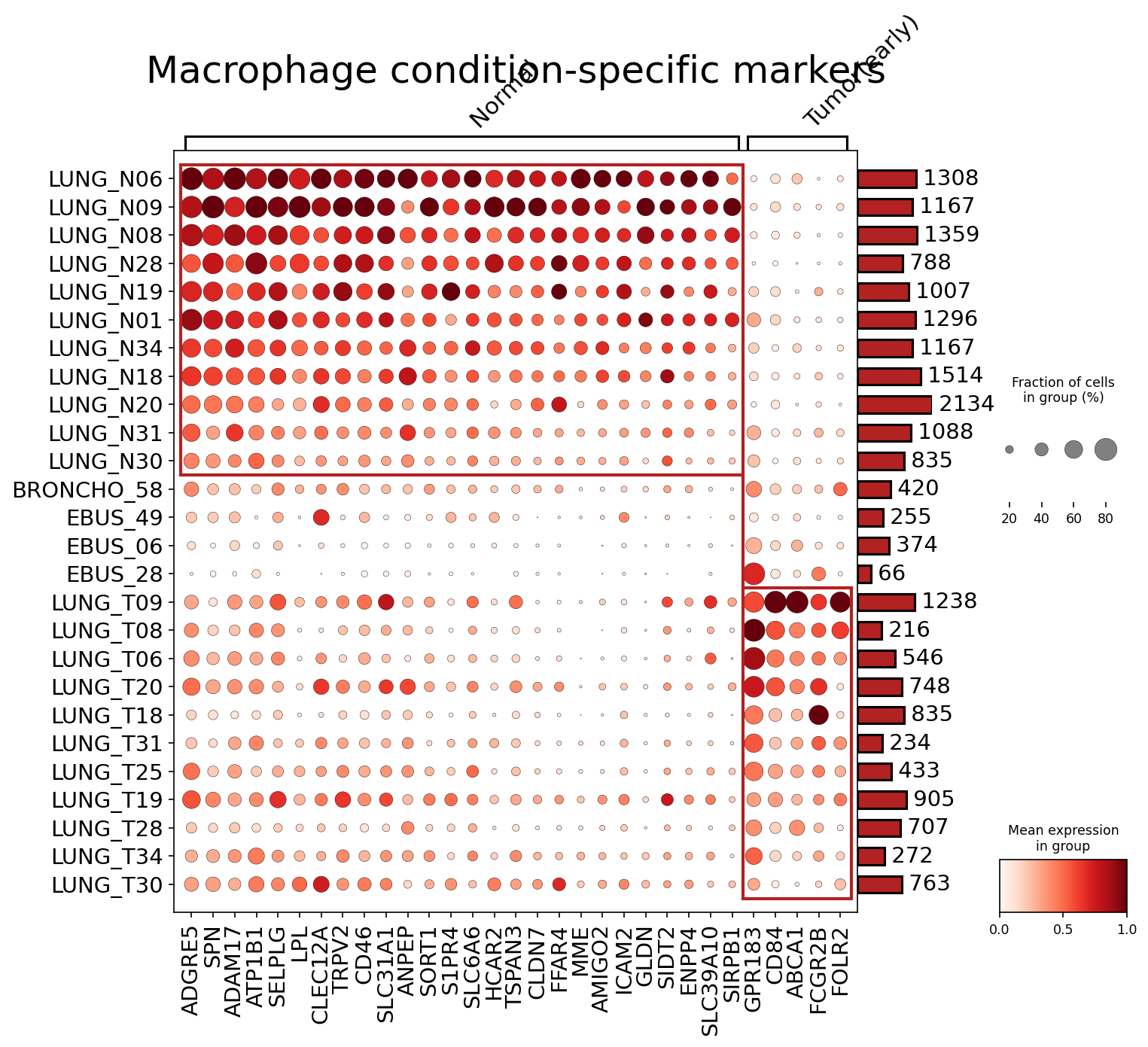
[Analysis Visualization Results]...
Analysis Overview
This analysis aimed to identify and visualize condition-specific surfaceome markers for Macrophage cells derived from single-cell RNA sequencing data of human lung tissue. The comparison focused on distinguishing macrophages from 'Normal' lung tissue versus those from 'Tumor(early)' lung tissue. The plot_markers_and_expression_dot tool was utilized, configured to identify up to 50 surfaceome-specific markers per condition with specific statistical and expression cutoffs.
Visual Summary
The dot plot effectively illustrates the differential expression patterns of surfaceome markers across individual samples, grouped by 'Normal' and 'Tumor(early)' conditions, for Macrophage cells.
- Clear Segregation: The most prominent feature is the distinct expression profile of markers separating 'Normal' samples from 'Tumor(early)' samples. A block of genes on the left side of the x-axis (e.g., ADGRE5, SPN, ADAM17, LPL, CLEC12A) shows high mean expression (darker red dots) and high prevalence (larger dot size) in the 'Normal' samples (top rows). Conversely, a set of genes on the right side (e.g., SIRPB1, GPR183, CD84, ABCA1, FCGR2B, FOLR2) are highly expressed and prevalent in the 'Tumor(early)' samples (bottom rows).
- Sample Heterogeneity: While there's a general trend, some samples within each condition show variability in marker expression intensity and cell fraction. For instance, some 'Normal' samples like LUNG_N34 and LUNG_N18 show slightly less intense expression of the 'Normal' markers compared to others. Similarly, within 'Tumor(early)' samples, LUNG_T25 and LUNG_T19 show particularly strong expression for the 'Tumor(early)' specific markers.
- Intermediate/Low Cell Count Samples: Samples such as BRONCHO_58, EBUS_49, EBUS_06, and EBUS_28, located between the clear 'Normal' and 'Tumor(early)' clusters, generally exhibit lower expression levels of most markers and often have fewer Macrophage cells (indicated by the smaller bars on the right), which might reflect technical factors or potentially represent transitional or less clearly defined states.
- Marker Abundance: The bar plot on the far right indicates the total number of Macrophage cells per sample, providing context for the observed expression levels and prevalence.
Biological Interpretation
The differential expression of surfaceome markers highlights a significant remodeling of macrophage identity and function in the early lung tumor microenvironment compared to normal tissue.
Macrophages in Normal Lung Tissue
The markers enriched in 'Normal' lung macrophages are indicative of a homeostatic, tissue-resident phenotype:
- ADGRE5 (CD97): A G-protein coupled receptor involved in cell adhesion and immune regulation, often expressed on myeloid cells, contributing to tissue integrity. https://www.genecards.org/cgi-bin/carddisp.pl?gene=ADGRE5
- SPN (CD43): A sialomucin that plays roles in cell adhesion, T-cell activation, and differentiation, contributing to general immune surveillance. https://www.genecards.org/cgi-bin/carddisp.pl?gene=SPN
- LPL (Lipoprotein Lipase): An enzyme critical for lipid metabolism, suggesting roles in energy homeostasis and lipid processing within the normal lung environment. https://www.genecards.org/cgi-bin/carddisp.pl?gene=LPL
- CLEC12A: A C-type lectin receptor found on myeloid cells, involved in immune recognition and regulation, potentially indicating a role in clearing cellular debris or pathogens in healthy tissue. https://www.genecards.org/cgi-bin/carddisp.pl?gene=CLEC12A
- S1PR4 (Sphingosine-1-Phosphate Receptor 4): Involved in immune cell trafficking and inflammatory responses, potentially modulating macrophage migration in normal tissue. https://www.genecards.org/cgi-bin/carddisp.pl?gene=S1PR4
These markers collectively point to macrophages that maintain tissue integrity, participate in metabolic homeostasis, and provide innate immune surveillance in the healthy lung.
Macrophages in Early Lung Tumor (Tumor-Associated Macrophages, TAMs)
The markers specifically upregulated in 'Tumor(early)' lung macrophages strongly suggest an immune-modulatory and pro-tumoral phenotype characteristic of Tumor-Associated Macrophages (TAMs):
- SIRPB1 (SIRP-beta-1): An activating receptor on myeloid cells that can promote pro-tumoral functions and contribute to T-cell suppression. It is closely related to SIRPα, which interacts with CD47, a common "don't eat me" signal on cancer cells. https://www.genecards.org/cgi-bin/carddisp.pl?gene=SIRPB1
- ABCA1 (ATP Binding Cassette Subfamily A Member 1): A cholesterol efflux pump. Upregulation in TAMs can indicate altered lipid metabolism, which is crucial for TAM polarization and function, often associated with an M2-like, immunosuppressive phenotype. https://www.genecards.org/cgi-bin/carddisp.pl?gene=ABCA1
- FCGR2B (CD32B): A low-affinity inhibitory IgG Fc receptor. Its expression on TAMs can dampen anti-tumor immune responses by inhibiting activating Fc receptors and antibody-dependent cellular cytotoxicity. https://www.genecards.org/cgi-bin/carddisp.pl?gene=FCGR2B
- FOLR2 (Folate Receptor Beta): Frequently expressed on activated, immunosuppressive macrophages, particularly TAMs, and is involved in folate uptake, which supports rapid cell proliferation, including that of cancer cells. https://www.genecards.org/cgi-bin/carddisp.pl?gene=FOLR2
The expression of these markers suggests that even in early tumor stages, lung macrophages adopt a phenotype that likely supports tumor growth and immune evasion, consistent with established roles of TAMs in cancer progression.
Clinical or Translational Implications
The distinct surfaceome marker profiles identified for macrophages in 'Normal' versus 'Tumor(early)' lung conditions have several important clinical and translational implications:
- Biomarker Discovery for Early Detection and Prognosis: The identified tumor-associated macrophage (TAM) specific markers, such as SIRPB1, ABCA1, FCGR2B, and FOLR2, could serve as potential biomarkers for the early detection of lung cancer or for assessing the degree of macrophage infiltration and polarization in the tumor microenvironment. These markers could be assessed in tissue biopsies, cytology samples (e.g., from bronchoalveolar lavage or EBUS aspirates), or even in liquid biopsies (e.g., circulating myeloid cells or extracellular vesicles) to aid in diagnosis and prognostication.
- Therapeutic Targets for Macrophage-Targeted Immunotherapy: The upregulated surfaceome markers on 'Tumor(early)' macrophages present attractive targets for novel immunotherapeutic strategies:
- SIRPB1: Targeting the SIRPB1-CD47 axis has shown promise in reprogramming macrophages from an immunosuppressive to an anti-tumoral state. Blocking SIRPB1 or CD47 (its ligand on tumor cells) aims to unleash macrophage phagocytic activity against cancer cells. PubMed search: CD47 SIRP alpha cancer immunotherapy
- FOLR2: Given its association with immunosuppressive TAMs, FOLR2 could be targeted with antibody-drug conjugates or other modalities to specifically deplete or reprogram these pro-tumoral macrophage populations. PubMed search: FOLR2 tumor associated macrophages therapy
- FCGR2B and ABCA1: Modulating the activity of these receptors could influence immune complex responses and lipid metabolism within TAMs, potentially shifting their phenotype towards an anti-tumoral role.
- Monitoring Therapeutic Response: Changes in the expression of these markers on macrophages could be monitored during treatment to assess the efficacy of immunotherapies or conventional cancer treatments in altering the tumor microenvironment.
- Experimental Validation: The identified markers provide a strong basis for further experimental validation using techniques such as multi-color flow cytometry, immunohistochemistry, or spatial transcriptomics on clinical samples. This would help confirm their diagnostic and therapeutic potential, investigate their functional implications in macrophage polarization and interaction with other immune cells, and explore their prognostic value in larger patient cohorts.
18. Fibroblast Condition-Specific Surfaceome Marker Analysis in Lung Tissue
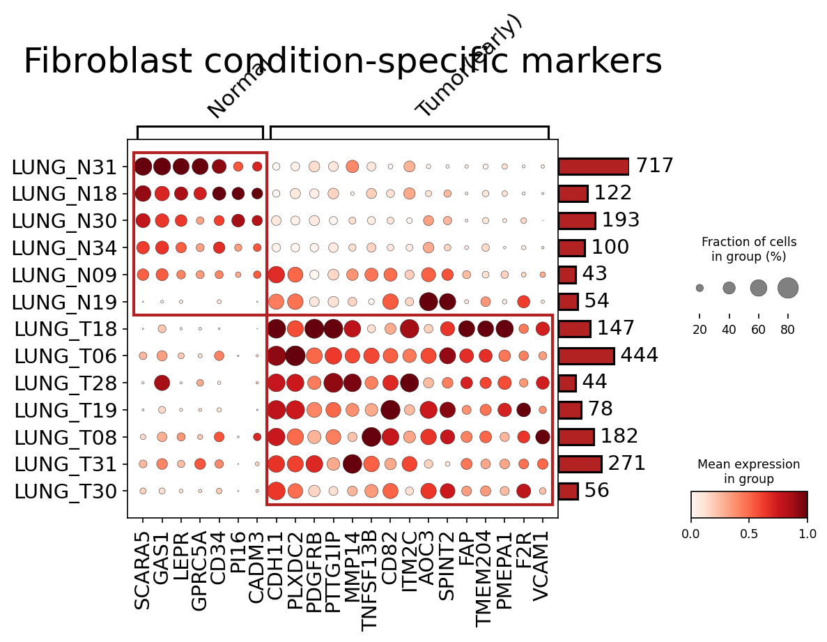
[Analysis Visualization Results]...
Analysis Overview
This analysis aimed to identify condition-specific surfaceome markers in Fibroblasts from single-cell RNA-seq data of human lung tissue. The comparison was performed between 'Normal' and 'Tumor (early)' conditions. The plot_markers_and_expression_dot tool was used to visualize the top 50 surfaceome markers per condition, filtered by expression characteristics to highlight differential expression. Surfaceome markers are particularly relevant for their accessibility to targeted therapies and cell-surface phenotyping.
Visual Summary
The dot plot effectively illustrates the differential expression of surfaceome markers in Fibroblast cells across various lung samples categorized as 'Normal' (LUNG_N samples) or 'Tumor (early)' (LUNG_T samples).
Distinct Gene Clusters: The plot clearly shows two main clusters of genes
- A cluster of genes (highlighted by the top red box) exhibiting high expression and prevalence in 'Normal' samples (LUNG_N31, LUNG_N18, LUNG_N30, LUNG_N34, LUNG_N09, LUNG_N19). These include SCARA5, GAS1, LEPR, GPRC5A, CD34, CD16, CADM3, and CDH11.
- Another prominent cluster of genes (highlighted by the bottom red box) showing elevated expression and higher fraction of expressing cells predominantly in 'Tumor (early)' samples (LUNG_T18, LUNG_T06, LUNG_T28, LUNG_T19, LUNG_T08, LUNG_T31, LUNG_T30). Key markers in this group include PLXDC2, PDGFRB, PTTG1IP, MMP14, TNFSF13B, CD82, ITM2C, AOC3, SPINT2, FAP, TMEM204, PMEPA1, F2R, and VCAM1.
Expression and Prevalence:
- For genes in the 'Normal' cluster, dot colors are predominantly dark red, indicating high mean expression, and dot sizes are large, suggesting a high fraction of fibroblasts in these samples express these markers.
- Conversely, for genes in the 'Tumor (early)' cluster, the same pattern of high expression and prevalence is observed within the 'Tumor (early)' samples, while expression is minimal or absent in 'Normal' samples.
- Sample-Specific Patterns: Some individual samples show nuances, for example, LUNG_N09 and LUNG_N19, despite being 'Normal' samples, show some overlap in expressing 'Tumor (early)' markers like FAP, albeit at lower levels or in fewer cells compared to true 'Tumor' samples. This could suggest early phenotypic shifts or heterogeneity within 'Normal' adjacent tissue.
- Cell Counts: The bar chart on the right indicates the number of Fibroblast cells per sample, providing context for the robustness of expression measurements in each group.
Biological Interpretation
The identified condition-specific surfaceome markers reveal significant phenotypic shifts in Fibroblasts during early lung tumorigenesis, transitioning from a quiescent or normal state to an activated, tumor-associated fibroblast (CAF) phenotype.
Markers Associated with Normal Fibroblasts:
- SCARA5 (Scavenger Receptor Class A Member 5): Involved in cell adhesion, growth inhibition, and suppression of angiogenesis. Its downregulation in tumor contexts suggests a loss of these homeostatic functions in CAFs.
- GAS1 (Growth Arrest Specific 1): Known to induce cell cycle arrest and apoptosis, acting as a tumor suppressor in various contexts. High expression in normal fibroblasts supports their quiescent state.
- LEPR (Leptin Receptor): Leptin signaling is complex in cancer, but its presence in normal fibroblasts may reflect metabolic regulation or a non-activated state.
- GPRC5A (G Protein-Coupled Receptor Class C Group 5 Member A): Reported as a tumor suppressor in the lung, its expression in normal fibroblasts aligns with healthy tissue function.
- CD34: Often associated with quiescent fibroblasts (e.g., in skin or bone marrow) and is typically lost upon activation into myofibroblasts. Its presence here reinforces a non-activated phenotype for normal lung fibroblasts. PubMed search: CD34 quiescent fibroblasts
- CDH11 (Cadherin 11): While involved in mesenchymal cell adhesion and can contribute to fibrosis and cancer progression, its expression in normal fibroblasts suggests a baseline role in tissue structure, with context-dependent pathological implications.
Markers Associated with Tumor (early) Fibroblasts:
The upregulation of these markers strongly indicates the transformation of normal fibroblasts into cancer-associated fibroblasts (CAFs), which are critical components of the tumor microenvironment (TME) and play pivotal roles in tumor growth, invasion, and metastasis.
- PDGFRB (Platelet-Derived Growth Factor Receptor Beta): A key receptor for PDGF, which stimulates fibroblast proliferation, migration, and extracellular matrix (ECM) production. Upregulation is a hallmark of activated fibroblasts and CAFs. GeneCards: PDGFRB
- MMP14 (Matrix Metalloproteinase 14, MT1-MMP): A membrane-bound MMP that degrades ECM components, facilitating cancer cell invasion and metastasis. Highly characteristic of aggressive CAFs. GeneCards: MMP14
- FAP (Fibroblast Activation Protein Alpha): Considered one of the most specific markers for activated fibroblasts in cancer and fibrotic diseases. FAP-expressing CAFs promote tumor growth, immune suppression, and ECM remodeling. GeneCards: FAP
- PLXDC2 (Plexin Domain Containing 2): Involved in angiogenesis and cell migration, its upregulation in CAFs can contribute to tumor vascularization and spread.
- PTTG1IP (PTTG1 Interacting Protein): Associated with cell proliferation, migration, and invasion, suggesting a pro-tumorigenic role.
- AOC3 (Amine Oxidase Copper Containing 3, VAP-1): Involved in leukocyte extravasation, inflammation, and angiogenesis. Its expression on CAFs could facilitate immune cell infiltration (or exclusion depending on context) and neo-angiogenesis.
- PMEPA1 (Prostate Transmembrane Protein, Androgen Induced 1): Plays a role in TGF-beta signaling, often promoting epithelial-mesenchymal transition (EMT) and cancer progression.
- VCAM1 (Vascular Cell Adhesion Molecule 1): Typically associated with endothelial cells, its expression on CAFs can mediate adhesion of tumor cells or immune cells, influencing metastasis and immune responses.
The distinct marker profiles highlight a clear functional reprogramming of fibroblasts in early lung tumorigenesis, with CAFs acquiring capabilities to remodel the ECM, promote angiogenesis, and support tumor cell survival and invasion.
Clinical or Translational Implications
The identification of specific surfaceome markers for tumor-associated fibroblasts in early lung cancer holds significant clinical and translational potential:
- Diagnostic and Prognostic Biomarkers: Genes like FAP, PDGFRB, and MMP14 could serve as early diagnostic markers for lung cancer, potentially detectable in tissue biopsies or even liquid biopsies if released vesicles carry these markers. Their expression levels might also correlate with disease aggressiveness or patient outcomes.
- Therapeutic Targets: The surface localization of these markers makes them ideal candidates for targeted therapies.
- FAP and PDGFRB: Therapies targeting FAP-positive CAFs (e.g., FAP-specific antibodies, CAR-T cells, or small molecule inhibitors) are already under investigation in various cancers. Similarly, PDGFRB inhibitors are used in some cancers and could be explored for lung CAFs. PubMed search: FAP targeted therapy cancer
- MMP14: Inhibitors of MMP14 could disrupt ECM remodeling, thereby hindering tumor invasion and metastasis.
- VCAM1 and AOC3: Targeting these adhesion molecules could potentially reduce inflammatory cell recruitment or block metastatic seeding.
- Patient Stratification: The specific markers could help stratify patients based on their tumor microenvironment composition, guiding personalized treatment strategies.
- Monitoring Treatment Response: Changes in the expression of these CAF markers could be used to monitor the effectiveness of anti-cancer therapies that aim to modulate the TME.
- Understanding Early Disease Progression: The markers identified for 'Tumor (early)' provide insights into the initial stages of CAF activation, which is crucial for developing interventions to prevent full-blown stromal desmoplasia and immune evasion.
19. Condition-Specific Surfaceome Markers in CD4+ T cells Across Lung Conditions
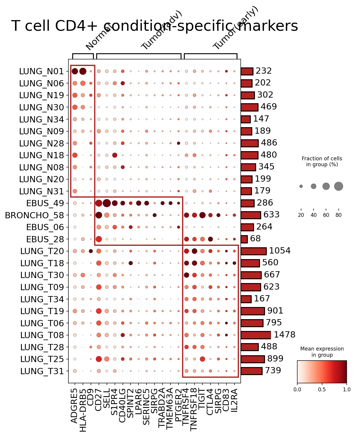
[Analysis Visualization Results]...
Analysis Overview
This analysis identifies condition-specific surfaceome markers in CD4+ T cells by comparing gene expression across Normal, Tumor (advanced), and Tumor (early) conditions within lung tissue. The dot plot visualizes the mean expression level (color intensity) and the fraction of cells expressing each gene (dot size) for selected surface markers across individual samples, which are grouped by condition. The goal is to highlight potential biomarkers and therapeutic targets relevant to lung cancer progression.
Visual Summary
The dot plot effectively displays distinct patterns of surfaceome marker expression on CD4+ T cells across different conditions and samples.
- Normal Condition Markers: Samples from the 'Normal' group (e.g., LUNG_N01, LUNG_N06) show high expression and prevalence of markers such as ADGRE5, HLA-DRB5, CD9, CD27L, SELL, and S1PRG. These genes appear to be highly characteristic of CD4+ T cells in the healthy lung environment.
- Tumor (Advanced) Condition Markers: The 'Tumor(adv)' samples (e.g., EBUS_49, BRONCHO_58) exhibit strong upregulation of markers including CD40LG, SPINT2, LPAR6, SERINC5, SIRPG, TRABD2A, and TMEM63A. These markers are notably absent or expressed at very low levels in the normal condition, suggesting their association with advanced tumor pathogenesis.
- Tumor (Early) Condition Markers: 'Tumor(early)' samples (e.g., LUNG_T20, LUNG_T18) display a unique set of highly expressed markers, particularly PTGER2, TNFRSF4 (OX40), TNFRSF18 (GITR), TIGIT, CTLA4, CD83, and IL2RA (CD25). Some markers like SIRPG are shared with the 'Tumor(adv)' group, indicating potential sustained relevance across tumor stages. The presence of well-known immune checkpoint molecules like TIGIT and CTLA4 is particularly striking in the early tumor microenvironment.
Overall, the plot reveals a clear shift in the surface protein landscape of CD4+ T cells as the tissue transitions from normal to early-stage and then to advanced-stage lung tumor.
Biological Interpretation
The differential expression of these surfaceome markers provides crucial insights into the functional states and roles of CD4+ T cells in the lung tumor microenvironment.
- Normal Lung CD4+ T cell Phenotype: Markers like SELL (CD62L) are classic homing receptors, suggesting the presence of naive or central memory T cells capable of recirculating through lymphoid organs. HLA-DRB5 indicates MHC class II expression, potentially related to antigen presentation or interactions with antigen-presenting cells in homeostatic conditions.
- CD4+ T cells in Advanced Tumors: The upregulation of CD40LG (CD154) suggests T cell activation and interaction with B cells or antigen-presenting cells, which could be part of an ongoing immune response, though its effectiveness needs further context. Other markers like SPINT2, LPAR6, SERINC5, and TRABD2A may reflect altered metabolic or signaling pathways within CD4+ T cells responding to the advanced tumor microenvironment, possibly contributing to tumor progression or immune modulation.
- Immunosuppressive Landscape in Early Tumors: The profile in early tumor samples is highly significant, characterized by the upregulation of several key immune checkpoint molecules and regulatory markers:
- TIGIT (T-cell immunoglobulin and ITIM domain) is an inhibitory receptor often associated with T cell exhaustion in cancer. PubMed search: TIGIT cancer immunotherapy
- CTLA4 (Cytotoxic T-lymphocyte associated protein 4) is a well-established immune checkpoint inhibitor, acting to downregulate T cell activation and proliferation, often expressed on regulatory T cells (Tregs) or exhausted T cells. GeneCards: CTLA4
- IL2RA (CD25), the alpha chain of the IL-2 receptor, is a marker for activated T cells but is also constitutively expressed at high levels on Tregs, which play a major role in immunosuppression within tumors. GeneCards: IL2RA
- The co-expression of TNFRSF4 (OX40) and TNFRSF18 (GITR), which are co-stimulatory receptors, alongside inhibitory checkpoints, suggests a complex interplay of activation and suppression mechanisms in the early tumor microenvironment. This might indicate an attempt by the immune system to respond that is simultaneously being countered by immunosuppressive pathways.
- PTGER2 (EP2) is a receptor for prostaglandin E2, which is frequently elevated in tumors and has immunosuppressive effects on T cells. Its expression suggests CD4+ T cells are sensing and responding to the immunosuppressive lipid environment.
The presence of a distinct immunosuppressive signature (TIGIT, CTLA4, IL2RA) in early tumor CD4+ T cells is a critical finding, indicating that the tumor establishes mechanisms to evade immune surveillance very early in its development.
Clinical or Translational Implications
The identified condition-specific surfaceome markers in CD4+ T cells have significant clinical and translational potential.
- Diagnostic and Prognostic Biomarkers: The distinct marker profiles could serve as biomarkers to differentiate between healthy lung tissue and early or advanced lung tumors. For instance, high expression of TIGIT and CTLA4 on CD4+ T cells might indicate early tumor presence or a pro-tumorigenic immune environment.
- Therapeutic Targets for Immunotherapy: The strong upregulation of immune checkpoint molecules such as TIGIT and CTLA4 in tumor-associated CD4+ T cells, particularly in early-stage disease, positions them as promising therapeutic targets for immunotherapy. Blocking these receptors could reinvigorate anti-tumor immune responses. Strategies combining anti-CTLA4 or anti-TIGIT therapies could be explored, especially given their co-expression. PubMed search: TIGIT CTLA4 lung cancer immunotherapy
- Targeting Immunosuppressive Pathways: The presence of IL2RA (CD25) on CD4+ T cells in tumors points to the potential role of Tregs. Therapies targeting CD25-expressing Tregs could reduce immunosuppression. Similarly, targeting PTGER2 could counteract PGE2-mediated immunosuppression.
- Modulating Co-stimulatory Pathways: The expression of OX40 (TNFRSF4) and GITR (TNFRSF18) suggests that agonists for these receptors could potentially be used to enhance anti-tumor T cell responses, perhaps in combination with checkpoint blockade strategies.
- Experimental Validation: As these are surface markers, they are amenable to further validation using established techniques such as flow cytometry, immunohistochemistry, or immunofluorescence on patient biopsies, which could facilitate their translation into clinical practice.
20. Differential Expression of Cell Cycle Genes in Lung Epithelial Cells Across Tumor Stages
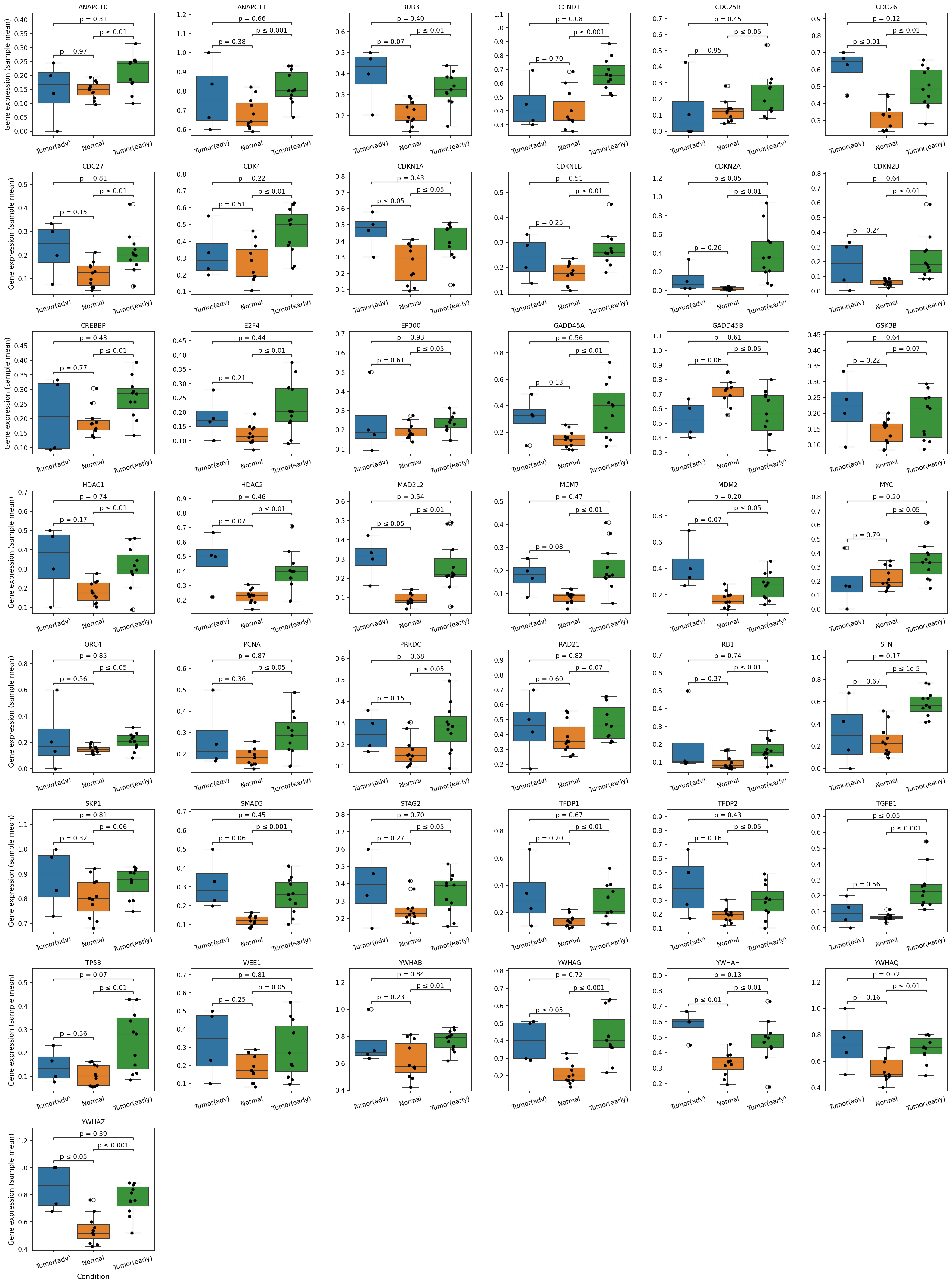
[Analysis Visualization Results]...
Analysis Overview
This analysis investigates the expression patterns of a curated set of cell cycle pathway-related genes within Lung Epithelial cells. The expression levels are compared across three conditions: advanced tumor (Tumor(adv)), early tumor (Tumor(early)), and normal lung tissue. The objective is to identify statistically significant differences in the expression of these crucial regulatory genes, providing insights into cell cycle dysregulation during lung cancer progression.
Visual Summary
The box plots display the gene expression (sample mean) for 24 selected cell cycle-related genes across the three conditions (Tumor(adv), Normal, Tumor(early)). Each dot represents the mean expression from an individual sample within that condition, providing a clear visualization of distribution and outliers. Pairwise statistical comparisons (p-values) are indicated above the boxes for Tumor(adv) vs. Normal, Tumor(early) vs. Normal, and Tumor(adv) vs. Tumor(early).
Key visual observations include:
- General Upregulation in Tumor Conditions: A predominant pattern observed is that many cell cycle-related genes show significantly higher expression in both Tumor(early) and Tumor(adv) conditions compared to Normal Lung Epithelial cells. This suggests an increased proliferative state in tumor cells.
- Higher Expression in Early Tumor Stage: For several genes, the expression appears highest in the Tumor(early) stage compared to both Normal and, in some cases, even Tumor(adv) conditions (e.g., ANAPC10, ANAPC11, BUB3, CCND1, CDC27, CDK4, CDKN1A, CDKN1B, CDC25B, CDC26, CDKN2A, CDKN2B, CREBBP, E2F4, EP300, GADD45A, GADD45B, GSK3B, HDAC1, HDAC2, MAD2L2, MCM7, MDM2, MYC, ORC4, PCNA, PRKDC, RAD21, RB1, SFN, SKP1, SMAD3, STAG2, TFDP1, TFDP2, TGFB1, TP53, WEE1, YWHAB, YWHAE, YWHAG, YWHAH, YWHAQ, YWHAZ).
Statistically Significant Differences
- A large number of genes, including core cell cycle drivers like CCND1 (Cyclin D1), CDK4 (Cyclin-dependent kinase 4), E2F4 (E2F transcription factor 4), MCM7 (Minichromosome Maintenance Complex Component 7), MYC, and PCNA (Proliferating Cell Nuclear Antigen), show significantly elevated expression in Tumor(early) compared to Normal (p ≤ 0.01 or p ≤ 0.05).
- Some genes also show significantly higher expression in Tumor(adv) compared to Normal, though for some the magnitude of difference or significance is less pronounced than for Tumor(early) vs. Normal.
- Interestingly, for many genes, while expression is higher in both tumor conditions than normal, the difference between Tumor(early) and Tumor(adv) is often not statistically significant or is less clear. However, some genes like CDKN1A (p21) show higher expression in Tumor(adv) compared to Normal (p <= 0.05) and also in Tumor(early) vs Normal (p <= 0.05).
- TP53 (Tumor Protein P53), a critical tumor suppressor, shows significantly higher expression in Tumor(early) compared to Normal (p ≤ 0.01), and a trend of higher expression in Tumor(adv) compared to Normal (p = 0.07).
Biological Interpretation
The observed widespread upregulation of cell cycle-related genes in Lung Epithelial cells from both early and advanced tumor conditions, especially when compared to normal tissue, strongly indicates a hyper-proliferative state characteristic of cancer. Given that Lung Epithelial cells are identified as the tumor origin cell type, these changes are directly relevant to tumorigenesis.
- Upregulation of Proliferative Markers: Genes like CCND1, CDK4, E2F4, MCM7, MYC, and PCNA are central to cell cycle progression, DNA replication, and cellular proliferation. Their increased expression is a hallmark of uncontrolled cell growth, driving the transition from normal to cancerous states [1, 2].
- Cyclin D1 (CCND1) and CDK4 form a complex that promotes progression through the G1 phase of the cell cycle. Their overexpression is common in many cancers and can lead to unchecked cell division [1].
- MCM7 is part of the MCM complex essential for DNA replication initiation and elongation. Its elevated levels are often associated with aggressive tumor phenotypes [2].
- MYC is a proto-oncogene that plays a critical role in cell proliferation, growth, and apoptosis. Dysregulation of MYC is a driver in a vast majority of human cancers [3].
- PCNA is a cofactor for DNA polymerase and is widely used as a marker for cell proliferation [4].
- DNA Damage Response and Cell Cycle Checkpoints: Genes like GADD45A and GADD45B (Growth Arrest and DNA Damage Inducible Alpha/Beta) are often induced by cellular stress and DNA damage, mediating cell cycle arrest or apoptosis. Their upregulation in tumor cells could reflect ongoing genomic instability or an attempt by the cell to cope with oncogenic stress, though they can also paradoxically support tumor growth in certain contexts [5].
Dysregulation of Tumor Suppressors and Regulators
- TP53, a critical tumor suppressor, is upregulated in tumor conditions compared to normal. While TP53 mutations are common in cancer, increased wild-type TP53 expression can also occur in response to oncogenic stress or DNA damage as a protective mechanism, or mutated TP53 can accumulate [6]. Further investigation into the mutational status of TP53 would be crucial here.
- CDKN1A (p21) and CDKN2A/B (p16/p15) are cyclin-dependent kinase inhibitors that arrest the cell cycle. Their upregulation in tumor cells could indicate feedback mechanisms attempting to halt proliferation in response to oncogenic signals, or it could be a consequence of specific tumor pathways. For example, p21 can be induced by p53 in response to DNA damage [7].
- Significance in Early Tumor Stage: The observation that many genes show highly significant upregulation in Tumor(early) compared to Normal, and sometimes comparable or even higher levels than Tumor(adv), suggests that cell cycle dysregulation is an early and critical event in lung cancer development. This highlights the importance of these pathways in the initiation and establishment of the tumor phenotype within lung epithelial cells.
Clinical or Translational Implications
The pervasive dysregulation of cell cycle genes in Lung Epithelial cells points to several clinical implications:
- Early Detection and Prognosis: Genes consistently and significantly upregulated in early tumor stages (e.g., CCND1, CDK4, MCM7, MYC, PCNA) could serve as potential biomarkers for early lung cancer detection or as prognostic indicators for disease aggressiveness.
- Therapeutic Targets: The identified cell cycle components represent potential therapeutic targets. Inhibitors of CDKs (e.g., CDK4/6 inhibitors) are already approved for various cancers, and their efficacy in lung cancer, particularly against specific epithelial cell populations, warrants further investigation [8]. Targeting MYC or MCM complexes could also be viable strategies.
- Understanding Tumor Progression: The distinct patterns between early and advanced stages, even if not universally significant, can help differentiate specific biological processes or vulnerabilities that emerge or are sustained during tumor evolution. This knowledge could inform stage-specific treatment approaches.
References
- CCND1 & CDK4 in Cancer: Genecards: CCND1, CDK4.
- MCM7 as a Cancer Biomarker: PubMed search: MCM7 cancer biomarker.
- MYC in Cancer: Genecards: MYC.
- PCNA in Cancer: PubMed search: PCNA proliferation cancer.
- GADD45 in Cancer: PubMed search: GADD45 cancer role.
- TP53 in Cancer: Genecards: TP53.
- CDKN1A (p21) function: Genecards: CDKN1A.
- CDK4/6 inhibitors: PubMed search: CDK4/6 inhibitors lung cancer.
21. 폐 상피세포의 플로이드 상태 및 질병 단계별 유전자 온톨로지(GSA) 분석 결과
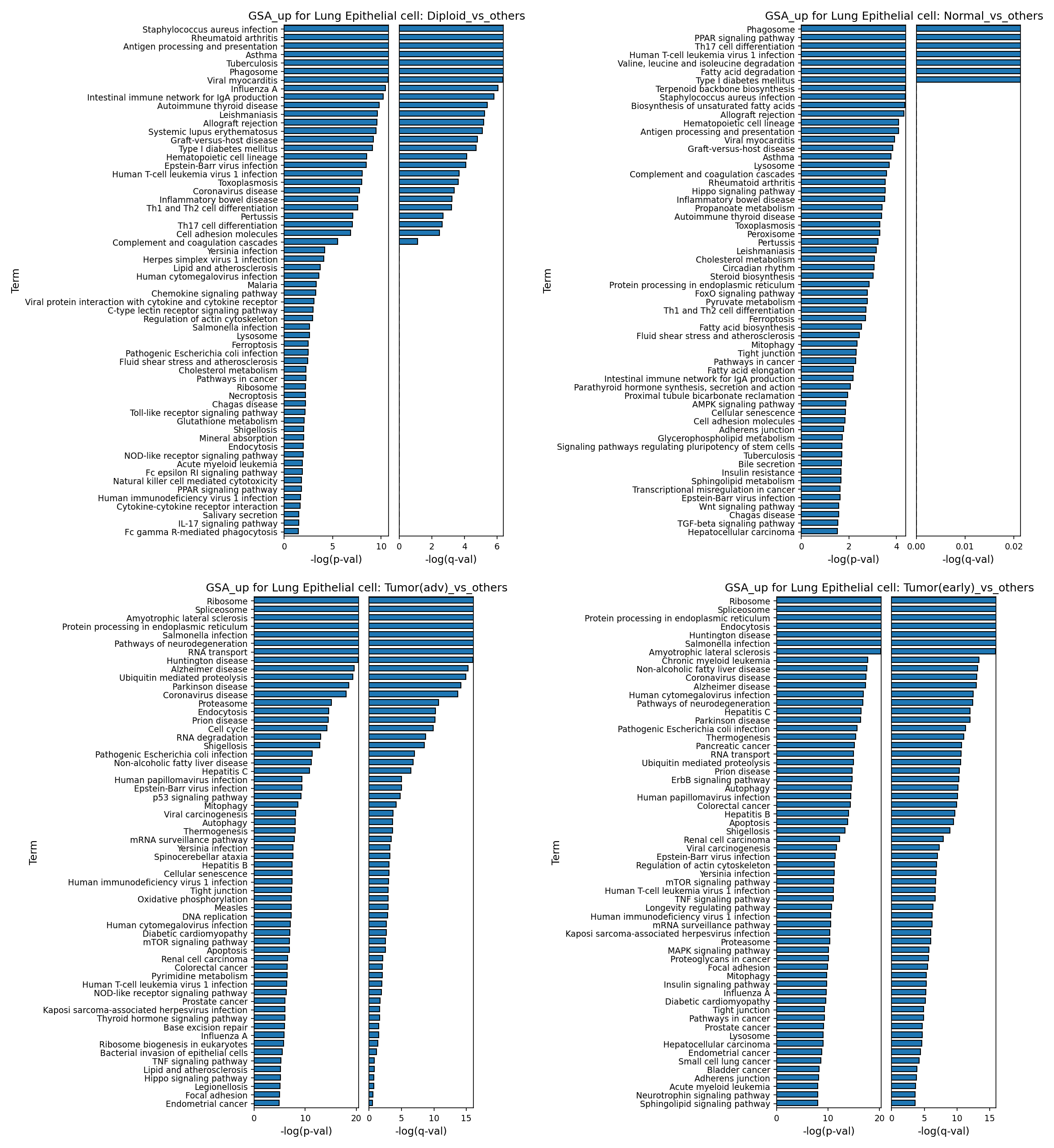
[Analysis Visualization Results]...
Analysis Overview
본 분석은 단일 세포 RNA 시퀀싱(scRNA-seq) 데이터를 활용하여 폐 상피세포(Lung Epithelial cell)에서 발현이 상향 조절되는 유전자들을 기반으로 유전자 온톨로지(Gene Ontology, GO) 경로 농축 분석(GSA)을 수행한 결과입니다. 각 패널은 폐 상피세포를 다음 조건별로 분류하여 '해당 조건 대 나머지 모든 조건(one-vs-rest)'으로 비교했을 때 상향 조절된 유전자가 농축된 GO 경로를 막대 그래프로 보여줍니다:
- Diploid_vs_others: 이배체(Diploid) 핵형을 가진 폐 상피세포 대 나머지 모든 폐 상피세포.
- Normal_vs_others: 정상(Normal) 조직 유래 폐 상피세포 대 나머지 모든 폐 상피세포.
- Tumor(adv)_vs_others: 진행성 종양(Tumor(adv)) 조직 유래 폐 상피세포 대 나머지 모든 폐 상피세포.
- Tumor(early)_vs_others: 초기 종양(Tumor(early)) 조직 유래 폐 상피세포 대 나머지 모든 폐 상피세포.
그래프의 x축은 경로 농축의 통계적 유의성을 나타내는 -log(p-val) 및 -log(q-val) 값을 보여주며, 값이 높을수록 더 유의미한 농축을 의미합니다.
Visual Summary
- Diploid 및 Normal 폐 상피세포 (vs_others): 이 두 그룹의 폐 상피세포는 주로 면역 반응, 항원 제시, 식세포 작용(Phagosome) 및 다양한 감염성 질환(예: *Staphylococcus aureus infection*, *Tuberculosis*, *Influenza A*) 관련 경로가 강하게 농축되어 있습니다. 이는 폐 상피세포가 숙주 방어 및 면역 조절에 중요한 역할을 함을 시사합니다. Normal 세포에서는 지방산 및 아미노산 대사, PPAR signaling pathway와 같은 대사 경로도 함께 관찰됩니다.
- Tumor(early) 및 Tumor(adv) 폐 상피세포 (vs_others): 종양 세포에서는 이배체 또는 정상 세포와는 확연히 다른 경로들이 농축되어 있습니다. "Ribosome", "Spliceosome", "Protein processing in endoplasmic reticulum", "RNA transport/degradation" 등 기본적인 세포 내 단백질 합성 및 처리, RNA 대사 관련 경로가 매우 강하게 농축되어 있습니다. 이는 암세포의 빠른 증식과 성장을 위한 생체 합성 요구 증가를 반영합니다.
- 종양 진행에 따른 변화: 초기 종양과 진행성 종양 세포 간의 농축 경로는 매우 유사한 패턴을 보이며, 많은 핵심적인 세포 기능 이상이 종양의 초기 단계부터 확립됨을 시사합니다. 진행성 종양에서는 "Cell cycle" 경로가 명시적으로 농축되고, "p53 signaling pathway"의 유의성이 더 높아지는 경향이 관찰됩니다.
- 특이 경로: 종양 세포(초기 및 진행성)에서는 "Amyotrophic lateral sclerosis", "Huntington disease", "Alzheimer disease" 등 신경퇴행성 질환과 관련된 경로들이 일관되게 농축되는 특이한 현상이 관찰됩니다. 또한, ErbB signaling pathway는 초기 종양에서, p53 signaling pathway는 진행성 종양에서 두드러집니다.
Biological Interpretation
폐 상피세포는 폐 조직의 구조적 무결성을 유지하고 외부 병원체로부터 보호하는 중요한 역할을 합니다. 본 GSA 결과는 이러한 기능적 차이를 플로이드 상태 및 질병 단계에 따라 명확히 보여줍니다.
- 건강한 폐 상피세포의 면역 및 대사 기능: Diploid 및 Normal 폐 상피세포에서 관찰되는 면역 관련 경로의 농축은 폐 상피세포가 단순한 물리적 장벽을 넘어 항원 제시 세포와 상호작용하며 선천 및 적응 면역 반응에 적극적으로 참여함을 보여줍니다. 이는 폐 상피세포가 감염 및 염증에 대한 최전선 방어자 역할을 한다는 기존 지식과 일치합니다. 정상 세포에서 활발한 대사 경로의 농축은 건강한 세포의 에너지 항상성 유지 및 기능 수행에 필수적인 대사 활동을 반영합니다. PubMed search: lung epithelial cell immune function
- 종양 세포의 생체 합성 재편: 종양 세포(초기 및 진행성)에서 리보솜, 스플라이소좀, ER 내 단백질 처리 등 생체 합성 및 단백질 항상성 관련 경로가 강하게 상향 조절되는 것은 암세포가 통제되지 않는 증식을 위해 필요한 막대한 양의 단백질과 세포 구성 요소를 생산하기 위한 대사 재편성을 겪고 있음을 나타냅니다. 이러한 경로는 암세포의 "성장 욕구(growth imperative)"를 직접적으로 반영합니다.
- 암 발생 및 진행의 핵심 신호 경로: 초기 종양 세포에서 ErbB signaling pathway의 활성화는 상피세포 성장 인자 수용체(EGFR) 계열의 활성화를 시사하며, 이는 폐암에서 흔히 관찰되는 발암 경로입니다 (데이터 컨텍스트에 EGFR_mutation 정보 포함). 진행성 종양에서 p53 signaling pathway의 농축은 종양 억제 유전자인 p53의 기능 이상 또는 스트레스 반응으로 인한 활성화를 나타내며, 이는 암 진행의 중요한 특징입니다. GeneCards: EGFR, GeneCards: TP53
- 종양 세포의 독특한 스트레스 반응: 초기 및 진행성 종양 세포에서 공통적으로 신경퇴행성 질환 관련 경로들이 농축되는 것은 흥미로운 발견입니다. 이들 경로는 단백질 오접힘(protein misfolding), 응집(aggregation), 프로테아좀 기능 이상 및 미토콘드리아 기능 장애와 같은 세포 스트레스 반응과 연관되어 있습니다. 이는 암세포가 겪는 높은 대사 스트레스와 불안정한 유전체 환경이 신경퇴행성 질환과 유사한 세포 스트레스 반응 기전을 유도할 수 있음을 시사합니다.
Clinical or Translational Implications
- 바이오마커 및 조기 진단: 정상 폐 상피세포와 종양 폐 상피세포 간의 면역 및 대사, 그리고 생체 합성 경로의 뚜렷한 차이는 폐암의 조기 진단 및 정상 조직과 암 조직 구별을 위한 새로운 바이오마커 개발의 가능성을 제시합니다.
치료 표적 발굴:
- 종양 세포에서 리보솜, 스플라이소좀, 단백질 처리 경로의 과도한 활성화는 이러한 기본적인 세포 기구를 표적으로 하는 항암제 개발의 근거를 제공할 수 있습니다. 예를 들어, 단백질 합성 억제제나 스플라이소좀 억제제 등은 암세포의 높은 생체 합성 수요를 차단하여 치료 효과를 낼 수 있습니다.
- 초기 종양에서 ErbB signaling pathway의 활성화는 EGFR 돌연변이 양성 폐암 환자에게 사용되는 EGFR 티로신 키나아제 억제제(TKI)의 효능을 뒷받침하며, 해당 환자군을 위한 표적 치료 전략의 중요성을 강조합니다.
- 진행성 종양의 p53 signaling pathway 변화는 p53 복구 또는 활성화 전략이 진행성 폐암 치료에 잠재적인 이점을 가질 수 있음을 시사합니다.
- 신규 치료 접근법: 신경퇴행성 질환과 공유하는 세포 스트레스 반응 경로의 발굴은 기존 신경퇴행성 질환 치료제를 암 치료에 재활용(drug repurposing)하거나, 단백질 항상성 및 세포 스트레스 반응을 조절하는 새로운 치료 전략을 탐색할 수 있는 기회를 제공합니다.
22. Gene Set Enrichment Analysis Reveals Distinct Pathway Deregulation Across Lung Cancer Cell Types
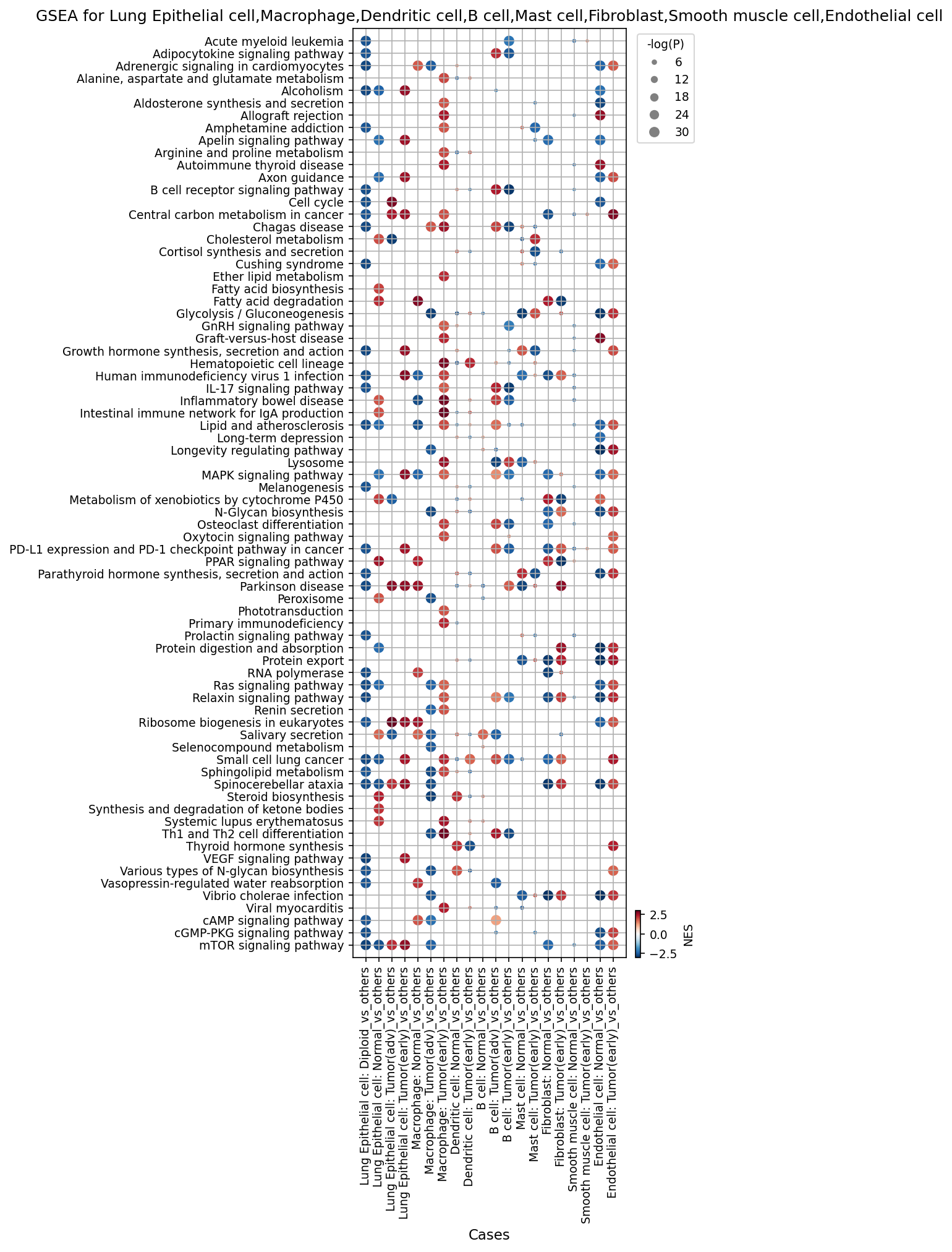
[Analysis Visualization Results]...
Analysis Overview
This analysis presents Gene Set Enrichment Analysis (GSEA) results for key major cell types found in human lung tissue, comparing specific conditions (Diploid, Normal, Tumor(early), Tumor(adv)) against all other conditions within the same cell type (e.g., Lung Epithelial cell: Tumor(early) vs. all other Lung Epithelial cells across other conditions). The goal is to identify significantly altered biological pathways across different cell types in the context of lung cancer progression. The results are visualized as a dot plot, where the color of each dot represents the Normalized Enrichment Score (NES) – red for positive enrichment (upregulation) and blue for negative enrichment (downregulation) – and the size of the dot indicates the statistical significance (-log(p-value)).
Visual Summary
The dot plot effectively visualizes pathway enrichment patterns across nine major cell types: Lung Epithelial cell, Macrophage, Dendritic cell, B cell, Mast cell, Fibroblast, Smooth muscle cell, and Endothelial cell, across various conditions (Diploid, Normal, Early Tumor, Advanced Tumor).
- Dominant Upregulation in Tumor Conditions: A striking observation is the prevalence of red dots (positive NES, indicating upregulation) in the columns representing "Tumor(early)_vs_others" and "Tumor(adv)_vs_others" across most cell types, especially Lung Epithelial cells, Macrophages, Dendritic cells, and Endothelial cells. This suggests a widespread activation of numerous biological pathways in the tumor microenvironment.
- Downregulation in Normal/Diploid Conditions: Conversely, "Normal_vs_others" and "Diploid_vs_others" comparisons often show blue dots (negative NES), particularly for proliferative and metabolic pathways. This indicates these pathways are relatively less active in normal or diploid cells compared to the tumor-associated cells.
- Consistent Cancer-Associated Pathways: Pathways related to cell cycle, proliferation (e.g., "Cell cycle"), metabolism (e.g., "Glycolysis / Gluconeogenesis", "Ribosome biogenesis in eukaryotes"), and key signaling pathways (e.g., "MAPK signaling pathway", "mTOR signaling pathway", "VEGF signaling pathway") are frequently and significantly upregulated in tumor conditions across multiple cell types.
- Immune Evasion Pathways: "PD-L1 expression and PD-1 checkpoint pathway in cancer" is notably upregulated in tumor-associated Lung Epithelial cells, Macrophages, Dendritic cells, and B cells, highlighting a common mechanism of immune suppression.
- Stromal and Endothelial Remodeling: Fibroblasts and Endothelial cells in tumor conditions show upregulation of metabolic and signaling pathways, consistent with their active roles in remodeling the tumor microenvironment and angiogenesis.
Biological Interpretation
The GSEA results provide clear insights into the perturbed biological processes within different cellular compartments during lung cancer development:
- Epithelial Oncogenesis and Metabolic Reprogramming: Lung Epithelial cells, the presumed cell of origin for lung cancer, exhibit robust upregulation of pathways critical for proliferation ("Cell cycle"), altered metabolism ("Glycolysis / Gluconeogenesis", "Ribosome biogenesis in eukaryotes", "Fatty acid degradation", "Cholesterol metabolism"), and survival signaling ("MAPK signaling pathway", "mTOR signaling pathway"). This metabolic rewiring (known as the Warburg effect and increased anabolism) is a hallmark of cancer, supporting rapid cell division and biomass accumulation PubMed: Warburg effect cancer metabolism. The strong activation of "VEGF signaling pathway" in tumor epithelial cells suggests their direct contribution to an angiogenic environment, fostering tumor vascularization GeneCards: VEGF. The downregulation of these pathways in normal and diploid epithelial cells underscores the drastic functional shift in cancerous cells.
- Tumor-Associated Macrophage (TAM) Activity: Macrophages in both early and advanced tumor conditions show significant enrichment for pathways involved in immune modulation ("PD-L1 expression and PD-1 checkpoint pathway in cancer", "IL-17 signaling pathway"), inflammation ("Adipocytokine signaling pathway"), and metabolic reprogramming. This phenotype is characteristic of M2-like TAMs, which promote tumor growth, angiogenesis, and immune suppression within the tumor microenvironment PubMed: Tumor associated macrophages cancer.
- Dendritic Cell and B Cell Immune Evasion: Dendritic cells and B cells in the early tumor microenvironment exhibit upregulation of "PD-L1 expression and PD-1 checkpoint pathway in cancer", implying their shift towards an immunosuppressive role rather than immune activation. This indicates that multiple immune cell types are co-opted by the tumor to evade host immunity. B cell receptor signaling pathway upregulation in early tumor B cells suggests their activation, possibly leading to the production of tumor-promoting antibodies or acting as antigen-presenting cells in an altered manner PubMed: B cells tumor microenvironment.
- Stromal Support and Angiogenesis: Fibroblasts and Endothelial cells in early tumor conditions show upregulated metabolic pathways (e.g., "Glycolysis / Gluconeogenesis", "Ribosome biogenesis in eukaryotes") and signaling pathways like MAPK and mTOR. Endothelial cells also show strong activation of "VEGF signaling pathway", directly supporting tumor angiogenesis—the formation of new blood vessels essential for tumor growth and metastasis PubMed: Angiogenesis cancer. Fibroblasts, likely transitioning to cancer-associated fibroblasts (CAFs), contribute to extracellular matrix remodeling and provide growth factors that support tumor progression PubMed: Cancer associated fibroblasts.
- Pathway Interconnectivity: The recurring enrichment of "MAPK signaling pathway" and "mTOR signaling pathway" across various tumor-associated cell types highlights their central roles as master regulators of cell proliferation, survival, and metabolism, often aberrantly activated in cancer.
Clinical or Translational Implications
These findings have significant clinical and translational implications for lung cancer:
- Immunotherapy Targets: The widespread upregulation of "PD-L1 expression and PD-1 checkpoint pathway in cancer" across Lung Epithelial cells, Macrophages, Dendritic cells, and B cells in tumor conditions strongly supports the rationale for PD-1/PD-L1 blockade immunotherapy in lung cancer. It also suggests that targeting PD-L1 on non-tumor cells in the TME could be crucial for therapeutic efficacy PubMed: PD-1 PD-L1 lung cancer immunotherapy.
- Metabolic Targeting: The observed metabolic reprogramming, particularly enhanced glycolysis and ribosome biogenesis in tumor and associated stromal cells, presents opportunities for novel therapeutic strategies that target cancer metabolism to inhibit tumor growth and improve drug sensitivity.
- Combination Therapies: Given the involvement of multiple signaling pathways (MAPK, mTOR, VEGF) and cell types, combination therapies that simultaneously target these pathways or the interactions between tumor and stromal cells could be more effective than single-agent approaches.
- Biomarkers for Progression: The distinct pathway signatures identified could serve as potential biomarkers for distinguishing early from advanced disease or for predicting treatment response. For instance, specific metabolic pathway signatures in TAMs might indicate their pro-tumorigenic polarization, guiding treatment selection.
- Understanding Disease Heterogeneity: The cell-type-specific pathway enrichments underscore the heterogeneous nature of the tumor microenvironment and the need for precision medicine approaches that consider the unique contributions of each cell type to disease progression.
23. Discussion
The integrated single-cell analysis of human lung tissue across normal, early, and advanced tumor stages provides a high-resolution view of the dynamic cellular and molecular landscape of lung cancer progression. A central finding is the robust identification of malignant lung epithelial cells, characterized by clear aneuploidy and extensive copy number variations (CNVs), which are largely confined to tumor samples. Recurrent amplifications of key oncogenes like EGFR (7p12.1:7q21.11) and EIF3E (8q22.1:8q24.12) were identified within these tumor-origin cells, underscoring their potential as drivers of tumorigenesis and progression (Sections 4, 11). These malignant epithelial cells exhibit a hyper-proliferative state, with significant upregulation of core cell cycle genes (e.g., CCND1, CDK4, MYC, PCNA) from early tumor stages, alongside metabolic reprogramming (glycolysis, ribosome biogenesis) and activation of survival signaling pathways (MAPK, mTOR, VEGF) (Sections 20, 21, 22).
The tumor microenvironment (TME) undergoes significant remodeling from early to advanced stages, profoundly impacting immune and stromal cell compartments. Immune populations show a progressive shift towards immunosuppression: a general reduction in cytotoxic T cells and NK cells, alongside a subtle but consistent increase in regulatory T cells (Tregs), is observed (Sections 6, 7). Statistically, early tumors show elevated Th1, Th2, Th17, and Treg cells, while advanced tumors exhibit a decrease in Th1, Th2, Th17, ILC1, ILCreg, and LTI cells, indicating a progressive weakening of anti-tumor immunity (Section 8). CD4+ T cells in early tumors express a distinct immunosuppressive signature with upregulated TIGIT, CTLA4, and IL2RA (CD25) (Section 19). Particularly striking is the reprogramming of macrophages, transitioning from a balanced M1/M2A profile in normal tissue to a dominance of M2A and M2B phenotypes, with a significant expansion of pro-tumorigenic M2B macrophages in advanced tumors (Sections 9, 10). Condition-specific surface markers on macrophages, such as upregulation of SIRPB1, ABCA1, FCGR2B, and FOLR2 in tumor conditions, further define their immunosuppressive, pro-tumorigenic state (Section 17). Similarly, fibroblasts transform into cancer-associated fibroblasts (CAFs), marked by increased expression of FAP, PDGFRB, and MMP14, actively contributing to extracellular matrix remodeling and creating a supportive niche for tumor growth (Section 18).
Complex cell-cell interaction (CCI) networks orchestrate these microenvironmental changes. Normal tissue CCIs reflect tissue homeostasis, while early tumors show emerging interactions involving aneuploid cells, WNT signaling, and early angiogenesis (VEGFA-NRP2). Advanced tumors exhibit a profoundly altered and intensified CCI landscape, driven predominantly by interactions involving aneuploid tumor cells. Key pro-tumorigenic pathways include widespread Prostaglandin E2 (PGE2) signaling (via PTGES3-PTGERs), extensive integrin-mediated interactions (e.g., FN1, TNC, SPP1 with integrins) critical for ECM remodeling and invasion, and sustained angiogenesis (VEGFA-NRP2) (Sections 12, 13, 15). Furthermore, a detailed analysis of immune checkpoint and cell cycle related genes in CCIs reveals sustained EGFR pathway activation and, notably, a pervasive upregulation of TGF-beta signaling (TGFB1_TGFbeta_receptor1/2 and TGFB1_integrin_avb6_complex) in advanced tumors, promoting immunosuppression and tumor progression across various cell types (Section 14). Overall, this multi-faceted analysis provides a comprehensive understanding of how lung cancer cells rewire their genomic, transcriptional, and microenvironmental landscapes to drive progression.
Hypotheses:
- Aneuploidy in lung epithelial cells is a primary driver of tumor progression, correlating with the activation of oncogenic pathways (EGFR, MYC) and profound genomic instability from early stages of lung cancer.
- Lung cancer progression is characterized by a dynamic remodeling of the immune microenvironment towards an immunosuppressive state, driven by a decline in anti-tumor T cells and NK cells, an increase in Tregs, and a polarization of macrophages towards pro-tumorigenic M2-like phenotypes, mediated by specific cell-cell interactions (e.g., SIRPA-CD47, PGE2 signaling).
- The tumor microenvironment actively supports tumor growth and invasion through comprehensive stromal reprogramming, involving activated fibroblasts (CAFs) that remodel the extracellular matrix (e.g., via MMP14, SPP1-integrin interactions) and promote angiogenesis (e.g., VEGFA-NRP2 signaling) from early stages.
Potential therapeutic targets:
- EGFR/ERBB2 (HER2): Amplified and highly expressed in malignant lung epithelial cells, driving proliferation and survival. Established oncogenic drivers in lung cancer. Evidence: Sections 4, 14, 16 (CNV, CCI, Marker expression) Validation: Targeted therapies like EGFR TKIs and HER2-targeting antibodies are standard-of-care, but understanding specific ligand interactions or co-targets can refine their use.
- TGF-beta signaling (e.g., TGFB1-TGFbeta_receptor1/2, TGFB1-integrin_avb6_complex): Pervasively upregulated in advanced tumors across multiple cell types, driving immunosuppression, epithelial-mesenchymal transition (EMT), and fibrosis. Evidence: Section 14 (CCI) Validation: Clinical trials for TGF-beta inhibitors are ongoing; in vitro/in vivo studies can validate their role in reversing immunosuppression and inhibiting tumor progression.
- Immune Checkpoints (TIGIT, CTLA4, PD-1/PD-L1): TIGIT and CTLA4 are upregulated on CD4+ T cells in early tumors. PD-L1/PD-1 pathway is broadly upregulated across tumor-associated immune and epithelial cells, mediating immune evasion. Evidence: Sections 19, 22 (Marker expression, GSEA) Validation: Targeting these pathways with checkpoint inhibitors (e.g., anti-TIGIT, anti-CTLA4, anti-PD-1/PD-L1) is a proven strategy, with ongoing studies for combination therapies.
- Tumor-Associated Macrophage (TAM) markers (SIRPB1, FOLR2, FCGR2B): Upregulated on pro-tumorigenic M2-like macrophages in tumor microenvironment, contributing to immune suppression and tumor growth. Evidence: Section 17 (Marker expression) Validation: Developing antibody-drug conjugates or small molecule inhibitors to deplete or reprogram these specific TAM populations, or blocking their interactions (e.g., SIRPB1-CD47).
- Cancer-Associated Fibroblast (CAF) markers (FAP, PDGFRB, MMP14): Highly expressed on activated fibroblasts in tumor conditions, driving ECM remodeling, angiogenesis, and providing pro-tumorigenic support. Evidence: Section 18 (Marker expression) Validation: Targeting FAP-positive CAFs with specific antibodies or CAR-T cells, or inhibiting PDGFRB/MMP14 to disrupt stromal support and hinder invasion.
- Prostaglandin E2 (PGE2) pathway (PTGES3-PTGERs): Strong and pervasive signaling in advanced tumors, mediating immunosuppression, angiogenesis, and tumor growth. Evidence: Sections 12, 15 (CCI) Validation: Inhibitors of COX-2 (PGE2 synthesis) or PGE2 receptors (PTGERs) to disrupt pro-tumorigenic feedback loops and enhance anti-tumor immunity.
- SPP1-Integrin axis: Markedly upregulated in advanced tumors, crucial for tumor cell survival, proliferation, angiogenesis, and metastasis by mediating cell-ECM interactions. Evidence: Section 13 (CCI) Validation: Developing inhibitors against SPP1 or specific integrin receptors (e.g., αvβ3, αvβ1) to disrupt tumor cell adhesion, migration, and survival.
- VEGFA-NRP2/VEGFRs: Increased activity in tumor conditions, indicating enhanced angiogenesis crucial for tumor growth and metastasis. Evidence: Sections 13, 15, 22 (CCI, GSEA) Validation: Anti-VEGF/VEGFR therapies are established, but specific targeting of NRP2 or combination with other agents may enhance efficacy.
- Cell Cycle Regulators (e.g., CDK4/6, MYC): Upregulated in malignant lung epithelial cells, driving hyper-proliferation from early stages of tumorigenesis. Evidence: Section 20 (Gene expression) Validation: CDK4/6 inhibitors are effective in other cancers; evaluate their efficacy in specific lung cancer patient subsets, potentially combined with other therapies.
Follow-up validation ideas:
- Confirm EGFR and EIF3E amplifications and expression in larger patient cohorts using FISH, IHC, or targeted sequencing. Conduct functional studies (e.g., CRISPR knockout/knockdown) in lung cancer cell lines or organoids to validate the impact of these amplifications on cell cycle dysregulation, proliferation, and tumor growth.
- Utilize flow cytometry or multiplex immunohistochemistry on patient lung tumor biopsies to quantify shifts in T cell, NK cell, Treg, and macrophage populations and their polarization markers. Perform in vitro co-culture assays with tumor cells, macrophages, and T cells to functionally validate the roles of identified CCI pairs (e.g., SIRPA-CD47, PGE2 receptors) in mediating immune suppression or macrophage polarization.
- Validate CAF markers (FAP, PDGFRB, MMP14) and their spatial distribution in tumor sections using IHC/immunofluorescence. Employ 3D co-culture models of tumor cells and fibroblasts to study ECM remodeling, invasion, and angiogenesis mediated by specific CCI (e.g., SPP1-integrin, VEGFA-NRP2) and test the efficacy of inhibitors targeting these pathways.
Limitations:
The single-cell RNA-seq data inherently lacks direct spatial information, limiting a precise understanding of cell-cell interactions within the complex tissue architecture. While computational inference of cell-cell interactions is provided, direct validation of cell proximity and contact in situ would be beneficial. The current findings are largely correlative; functional validation through in vitro and in vivo models is required to establish causality. Inter-patient heterogeneity, observed in cell populations and marker expression, implies that broad conclusions should be carefully applied to individual patients. The 'unassigned' cell populations, particularly prominent in advanced tumors, suggest the presence of highly aberrant or novel cell states that warrant further characterization. Ploidy status, although a robust indicator of malignancy, is inferred rather than directly measured for every single cell, which has inherent limitations. Furthermore, while the dataset covers diverse conditions, the number of individual samples per condition, especially for 'Tumor(adv)', is relatively small, which might affect the statistical power for some comparisons and the generalizability of certain findings.
24. Query List
- Show UMAP with condition, sample, celltype_major, celltype_minor, ploidy_dec, and celltype_subset in 2 columns and save it.
- Show major cell type scores on UMAP and save it.
- Show the overall Celltype_subset marker expression dot plot and save it. Keep target_cell as None and keep the other arguments at their default values.
- Select tumor-origin cells and unassigned cells, group them by sample, show CNV heatmap, include a summary of significantly amplified copy number regions, and save it.
- Show CNV patterns as UMAP, including major cell type, minor cell type, ploidy results, condition, and sample in 2 columns, and save it.
- Show population bar plot for minor cell types and save it.
- Show subset population bar plot for T cells and save it.
- Show box plots for statistically significant differences in T cell subset populations between conditions, and save it. Set ncols appropriately based on the total number of panels.
- Show subset population bar plot for macrophages and save it.
- Show box plots for statistically significant differences in macrophage subset populations between conditions, and save it. Set ncols appropriately based on the total number of panels.
- Select tumor-origin cells and unassigned cells, show their ploidy population as a bar plot, and save it.
- Show cell-cell interaction patterns including Lung Epithelial cell, Fibroblast, Macrophage, and T cell, grouped by condition, and save it. Select up to 80 cell-cell interactions per condition.
- Show cell-cell interactions by condition and save the result. Select at most 80 cell-cell interactions for each condition.
- Select only genes related to immune checkpoint and cell cycle pathways, show cell-cell interactions for these genes, and save it.
- Find statistically significant differences in cell-cell interactions for major immune and stromal cells by condition, show them as a dot plot, and save it. Set max_n_items_per_group = 25.
- Show the condition-specific markers for tumor-origin cells (Lung Epithelial cell) as a dot plot and save the result. Use only surfaceome markers, up to 50 markers per condition.
- Extract condition-specific markers for Macrophage, show them as a dot plot, and save it. Show only surfaceome markers, up to 50 per condition.
- Extract condition-specific markers for Fibroblast, show them as a dot plot, and save it. Show only surfaceome markers, up to 50 per condition.
- Extract condition-specific markers for T cell CD4+, show them as a dot plot, and save it. Show only surfaceome markers, up to 50 per condition.
- Select cell cycle pathway-related genes, find statistically significant differences in their expression by condition for Lung Epithelial cell, show them as a box plot, and save it. Set max_n_items_to_plot = 24, and set ncols appropriately to achieve a 2x3 aspect ratio based on the total number of panels.
- Show Gene Ontology (GSA) analysis results for epithelial cells as bar plots and save the result.
- Show Gene set enrichment analysis results as a dot plot for major cell types, and save it. Set color map to RdBu_r and n_pws_to_show = 80.