Single-Cell Transcriptomic Analysis Reveals Cellular Reprogramming and Dysregulated Communication in Human Liver Cirrhosis
This report details a single-cell RNA sequencing analysis of human liver tissue, comparing healthy controls with cirrhotic conditions. Key findings include dramatic shifts in cellular composition, particularly a reduction in hepatocytes and an expansion of immune and fibrogenic cells. Intercellular communication is profoundly altered, characterized by increased pro-inflammatory, pro-fibrotic, and pro-angiogenic signaling. Furthermore, specific cell types exhibit distinct transcriptional reprogramming, pointing to dysregulated metabolic, immune, and proliferative pathways that drive disease progression.
Contents
- Dataset overview
- UMAP Visualization of Single-Cell Liver Data Across Conditions and Cell Types
- Major Cell Type Score UMAP Visualization and Annotation Assessment
- Marker Expression Dot Plot for Cell Subsets
- Cell Type Population Analysis in Healthy and Cirrhotic Liver
- Population Dynamics of T cell Subsets in Cirrhotic vs. Healthy Liver
- Alterations in Liver T Cell Subset Proportions in Cirrhosis
- Macrophage Subset Population Analysis in Cirrhosis
- Macrophage Subset Population Shifts in Cirrhosis
- Cell-Cell Interaction Patterns in Liver Cirrhosis and Healthy Conditions
- Condition-Specific Cell-Cell Interaction Patterns in Liver Cirrhosis
- Cholangiocyte Condition-Specific Surfaceome Markers in Liver Cirrhosis
- Macrophage Condition-Specific Surfaceome Markers in Liver Cirrhosis
- Hepatic Stellate Cell Condition-Specific Surfaceome Markers in Cirrhosis
- T cell CD4+ Condition-Specific Surfaceome Markers in Liver Cirrhosis
- Gene Ontology (GSA) Analysis of Cholangiocytes in Liver Cirrhosis
- Gene Set Enrichment Analysis of Cell Type-Specific Pathways in Cirrhosis
- Discussion
- Query List
0. Dataset overview
Dataset Summary
- Data Type: Single-cell RNA-seq data, processed into an AnnData object.
- Dimensions: Contains 60,503 cells and 22,784 genes.
- Species & Tissue: Data is from human liver tissue.
- Conditions: Includes samples from 'healthy' and 'cirrhosis' conditions.
Cell Type Annotations:
- Major: Myeloid cell, T cell, B cell, Plasma cell, Stromal cell, Liver Epithelial cell, Mast cell, Endothelial cell, unassigned.
- Minor: Macrophage, T cell CD8+, NK cell, B cell, T cell CD4+, Dendritic cell, Plasma cell, Hepatic stellate cell, Hepatocyte, Mast cell, Endothelial cell, Cholangiocyte, ILC, Smooth muscle cell, unassigned, Fibroblast.
- Subset: Detailed sub-types like Macrophage (M1), T cell (Cytotoxic), etc.
- Computed Results: Precomputed results available for Cell-Cell Interaction (CCI), Differential Gene Expression (DEG), Gene Set Enrichment Analysis (GSEA), and Gene Ontology (GSA) for various cell types and conditions.
- Reference Condition: 'healthy' is used as the reference for DEG, GSEA, and GSA analyses.
- CNV Estimates: Copy Number Variation estimates are not available in this dataset.
- Tumor Identification: No tumor identification has been performed or is available.
Inferred Biological Context
- The primary disease context being investigated is Cirrhosis, directly stated in the dataset's condition metadata. This aligns with the dataset originating from liver tissue.
- Analyses such as DEG, GSEA, and GSA are set up to compare conditions against the 'healthy' reference, further emphasizing the focus on understanding cirrhosis.
- Uncertainty: Low, as the condition 'cirrhosis' is explicitly present in the metadata. However, specific details on etiology, severity, or stage of cirrhosis are not provided.
1. UMAP Visualization of Single-Cell Liver Data Across Conditions and Cell Types
[Analysis Visualization Results]...
Analysis Overview
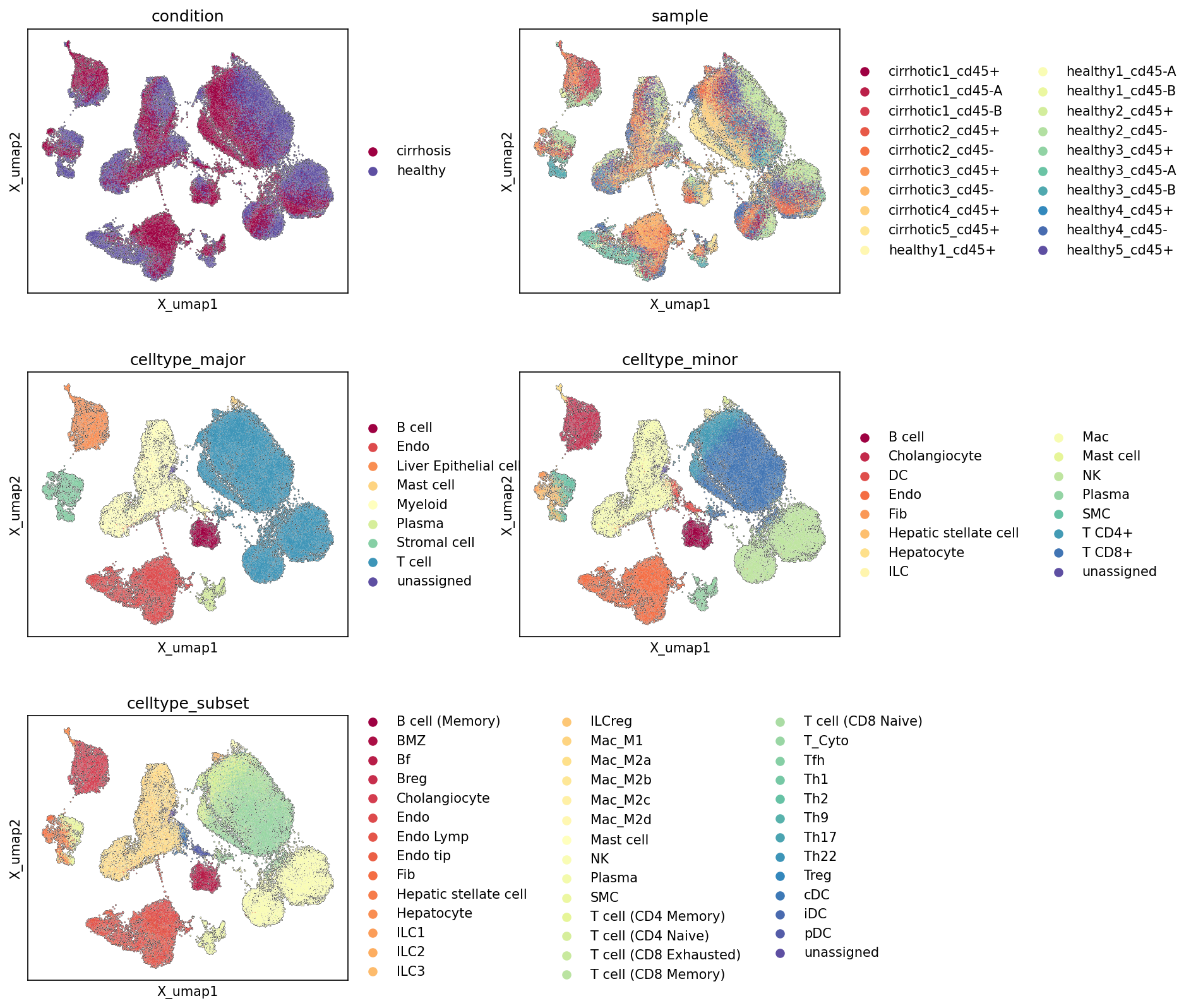
This analysis presents Uniform Manifold Approximation and Projection (UMAP) plots, visualizing the global transcriptional landscape of single-cell RNA-sequencing data from human liver tissue. The UMAPs are colored by various metadata attributes: condition (healthy vs. cirrhosis), sample, celltype_major, celltype_minor, and celltype_subset. The primary objective is to inspect the overall data structure, assess the quality of cell type annotations, identify potential batch effects, and observe the distribution of cells from different conditions within the embedding space.
Visual Summary
Condition and Sample Distribution
The UMAP plot colored by condition shows a clear separation between healthy and cirrhosis cells, particularly in several distinct regions of the embedding. Cirrhosis cells (red) appear to form several dominant clusters that are either distinct from or largely intermingled with healthy cells (blue) in other clusters. This indicates that transcriptomic differences between the healthy and cirrhotic liver are substantial and drive the overall separation in the UMAP space.
The sample UMAP plot reveals that cells from individual samples generally mix well within their respective conditions, without strong clustering of cells purely based on the original sample identifier. While some samples show localized enrichment, the overall structure largely reflects the condition-driven separation, suggesting that major batch effects originating from individual samples are not overtly dominating the UMAP projection. The variety of colors for different samples helps confirm broad representation of cells across the UMAP space, supporting the robust nature of the embedding.
Cell Type Clustering and Annotation Quality
The UMAPs colored by celltype_major, celltype_minor, and celltype_subset demonstrate a high degree of coherence and separation for most cell populations.
- celltype_major: Major cell types such as Myeloid cells, T cells, Liver Epithelial cells, Stromal cells, and B cells form well-defined, distinct clusters. This indicates that major cell lineages are transcriptionally unique and clearly distinguishable in the dataset.
- celltype_minor: Further refinement into minor cell types (e.g., Macrophage, Hepatocyte, Hepatic stellate cell, Cholangiocyte, T cell CD4+, T cell CD8+) also shows excellent separation. Specific immune cell populations (e.g., Macrophage, T cells, B cells) form large, dense clusters, while parenchymal and stromal cells (Hepatocyte, Hepatic stellate cell, Cholangiocyte) also occupy distinct regions.
- celltype_subset: At the highest resolution, celltype_subset annotations reveal fine-grained populations like different macrophage subtypes (M1, M2A, M2B, M2C, M2D), T cell subtypes (CD4 Naive, CD8 Memory, Cytotoxic, Treg, Exhausted), and B cell subtypes (Follicular, MZ, Memory, Breg). These subsets generally maintain their unique cluster identities, although some closely related subsets show more intermingling, which is expected given their shared lineage and potentially overlapping transcriptional states. For example, various macrophage subsets might show some continuum, and T cell subsets derived from the same major lineage are clustered in proximity.
Biological Interpretation
The observed separation of cirrhosis and healthy cells in the UMAP suggests significant transcriptomic reprogramming occurring in the liver microenvironment during cirrhosis. This differential clustering is likely driven by condition-specific gene expression changes impacting multiple cell types and their proportions.
The clear and distinct clustering of most celltype_major, celltype_minor, and celltype_subset annotations indicates that the cell type assignments are robust and biologically meaningful. This strong cellular identity separation provides a solid foundation for downstream analyses, such as differential gene expression or cell-cell interaction studies, ensuring that comparisons are made between genuinely distinct cell populations.
For instance, the prevalence of certain cell types in the "cirrhosis-dominant" regions of the UMAP (e.g., potentially enriched immune or stromal cell clusters, or altered liver epithelial populations) might point to their involvement in disease pathology. Conversely, "healthy-dominant" clusters would represent populations or states more characteristic of a non-diseased liver. The granularity of the celltype_subset annotations allows for the investigation of specific immune cell activation states or dysfunctional parenchymal/stromal cells in the context of cirrhosis. For example, specific macrophage polarization states (M1, M2 subtypes) or T cell exhaustion (T cell (CD8 Exhausted)) could be differentially enriched in cirrhotic samples, aligning with known immune responses in chronic liver disease (e.g., PubMed search for "liver cirrhosis immune cell infiltration").
Annotation Notes
The UMAP plots provide strong evidence for high-quality cell type annotations across major, minor, and subset levels. The distinct clustering for most cell populations validates the accuracy of the cell assignments, which is crucial for reliable interpretation of biological processes. The lack of severe sample-specific batch effects dominating the overall structure is also a positive indicator for the quality and generalizability of the dataset. These robust annotations and dataset structure are well-suited for subsequent in-depth analyses, such as identifying differentially expressed genes within specific cell types or investigating cell-cell communication changes in cirrhosis.
2. Major Cell Type Score UMAP Visualization and Annotation Assessment
[Analysis Visualization Results]...
Analysis Overview
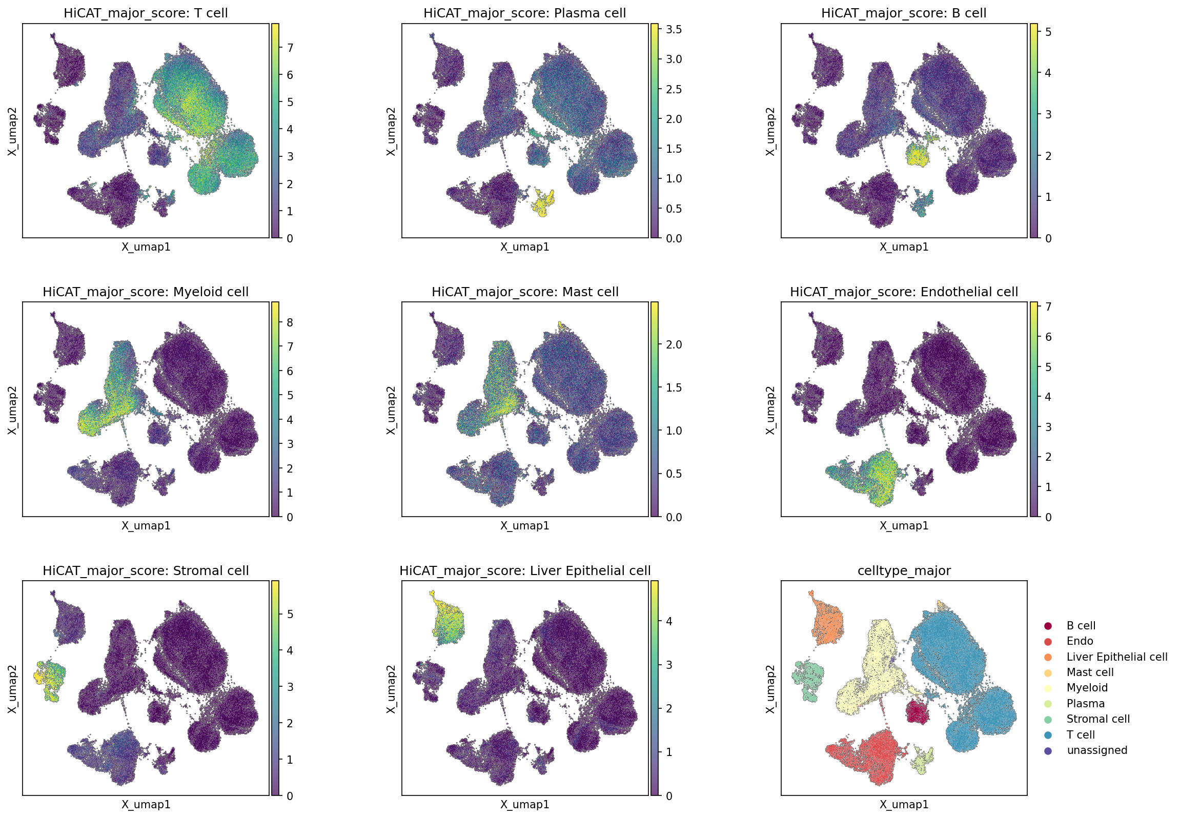
This analysis visualizes the distribution of HiCAT major cell type scores across the UMAP embedding for individual cell types, alongside the final assigned celltype_major labels. The purpose is to assess the quality and distinctness of the major cell type annotations derived from the single-cell RNA-seq data. Each plot displays the UMAP embedding, with cells colored according to their score for a specific major cell type, where warmer colors (yellow) indicate higher scores and cooler colors (purple) indicate lower scores. The final plot shows the UMAP colored by the assigned celltype_major to serve as a reference.
Visual Summary
The UMAP plots clearly demonstrate distinct clustering of cells corresponding to high scores for each major cell type.
- T cells: A large, well-defined cluster shows high T cell scores, consistent with the celltype_major annotation.
- Myeloid cells: Another large, central cluster exhibits high Myeloid cell scores, aligning well with its assigned region.
- B cells and Plasma cells: These show distinct, smaller clusters with high scores, clearly separated from T cells and each other, indicating successful differentiation between B lineage populations.
- Liver Epithelial cells: A prominent, distinct cluster with high scores is observed, representing hepatocytes and cholangiocytes.
- Endothelial cells, Stromal cells, and Mast cells: Each of these cell types also forms well-demarcated clusters with high specific scores, indicating their unique transcriptional identities are captured in the embedding.
- Comparison to celltype_major: The regions of high scores for each specific cell type consistently align with the corresponding colored clusters in the celltype_major UMAP plot (bottom right). This indicates that the automated scoring correctly identifies and localizes the major cell populations.
Biological Interpretation
The robust and distinct segregation of cells based on their major cell type scores, as observed on the UMAP embedding, provides strong confidence in the quality and accuracy of the cell type annotations. The clear spatial separation of clusters for T cells, Myeloid cells, B cells, Plasma cells, Liver Epithelial cells, Endothelial cells, Stromal cells, and Mast cells suggests that each major cell type possesses a unique and well-defined transcriptional signature. This distinctness is crucial for downstream analyses, such as differential gene expression or cell-cell interaction studies, ensuring that biological findings are attributed to correctly identified cell populations. The pattern indicates that the initial clustering and subsequent major cell type assignment successfully captured the underlying biological heterogeneity of the liver tissue.
Annotation Notes
The visualization of major cell type scores on the UMAP largely confirms the quality of the cell type annotations. The strong correspondence between high-score regions for individual cell types and their final assigned clusters in the celltype_major plot indicates that:
- High Annotation Confidence: The major cell types are well-separated and distinctly identifiable based on their gene expression profiles.
- Robust Clustering: The UMAP embedding effectively organizes cells into biologically meaningful groups, which are then accurately captured by the cell type scoring and annotation process.
- Minimal Ambiguity: There is minimal overlap in high scores for different major cell types across the UMAP, suggesting low ambiguity in assignments at this resolution.
This level of robust annotation is essential for subsequent in-depth analyses, particularly when investigating disease-specific changes in cirrhosis, as it ensures that observed molecular alterations are genuinely associated with specific cellular compartments.
3. Marker Expression Dot Plot for Cell Subsets
[Analysis Visualization Results]...
Analysis Overview
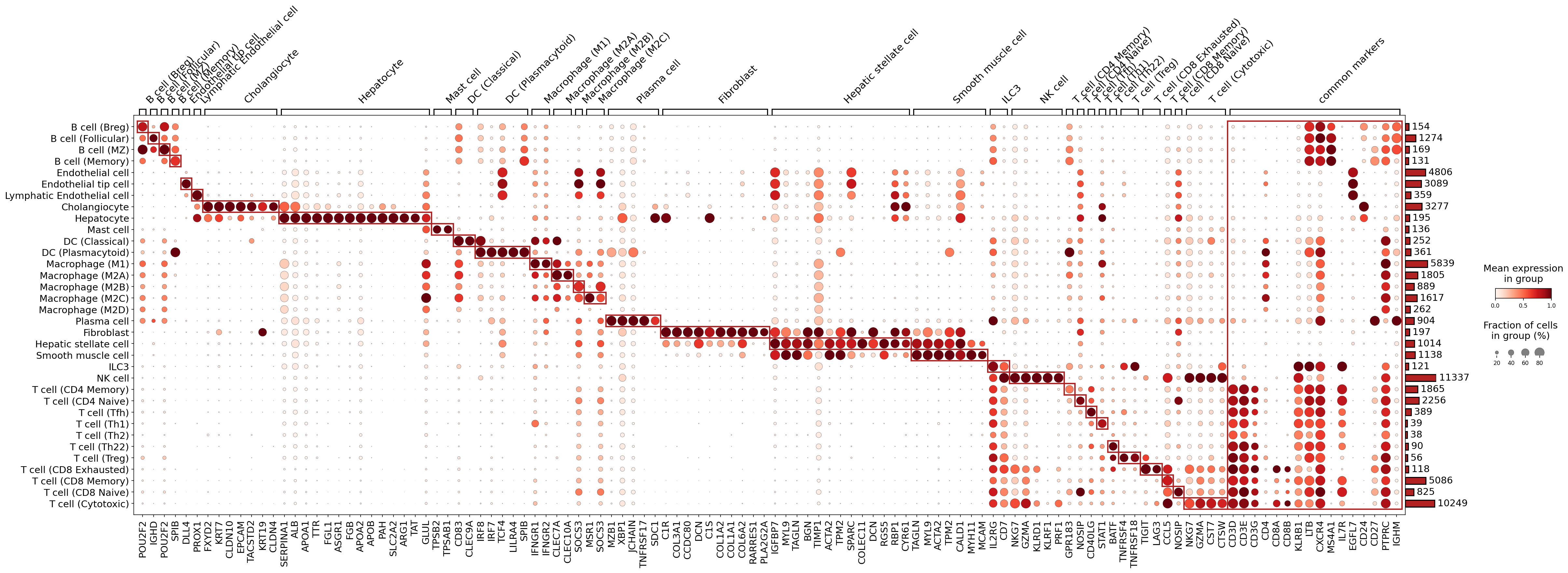
This analysis presents a marker expression dot plot generated for various celltype_subset populations from human liver single-cell RNA-seq data. The plot visualizes the expression patterns of key marker genes across these cell subsets, providing an essential check for the quality and specificity of cell type annotations. The size of each dot corresponds to the fraction of cells within a given subset expressing a particular gene, while the color intensity indicates the mean expression level of that gene. Genes are grouped by expected cell type association, with red boxes highlighting clusters of specific markers. The primary goal of this analysis is to confirm the identity and validate the annotations of the identified celltype_subset populations based on their canonical marker gene expression.
Visual Summary
The dot plot effectively displays differential gene expression across 44 distinct celltype_subset populations. Key observations include:
- Distinct Cell Type Signatures: Most celltype_subset populations exhibit clear and highly specific marker gene expression patterns, with distinct clusters of genes strongly and uniquely expressed within their assigned groups. This is visually reinforced by the red boxes encompassing sets of markers primarily expressed in a single cell type or closely related subsets.
- High Specificity of Markers: For many cell types, the identified markers show very little expression in other, unrelated cell populations, indicating high specificity. For instance, Hepatocytes show robust expression of liver-specific metabolic genes, while T cell subsets distinctly express T-cell receptor components and lineage-specific co-receptors.
- Granular Subset Resolution: The plot successfully delineates nuanced differences between closely related subsets, such as various B cell, T cell, Macrophage, and Endothelial cell subsets. For example, Lymphatic Endothelial cells are clearly distinguished from general Endothelial cells by markers like PROX1.
- "Common Markers" Column: A dedicated column for "common markers" highlights genes like PTPRC (CD45), which are broadly expressed across immune cells, as expected, serving as general leukocyte markers rather than specific cell type identifiers.
Biological Interpretation
The marker gene expression patterns observed in the dot plot strongly validate the assigned celltype_subset annotations and provide robust biological support for the identification of specific cell populations within the liver tissue.
Immune Cell Lineages:
- B cells: Subsets such as B cell (Follicular), B cell (MZ), B cell (Memory), and B cell (Breg) are clearly defined by general B cell markers like POU2F2, MS4A1 (CD20), CD79A, CD79B, and further distinguished by subset-specific markers such as CD27 for B cell (Memory) and IGHM for B cell (MZ).
- T cells: Pan-T cell markers (CD3D, CD3E, CD3G) are universally expressed in all T cell subsets. CD4+ T cells and CD8+ T cells are clearly separated by CD4 and CD8A/CD8B expression, respectively. Further differentiation within these lineages is supported by canonical markers: FOXP3 for T cell (Treg) GeneCards: FOXP3, LAG3 and TIGIT for T cell (CD8 Exhausted) PubMed search: T cell exhaustion markers, and cytotoxic markers like GZMA and PRF1 for T cell (Cytotoxic) and NK cells.
- Macrophage Subsets: General macrophage markers are observed, with subtle but discernible differences in expression of markers like MRC1 (CD206) and CD163 in M2-like subsets and inflammatory markers in M1-like macrophages, supporting the M1/M2 categorization.
- Dendritic Cells: Classical DCs and Plasmacytoid DCs show distinct marker profiles, with CLEC9A for DC (Classical) and LILRA4, IRF7, TCF4 for DC (Plasmacytoid) GeneCards: LILRA4.
- Plasma Cells: Characterized by strong expression of MZB1, XBP1, JCHAIN, and SDC1 (CD138), consistent with their antibody-producing function.
Non-Immune Liver Resident Cells:
- Hepatocytes: Exhibit robust and specific expression of crucial liver metabolic genes such as ALB (Albumin), APOA1, TTR, and enzymes like PAH, confirming their identity as the primary functional cells of the liver GeneCards: ALB.
- Cholangiocytes: Identified by canonical biliary epithelial markers KRT7 and KRT19, along with EPCAM, reinforcing their role in bile duct formation and function GeneCards: KRT19.
- Endothelial Cells: Distinct markers define general Endothelial cells (CDH5, PECAM1), Endothelial tip cells (DLL4), and Lymphatic Endothelial cells (PROX1, LYVE1), highlighting the vascular diversity within the liver.
- Stromal Cells: Hepatic stellate cells show expression of ACTA2 (alpha-SMA), DCN, and collagen genes (COL1A1, COL3A1), consistent with their quiescent state and potential activation into myofibroblast-like cells in liver fibrosis. Fibroblasts and Smooth muscle cells also exhibit expected ECM and contractile protein signatures, respectively.
Annotation Notes
The comprehensive marker expression patterns observed across all celltype_subset populations provide strong evidence for the accuracy and quality of the cell type annotations in the AnnData object. The clear segregation of distinct marker profiles for each cell subset, coupled with the consistency of these markers with established biological knowledge, validates the cellular identities at a high resolution. This analysis confirms that the celltype_subset annotations are well-supported by the underlying gene expression data, laying a solid foundation for further downstream biological investigations into liver health and disease, such as cirrhosis, which is the inferred disease context for this dataset.
4. Cell Type Population Analysis in Healthy and Cirrhotic Liver
[Analysis Visualization Results]...
Analysis Overview
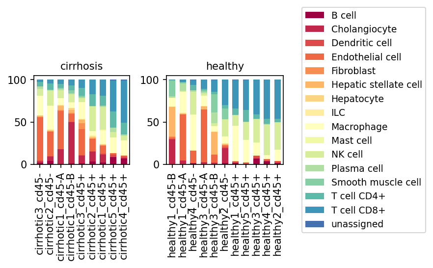
This stacked bar plot visualizes the relative abundance of minor cell types across individual samples, separated by disease condition (cirrhosis vs. healthy). Samples are further stratified by their CD45 expression status, indicated by _cd45- (likely non-hematopoietic cells) and _cd45+ (likely hematopoietic immune cells), providing insights into compositional shifts within distinct cellular compartments of the liver.
Visual Summary
The plot reveals significant alterations in liver cell type composition between healthy and cirrhotic conditions, particularly when distinguishing between CD45- and CD45+ cell fractions.
Healthy Liver Samples:
- CD45- fractions (e.g., healthy1_cd45-B, healthy3_cd45-A): These samples are largely dominated by Hepatocytes (light yellow/cream), which constitute the vast majority of the cellular population. Other significant cell types include Endothelial cells (orange-red), Hepatic stellate cells (light orange), and to a lesser extent, Fibroblasts (dark orange) and Cholangiocytes (red).
- CD45+ fractions (e.g., healthy1_cd45+, healthy5_cd45+): These samples show a predominance of immune cells. T cells (CD4+ and CD8+) (light and dark blue) and Macrophages (very light yellow) are the most abundant, with smaller contributions from NK cells (light green) and other immune cell types.
Cirrhotic Liver Samples:
- CD45- fractions (e.g., cirrhotic3_cd45-, cirrhotic2_cd45-): There is a striking and substantial reduction in Hepatocytes. Concurrently, there is a clear increase in the relative proportions of Cholangiocytes (red), Hepatic stellate cells (light orange), Fibroblasts (dark orange), and Endothelial cells (orange-red). The proportion of 'unassigned' cells (dark blue) also appears elevated in some cirrhotic CD45- samples compared to healthy.
- CD45+ fractions (e.g., cirrhotic3_cd45+, cirrhotic2_cd45+): These samples show a prominent expansion of several immune cell types. Macrophages (very light yellow) become a dominant cell type, showing a marked increase compared to healthy CD45+ samples. T cells (CD4+ and CD8+) (light and dark blue) also appear to be increased. Other immune cells, such as Plasma cells (light blue-green) and Dendritic cells (dark orange/red), also contribute more significantly to the overall composition.
Biological Interpretation
The observed shifts in cell populations are highly consistent with the known pathophysiology of liver cirrhosis:
- Hepatocyte Loss and Liver Damage: The drastic reduction in Hepatocytes in cirrhotic CD45- samples reflects the progressive loss of functional parenchymal cells due to chronic injury and fibrotic replacement, which is a hallmark of cirrhosis.
- Cholangiocyte Proliferation: The increase in Cholangiocytes indicates a "ductular reaction," a common feature in chronic liver diseases where biliary epithelial cells proliferate in response to injury and inflammation. This often contributes to the fibrotic process.
- Fibrogenic Cell Activation and Expansion: The increased proportions of Hepatic stellate cells and Fibroblasts in cirrhotic CD45- fractions are critical indicators of ongoing liver fibrosis. These cells are the primary producers of extracellular matrix components, leading to scar tissue formation and architectural distortion characteristic of cirrhosis. PubMed search: hepatic stellate cell fibrosis liver
- Immune Cell Infiltration and Inflammation: The significant expansion of Macrophages, T cells, and other immune cells (e.g., Plasma cells, Dendritic cells) in cirrhotic CD45+ fractions highlights the persistent inflammation that drives cirrhosis progression. Macrophages, in particular, play diverse roles in liver injury and repair, contributing to both inflammation and fibrosis. PubMed search: macrophages liver cirrhosis
- Distinct Compartmental Changes: The clear differentiation between CD45- (non-hematopoietic) and CD45+ (hematopoietic) fractions allows for precise observation of changes specific to the structural/parenchymal compartment versus the immune compartment, providing a more detailed picture of disease-associated cellular remodeling.
Clinical or Translational Implications
These findings have several potential clinical and translational implications:
- Biomarkers of Disease Progression: The altered proportions of specific cell types, such as the decline in hepatocytes and increases in fibrogenic cells (hepatic stellate cells, fibroblasts) and inflammatory cells (macrophages, T cells), could serve as prognostic biomarkers for cirrhosis progression or indicators of disease severity.
- Therapeutic Targets: Identifying the specific cell populations that expand or become activated in cirrhosis (e.g., activated hepatic stellate cells, pro-fibrotic macrophages) provides potential targets for novel anti-fibrotic or anti-inflammatory therapies.
- Monitoring Treatment Response: Quantifying these cellular shifts could be used to monitor the effectiveness of therapeutic interventions aimed at halting or reversing cirrhosis. A reduction in fibrogenic or inflammatory cell populations, coupled with stabilization of hepatocyte numbers, might indicate a positive treatment response.
5. Population Dynamics of T cell Subsets in Cirrhotic vs. Healthy Liver
[Analysis Visualization Results]...
Analysis Overview
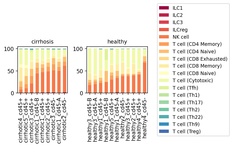
This analysis presents a population bar plot visualizing the relative proportions of various T cell subsets, NK cells, and Innate Lymphoid Cells (ILCs) within the major 'T cell' population across individual samples from both healthy and cirrhotic human liver tissues. Each bar represents a single sample, and the stacked segments illustrate the percentage contribution of each specific cell type (e.g., T cell (Cytotoxic), NK cell, T cell (Treg), ILCs). The primary goal is to identify shifts in immune cell composition associated with cirrhosis.
Visual Summary
The stacked bar plots display the relative proportions of T cell subsets, NK cells, and ILCs for each sample, grouped by condition (cirrhosis vs. healthy).
- Dominant Populations: In both healthy and cirrhotic samples, NK cells (orange) and T cell (Cytotoxic) (light yellow) constitute a significant portion of the analyzed immune cell population.
- NK Cell Expansion in Cirrhosis: A striking observation is the increased proportional representation of NK cells (orange) in most cirrhotic samples compared to healthy samples. This suggests a notable expansion or relative enrichment of NK cells in the cirrhotic liver microenvironment.
- T cell (Cytotoxic) Proportion: Correspondingly, the proportion of T cell (Cytotoxic) cells (light yellow) appears to be relatively reduced in many cirrhotic samples compared to healthy controls, potentially due to the proportional increase in NK cells.
- T cell (CD8 Exhausted) Presence: T cell (CD8 Exhausted) cells (lightest yellow) show a more noticeable presence in some cirrhotic samples, indicating a potential accumulation of exhausted CD8+ T cells in the disease state.
- Other Subsets: T cell (CD8 Naive), T cell (CD8 Memory), T cell (CD4 Naive), T cell (CD4 Memory), and various Th subsets (Th1, Th2, Th9, Th17, Th22, Tfh) are present in smaller proportions. While subtle shifts might occur, no dramatic changes are immediately apparent for these smaller populations across conditions from this visualization alone. T cell (Treg) (dark blue) also represents a minor population, showing no obvious overall trend.
- ILCs: ILCs (ILC1, ILC2, ILC3, ILCreg) are consistently present in very low proportions in both conditions.
- Sample Variability: There is some variability in cell subset proportions among individual samples within both the cirrhotic and healthy groups.
Biological Interpretation
The observed shifts in immune cell populations within the liver parenchyma provide insights into the immune dysregulation characteristic of cirrhosis.
- NK Cell Reactivity in Cirrhosis: The prominent increase in NK cell proportion in cirrhotic livers is biologically significant. NK cells are critical innate immune cells abundant in the liver, playing roles in anti-viral immunity, anti-tumor surveillance, and regulation of liver fibrosis. In chronic liver diseases like cirrhosis, NK cells can exhibit diverse functions, from contributing to liver damage through cytotoxicity to secreting cytokines that influence stellate cell activation and fibrogenesis [1, 2]. Their expansion may represent a compensatory mechanism against chronic injury or a contributor to the ongoing inflammatory and fibrotic processes.
- T cell Exhaustion in Chronic Disease: The potential increase in T cell (CD8 Exhausted) cells in cirrhosis aligns with the concept of immune exhaustion frequently observed in chronic infections and cancer, which can also be relevant in chronic liver diseases. T cell exhaustion is characterized by the progressive loss of effector functions, driven by persistent antigen stimulation and inhibitory receptor expression, potentially impairing the immune system's ability to clear damaged hepatocytes or viral infections [3].
- Balance of Adaptive Immunity: The relative decrease in T cell (Cytotoxic) cells, alongside the NK cell expansion, suggests a potential shift in the balance of cytotoxic immune responses in the cirrhotic liver. This could imply a less effective adaptive cytotoxic response or a proportional change due to the influx of other immune cells.
- Liver Microenvironment: The liver possesses a unique immune environment that balances immune tolerance with active immune responses. In cirrhosis, this balance is disrupted, leading to chronic inflammation, fibrosis, and impaired immune surveillance. The observed changes reflect the complex immune remodeling occurring during disease progression.
Clinical or Translational Implications
The changes in T cell subsets and NK cell populations have potential clinical implications for understanding disease progression and identifying therapeutic targets in cirrhosis.
- Biomarker Potential: The altered proportions of NK cells and T cell (CD8 Exhausted) could serve as potential biomarkers for cirrhosis severity or progression.
- Therapeutic Targets: Understanding the specific roles of expanded NK cells or exhausted T cells in cirrhosis could pave the way for targeted immunomodulatory therapies. For instance, strategies aimed at restoring T cell function or modulating NK cell activity might be explored to halt or reverse disease progression [4].
- Immune Surveillance: The shift in cytotoxic immune cell composition may impact the liver's ability to clear pre-malignant cells, potentially contributing to the increased risk of hepatocellular carcinoma (HCC) in cirrhotic patients [5].
---
References
- NK cell function in liver disease:
- PubMed search: "NK cells liver cirrhosis" https://pubmed.ncbi.nlm.nih.gov/?term=NK+cells+liver+cirrhosis
- Role of NK cells in fibrosis:
- PubMed search: "NK cells liver fibrosis" https://pubmed.ncbi.nlm.nih.gov/?term=NK+cells+liver+fibrosis
- T cell exhaustion:
- PubMed search: "T cell exhaustion chronic disease" https://pubmed.ncbi.nlm.nih.gov/?term=T+cell+exhaustion+chronic+disease
- Immunotherapy in liver disease:
- PubMed search: "immunotherapy liver cirrhosis" https://pubmed.ncbi.nlm.nih.gov/?term=immunotherapy+liver+cirrhosis
- Immune surveillance and HCC:
- PubMed search: "immune surveillance hepatocellular carcinoma" https://pubmed.ncbi.nlm.nih.gov/?term=immune+surveillance+hepatocellular+carcinoma
6. Alterations in Liver T Cell Subset Proportions in Cirrhosis
[Analysis Visualization Results]...
Analysis Overview
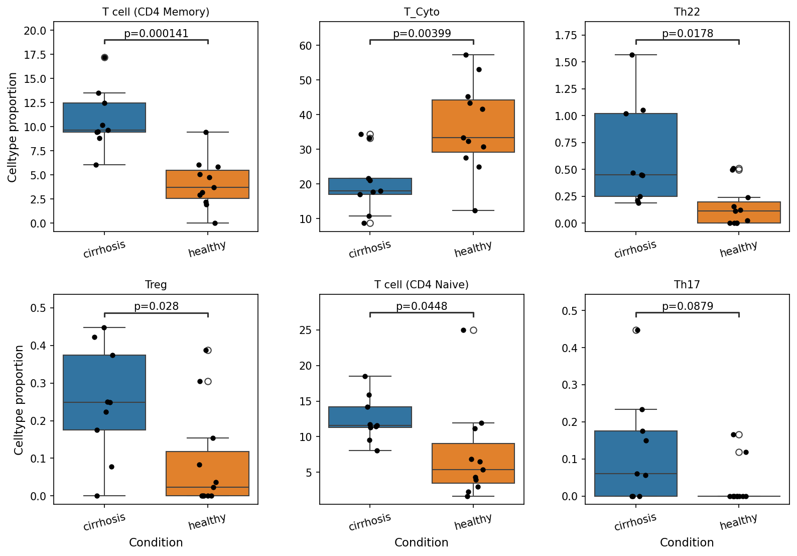
This analysis presents box plots illustrating the proportions of specific T cell subsets within the total T cell population, comparing healthy liver tissue to cirrhotic liver tissue. The plot_box_for_celltype_population_with_signif_difference tool was used to identify and visualize subsets showing statistically significant differences (p < 0.1) between conditions. These results provide insights into the T cell immune landscape shifts occurring in the liver during cirrhosis.
Visual Summary
The visualizations reveal distinct and statistically significant changes in the proportions of several T cell subsets in cirrhosis compared to healthy controls:
- T cell (CD4 Memory): The proportion of CD4 Memory T cells is significantly higher in cirrhotic livers compared to healthy livers (p = 0.000141).
- T_Cyto (Cytotoxic T cells): The proportion of Cytotoxic T cells is significantly lower in cirrhotic livers compared to healthy livers (p = 0.00399).
- Th22: The proportion of T helper 22 (Th22) cells is significantly higher in cirrhotic livers compared to healthy livers (p = 0.0178).
- Treg (Regulatory T cells): The proportion of Regulatory T cells is significantly higher in cirrhotic livers compared to healthy livers (p = 0.028).
- T cell (CD4 Naive): The proportion of CD4 Naive T cells is significantly higher in cirrhotic livers compared to healthy livers (p = 0.0448).
- Th17: The proportion of T helper 17 (Th17) cells also shows a trend towards being higher in cirrhotic livers, with borderline statistical significance (p = 0.0879).
In summary, cirrhotic livers exhibit an expansion of CD4 Memory, Th22, Regulatory T cells, and CD4 Naive T cells, alongside a reduction in Cytotoxic T cell proportions.
Biological Interpretation
The observed shifts in T cell subset populations in cirrhotic livers indicate a profound dysregulation of the local immune environment, consistent with the chronic inflammatory and fibrotic processes characteristic of cirrhosis.
- Increased CD4 Memory, Th22, Th17, and CD4 Naive T cells:
- The expansion of CD4 Memory T cells suggests persistent antigenic stimulation and chronic immune activation within the cirrhotic liver, potentially driven by liver damage, microbial products, or autoantigens.
- Th22 cells (producing IL-22) are known to play dual roles in liver disease. While IL-22 can be hepatoprotective and promote tissue repair, sustained elevation might also contribute to chronic inflammation and fibrogenesis in certain contexts [1].
- The trend towards increased Th17 cells (producing IL-17) is notable. Th17 cells are potent pro-inflammatory cells implicated in various autoimmune diseases and have been shown to contribute to liver inflammation and fibrosis in chronic liver diseases [2].
- An increase in CD4 Naive T cells in a chronically diseased environment might seem counterintuitive but could reflect altered homing mechanisms, local proliferation, or systemic immune changes affecting recruitment to the liver.
- Increased Regulatory T cells (Tregs): The significant expansion of Tregs is a common feature in chronic inflammatory conditions and cancer, including cirrhosis. Tregs suppress immune responses to maintain tolerance and limit inflammation. While they may attempt to dampen excessive inflammation in cirrhosis, an overabundance can also contribute to immune evasion, impair anti-viral or anti-tumor immunity, and potentially promote persistent tissue damage and fibrosis by dampening beneficial immune responses [3].
- Decreased Cytotoxic T cells (T_Cyto): The significant reduction in the proportion of Cytotoxic T cells (primarily CD8+ T cells) in cirrhotic livers is a critical finding. These cells are essential for clearing virally infected cells and tumor cells. Their decrease could imply immune exhaustion, functional impairment, or preferential sequestration elsewhere, potentially leading to impaired anti-viral immunity (e.g., in viral hepatitis-induced cirrhosis) and reduced immune surveillance against hepatocellular carcinoma (HCC), a major complication of cirrhosis [4]. This reduction might contribute to the immune tolerant state often seen in advanced liver disease.
Collectively, these findings suggest a complex immune milieu in cirrhosis characterized by chronic activation (memory T cells), potentially both protective and pathological cytokine responses (Th22, Th17), an attempt at immune suppression (Tregs), and a concerning deficit in effector functions (Cytotoxic T cells).
Clinical or Translational Implications
The observed shifts in T cell populations have important clinical implications for understanding disease progression and potential therapeutic strategies in cirrhosis:
- Prognostic Markers: The proportions of these T cell subsets could serve as potential biomarkers for the stage or progression of cirrhosis, reflecting the inflammatory and immunoregulatory state of the liver.
- Therapeutic Targets: Modulating specific T cell subsets might offer novel therapeutic avenues. For instance, strategies to enhance cytotoxic T cell function or rebalance the Th17/Treg axis could potentially reduce fibrosis, control viral infections, or improve anti-tumor immunity in patients with cirrhosis.
- HCC Risk: The reduction in cytotoxic T cells is particularly concerning given the high risk of HCC in cirrhosis. Restoring or boosting anti-tumor immunity by targeting these specific T cell deficiencies could be a strategy for HCC prevention or treatment.
- Immune Tolerance: The increased Treg population, alongside decreased cytotoxic T cells, highlights an immune tolerant or exhausted state in the cirrhotic liver, which could hinder effective immune responses to new insults or vaccines.
These population-level changes underscore the need for further functional characterization of these T cell subsets to fully elucidate their roles in the pathogenesis of human liver cirrhosis.
---
References:
- IL-22 in liver disease:
PubMed Search: IL-22 liver cirrhosis fibrosis
- Th17 cells in liver fibrosis:
PubMed Search: Th17 liver fibrosis cirrhosis
- Regulatory T cells in liver cirrhosis:
PubMed Search: regulatory T cells liver cirrhosis
- Cytotoxic T cells in cirrhosis and HCC:
PubMed Search: cytotoxic T cells liver cirrhosis HCC
7. Macrophage Subset Population Analysis in Cirrhosis
[Analysis Visualization Results]...
Analysis Overview
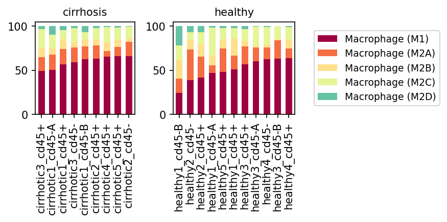
This analysis presents a population bar plot illustrating the relative proportions of various macrophage subsets, specifically Macrophage (M1), Macrophage (M2A), Macrophage (M2B), Macrophage (M2C), and Macrophage (M2D), across individual liver samples from both healthy individuals and patients with cirrhosis. The stacked bars for each sample represent the percentage contribution of each macrophage subset to the total macrophage population in that sample.
Visual Summary
The visualization clearly displays distinct patterns in macrophage subset composition between healthy and cirrhotic liver samples.
- Macrophage (M1) Dominance in Cirrhosis: In samples from cirrhotic livers, Macrophage (M1) (maroon) consistently represents the largest proportion of the macrophage population, frequently exceeding 50% and appearing notably higher than in most healthy samples.
- Reduced Macrophage (M2D) in Cirrhosis: Macrophage (M2D) (teal) is present in some healthy samples, contributing a small but noticeable percentage. However, its proportion is almost negligible or completely absent in nearly all cirrhotic samples.
- Macrophage (M2A) Contribution: Macrophage (M2A) (coral) is a significant component in both conditions, though its relative contribution appears more variable and potentially slightly reduced in some cirrhotic samples compared to its overall presence in healthy livers.
- Minor Subsets: Macrophage (M2B) (light yellow) and Macrophage (M2C) (pale yellow) contribute smaller proportions in both healthy and cirrhotic conditions, with no immediately apparent dramatic shifts between the two states.
- Inter-sample Variability: While clear trends emerge, there is also some sample-to-sample variability within both the healthy and cirrhotic groups, indicating potential heterogeneity in disease progression or individual immune responses.
Biological Interpretation
The observed shift in macrophage subset populations from healthy to cirrhotic liver tissues has significant biological implications for understanding liver disease progression.
- Pro-inflammatory Environment in Cirrhosis: The prominent increase in Macrophage (M1) cells in cirrhotic livers strongly suggests a highly activated pro-inflammatory state. M1 macrophages are typically characterized by the production of pro-inflammatory cytokines (e.g., TNF-α, IL-1β, IL-6) and reactive oxygen species, which contribute to ongoing hepatocyte damage, perpetuation of inflammation, and progression of fibrosis in chronic liver diseases.
- Impaired Resolution and Repair: Conversely, the near absence of Macrophage (M2D) in cirrhotic samples is notable. M2D macrophages are often associated with regulatory functions, angiogenesis, and tissue repair, contributing to immune tolerance and resolution of inflammation. Their reduction in cirrhosis could indicate a compromised ability of the liver to resolve inflammation and repair tissue damage effectively, favoring a chronic inflammatory and fibrotic environment.
- Macrophage Polarization in Liver Fibrosis: The balance between M1 and various M2 macrophage subsets is critical in liver pathology. While M2 macrophages are generally linked to pro-fibrotic processes and tissue remodeling, the specific polarization toward M1 dominance suggests that the inflammatory drive is a major characteristic of the macrophage landscape in cirrhosis. This imbalance likely contributes to the progressive nature of liver fibrosis and eventual liver failure PubMed search: macrophage polarization liver fibrosis.
Clinical or Translational Implications
The findings highlight the potential role of macrophage subset dysregulation in the pathogenesis of liver cirrhosis and suggest several clinical and translational avenues.
- Therapeutic Target Identification: The dominance of M1 macrophages in cirrhosis points to these cells as potential therapeutic targets. Strategies aimed at modulating macrophage polarization, for instance, by suppressing M1 activation or promoting a shift towards reparative M2 phenotypes, could help mitigate inflammation and slow fibrosis progression in cirrhotic patients.
- Biomarker for Disease Activity: The relative proportion of M1 macrophages, and perhaps the M1/M2D ratio, could serve as a novel biomarker for assessing disease activity, prognosis, or response to therapy in patients with cirrhosis.
- Understanding Disease Heterogeneity: Further research into the specific functions of these macrophage subsets in the cirrhotic microenvironment could uncover detailed mechanisms of disease progression and identify patient subgroups that might benefit from specific immunomodulatory treatments.
8. Macrophage Subset Population Shifts in Cirrhosis
[Analysis Visualization Results]...
Analysis Overview
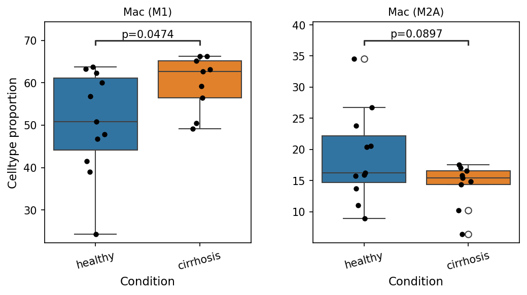
This analysis investigates the proportional changes of macrophage subsets within the liver, comparing healthy individuals to those with cirrhosis. Specifically, the proportions of Macrophage (M1) and Macrophage (M2A) subsets were quantified across conditions, and statistical significance was assessed using a p-value cutoff of 0.1. The results reveal statistically significant shifts in the relative abundance of these crucial immune cell populations in the context of cirrhosis.
Visual Summary
The box plots illustrate the celltype proportion (as a percentage) for two macrophage subsets, Mac (M1) and Mac (M2A), across healthy and cirrhotic liver samples.
Macrophage (M1) Population:
- There is a statistically significant increase in the proportion of Mac (M1) cells in cirrhotic samples compared to healthy samples (p=0.0474).
- The median proportion of Mac (M1) cells is visibly higher in the cirrhosis group, indicating an expansion of this subset.
Macrophage (M2A) Population:
- Conversely, there is a statistically significant decrease in the proportion of Mac (M2A) cells in cirrhotic samples compared to healthy samples (p=0.0897).
- The median proportion of Mac (M2A) cells is lower in the cirrhosis group, suggesting a reduction in this subset.
Biological Interpretation
Macrophages are critical immune cells in the liver, where they exist as resident Kupffer cells and monocyte-derived macrophages. Their polarization into distinct functional phenotypes, such as M1 (pro-inflammatory) and M2 (anti-inflammatory/pro-resolving/pro-fibrotic), plays a pivotal role in liver homeostasis, injury, and repair.
- Increased Mac (M1) in Cirrhosis: M1 macrophages are characterized by their pro-inflammatory functions, including the production of pro-inflammatory cytokines (e.g., TNF-α, IL-6, IL-1β) and reactive oxygen species, which contribute to tissue damage and perpetuate inflammatory responses. The observed increase in M1-like macrophages in the cirrhotic liver suggests an exacerbated inflammatory environment. Chronic inflammation is a hallmark of cirrhosis, driving hepatocellular injury and fibrosis progression [1].
- Decreased Mac (M2A) in Cirrhosis: M2A macrophages are generally associated with early stages of wound healing, tissue repair, and resolution of inflammation, often through the production of anti-inflammatory mediators and growth factors. A reduction in this specific M2 subtype might indicate an impaired capacity for beneficial tissue repair or an imbalance in the macrophage population that favors disease progression rather than resolution. While other M2 subtypes (e.g., M2B, M2C) can contribute to fibrogenesis, a decrease in M2A specifically points to a shift away from certain reparative processes [1].
Taken together, these findings suggest a significant reprogramming of the macrophage compartment in the cirrhotic liver, characterized by a shift towards a more pro-inflammatory (M1-skewed) and potentially less reparative (reduced M2A) phenotype. This imbalance in macrophage polarization is consistent with the chronic inflammation and ongoing tissue remodeling observed in liver cirrhosis, where macrophage activity significantly influences disease progression [2].
Clinical or Translational Implications
The distinct shifts in M1 and M2A macrophage proportions observed in cirrhosis could have several clinical and translational implications:
- Biomarkers: The proportions of Mac (M1) and Mac (M2A) could potentially serve as non-invasive biomarkers for the diagnosis, staging, or prognosis of cirrhosis, reflecting the underlying inflammatory and reparative status of the liver.
- Therapeutic Targets: Modulating macrophage polarization pathways offers a promising therapeutic strategy for cirrhosis. Interventions aimed at dampening the M1 pro-inflammatory response or promoting the beneficial aspects of M2-like macrophage functions could potentially mitigate liver inflammation, reduce fibrosis progression, and improve outcomes in patients with cirrhosis [2]. Further research into the specific functions of these macrophage subsets in human cirrhosis would be crucial for developing targeted therapies.
---
References:
[1] Macrophage polarization in liver disease (PubMed search): https://pubmed.ncbi.nlm.nih.gov/?term=macrophage+polarization+liver+cirrhosis
[2] Role of macrophages in liver fibrosis (PubMed search): https://pubmed.ncbi.nlm.nih.gov/?term=macrophage+liver+fibrosis
9. Cell-Cell Interaction Patterns in Liver Cirrhosis and Healthy Conditions
[Analysis Visualization Results]...
Analysis Overview
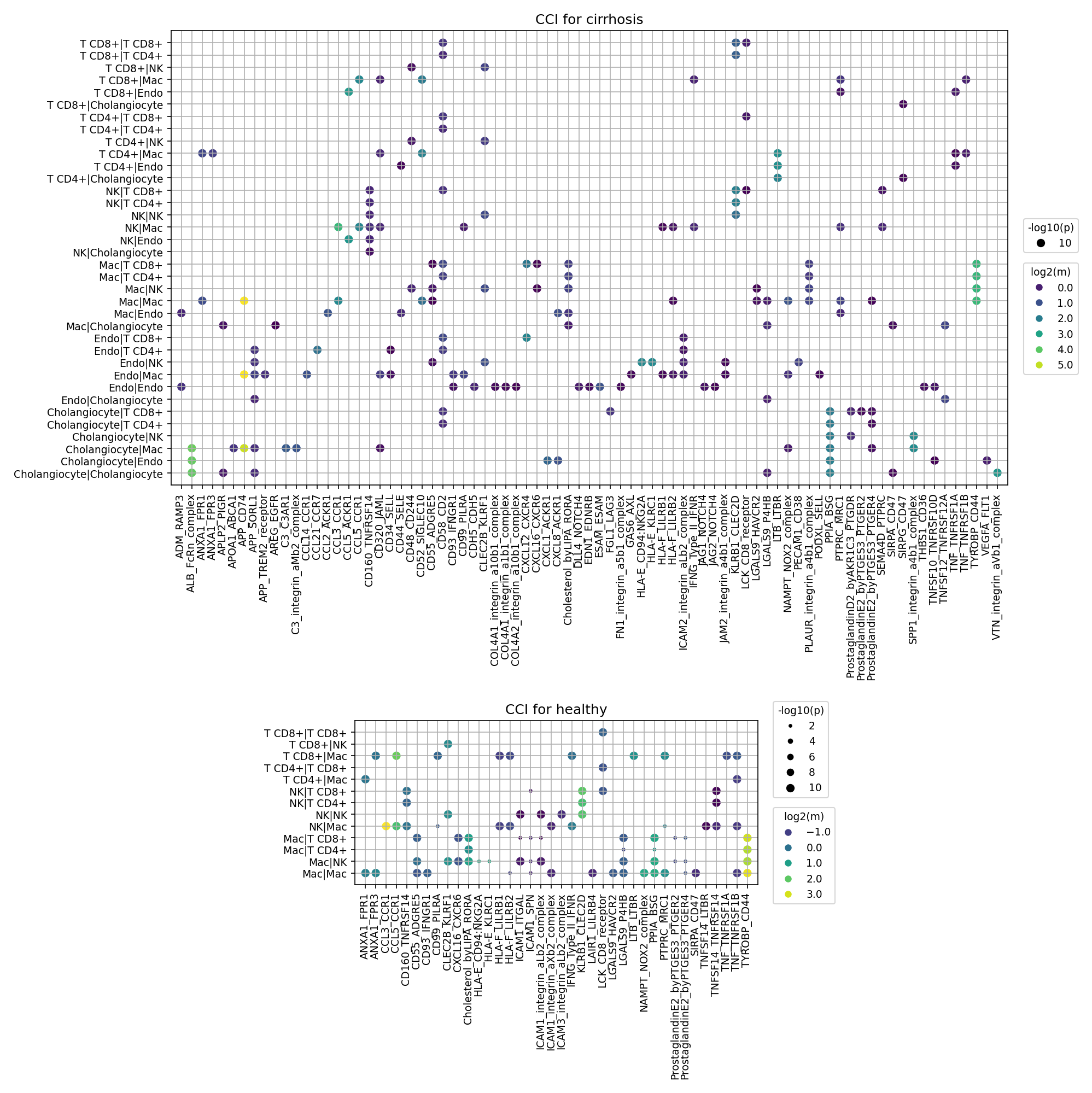
This analysis utilizes CellPhoneDB to investigate cell-cell interaction (CCI) patterns in human liver tissue, comparing healthy individuals with those affected by cirrhosis. The plot_cci_dots tool was used to visualize significant ligand-receptor interactions between different cell types, with a focus on identifying the top 80 interactions per condition based on significance. These dot plots display the statistical significance (-log10(p-value), represented by dot size) and the mean expression level (log2(mean), represented by dot color) of predicted ligand-receptor pairs for each interacting cell pair. The reference condition for comparison in downstream analyses is 'healthy', establishing cirrhosis as the primary disease context.
Visual Summary
The provided dot plots illustrate distinct cell-cell communication landscapes between healthy and cirrhotic liver conditions.
CCI for Cirrhosis
- Increased Complexity: The plot for cirrhosis exhibits a substantially greater number of significant cell-cell interactions compared to the healthy state, spanning a wider array of ligand-receptor pairs and cell-cell communication axes. This indicates a highly activated and reprogrammed cellular microenvironment.
- Key Interacting Cell Types: Macrophages, Endothelial cells, T cells (CD8+, CD4+, NK), and Cholangiocytes show extensive interactions, both among themselves and with other cell populations.
- Macrophages (Mac): Heavily involved in interactions, notably with other macrophages (Mac|Mac), Endothelial cells (Mac|Endo), and various T cell subsets.
- Endothelial Cells (Endo): Show frequent interactions with other Endothelial cells (Endo|Endo), Macrophages (Mac|Endo), and Cholangiocytes (Endo|Cholangiocyte).
- T cells (T CD8+, T CD4+, NK): Engage in numerous interactions, particularly with Macrophages and Endothelial cells, suggesting active immune cell recruitment and modulation.
- Cholangiocytes: Show significant interactions with Macrophages and Endothelial cells, indicative of their altered role in disease.
Prominent Ligand-Receptor Pairs: Several axes stand out as highly active
- Fibrosis and ECM Remodeling: SPP1-integrin_a5b1, SPP1-integrin_aVb3, SPP1-integrin_aVb5, FN1_integrin_a5b1, FN1_integrin_aVb3, FN1_integrin_aVb5, COL4A1_integrin_a1b1_complex, COL4A2_integrin_a2b1_complex. These represent interactions involving Osteopontin (SPP1), Fibronectin (FN1), and Collagen IV with various integrins, reflecting active extracellular matrix (ECM) deposition and remodeling.
- Inflammation and Immune Activation: TNF-TNFRSF1A, TNF-TNFRSF1B, CXCL12-CXCR4, ICAM1-ITGAL, CD40-CD40LG. These pathways are crucial for immune cell activation, recruitment, and inflammatory responses.
- Angiogenesis: VEGFA_FLT1 and VEGFA_VEGFR2 interactions are observed, suggesting active angiogenesis.
CCI for Healthy
- Reduced Interactions: The plot for healthy liver tissue displays fewer overall significant interactions and a narrower range of ligand-receptor pairs compared to the cirrhotic state. This signifies a more quiescent and homeostatic intercellular communication network.
- Key Interacting Cell Types: Interactions are present but less numerous, involving similar cell types such as T cells, NK cells, Macrophages, and Endothelial cells.
- Prominent Ligand-Receptor Pairs: Some interactions are shared with cirrhosis but often with lower strength or significance, while others appear to be more specific to the healthy state or represent basal functions:
- Immunoregulation and Homeostasis: ANXA1-FPR1, ANXA1-FPR2, ANXA1-FPR3 (Annexin A1), LGALS9-HAVCR2, LGALS9-CD44 (Galectin-9), ProstaglandinE2_byPTGES3_PTGER2, ProstaglandinE2_byPTGES3_PTGER4. These are often associated with maintaining immune tolerance and tissue homeostasis.
- Basal Immune Surveillance: CCL5-CCR1, CCL5-CCR3 (chemokine signaling) and ICAM1-ITGAL (adhesion) suggest baseline immune cell trafficking and adhesion.
- Basal TNF Signaling: TNF-TNFRSF1A, TNF-TNFRSF1B are present, likely reflecting low-level homeostatic signaling rather than a strong inflammatory burst.
Biological Interpretation
The differential cell-cell interaction patterns observed between healthy and cirrhotic livers highlight the profound changes in the tissue microenvironment during disease progression.
- Inflammation and Immune Cell Activation in Cirrhosis: The dramatic increase in interactions involving T cells, NK cells, and Macrophages, coupled with prominent TNF-TNFRSF and CXCL12-CXCR4 signaling, underscores a highly active inflammatory and immune response in cirrhosis.
- TNF-α signaling is a key driver of inflammation and cell death in liver injury, contributing to disease progression PubMed search: TNF alpha liver cirrhosis.
- The CXCL12-CXCR4 axis is critical for immune cell trafficking, recruitment of inflammatory cells, and activation of hepatic stellate cells (HSCs), promoting fibrosis GeneCards: CXCR4.
- Extracellular Matrix Remodeling and Fibrosis: The robust presence of interactions involving SPP1, FN1, and COL4A with various integrins in cirrhosis is a direct signature of active fibrogenesis.
- SPP1 (Osteopontin), a matricellular protein, plays a significant role in liver fibrosis by promoting inflammation, HSC activation, and ECM deposition PubMed search: SPP1 liver fibrosis. Its interaction with integrins facilitates cell adhesion and signaling crucial for fibrotic processes.
- Fibronectin (FN1) and Collagen (COL4A) are major components of the ECM. Their interactions with integrins are fundamental to cell-matrix adhesion, cellular migration, and tissue remodeling, all of which are dysregulated in fibrosis.
- Vascular Remodeling and Angiogenesis: The increased VEGFA interactions with its receptors (FLT1, VEGFR2) in cirrhotic liver point towards significant angiogenesis. Neovascularization is a common feature in chronic liver diseases, contributing to increased intrahepatic resistance and portal hypertension PubMed search: VEGFA liver cirrhosis angiogenesis.
- Macrophages as Central Regulators: Macrophages appear as key hubs of interaction in cirrhosis, engaging with multiple cell types through diverse pro-inflammatory and pro-fibrotic pathways. This highlights their pivotal role in orchestrating the pathological microenvironment.
- Cholangiocyte Involvement in Disease: Interactions of Cholangiocytes with Macrophages and Endothelial cells, coupled with pro-fibrotic signaling (e.g., SPP1-integrin), suggest their active participation in bile duct injury, proliferation, and contribution to liver fibrosis.
- Loss of Homeostatic Regulation: The reduced number of interactions in healthy tissue, with some involving molecules like Annexin A1 and Prostaglandin E2, suggests that homeostatic and immunoregulatory pathways are critical for maintaining liver health. These pathways appear to be overwhelmed or suppressed in the cirrhotic state.
Clinical or Translational Implications
The differential cell-cell interaction landscape provides critical insights for understanding cirrhosis pathogenesis and identifying potential therapeutic avenues.
- Therapeutic Target Prioritization: Ligand-receptor pairs that are highly active and specific to cirrhosis, such as SPP1-integrin axes, CXCL12-CXCR4, TNF-TNFRSF, and VEGFA-VEGFR pathways, represent promising therapeutic targets. Interventions aimed at blocking these specific interactions could attenuate inflammation, reduce fibrosis, and normalize vascular architecture in cirrhotic patients. For example, blocking SPP1 or its integrin receptors could directly impact fibrotic progression PubMed search: SPP1 inhibitor liver fibrosis therapy.
- Biomarker Discovery: The identified cell-cell interaction signatures could serve as novel biomarkers for disease progression, severity, or response to therapy. Monitoring the expression levels of key ligands or receptors involved in these pathological interactions might offer non-invasive ways to assess disease status.
- Understanding Disease Heterogeneity: The detailed mapping of cell-cell interactions can help dissect the complex interplay between different cell types in cirrhosis, potentially revealing specific cellular communication networks that drive distinct disease trajectories or patient subgroups.
- Experimental Validation: The identified crucial interactions provide a strong basis for further experimental validation in in vitro and in vivo models of liver cirrhosis, aiming to confirm their functional roles and therapeutic potential.
10. Condition-Specific Cell-Cell Interaction Patterns in Liver Cirrhosis
[Analysis Visualization Results]...
Analysis Overview
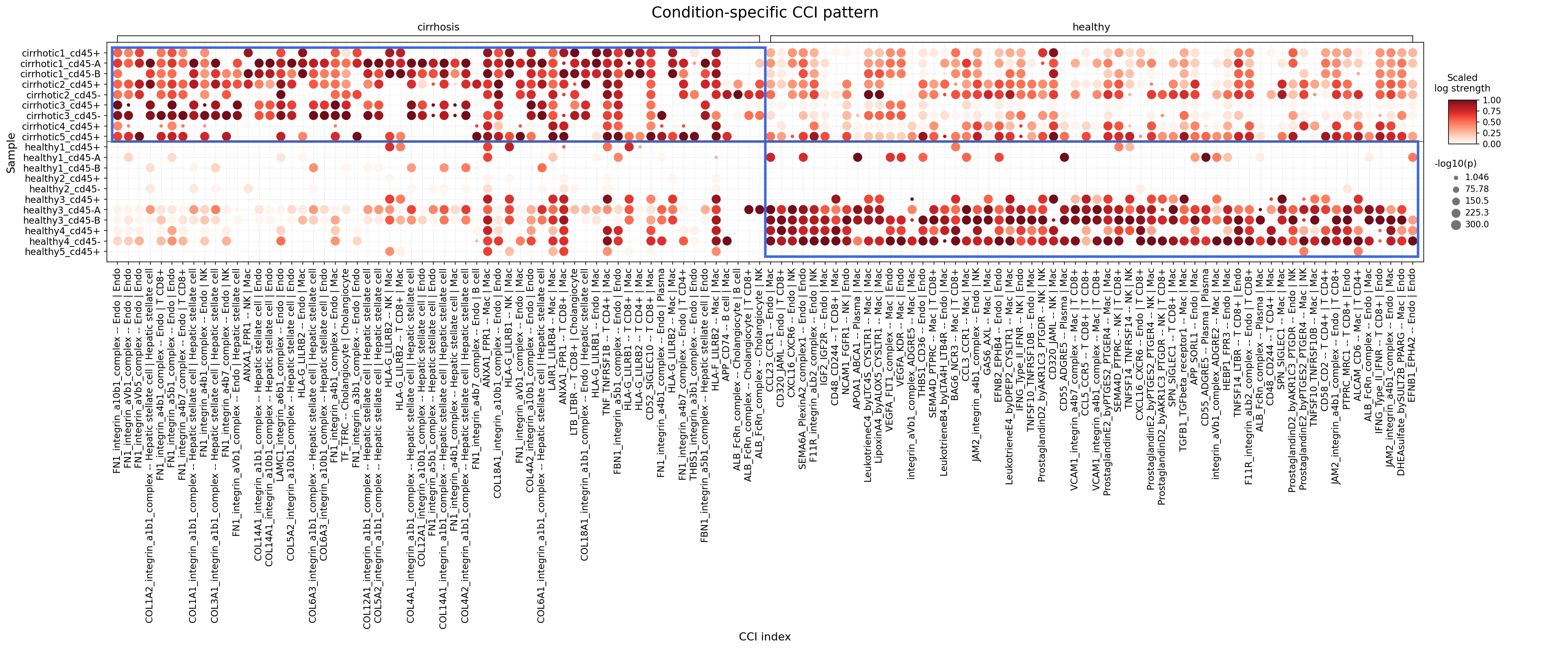
This analysis identifies cell-cell interactions (CCIs) that significantly differ between healthy and cirrhotic liver conditions, focusing on major tissue cells, immune cells, and stromal cells. The dot plot visualizes the scaled log strength and significance (-log10(p-value)) of specific ligand-receptor interactions between defined cell types across individual samples for both conditions. A t-test with an alternative='greater' setting was applied to detect interactions that are significantly stronger in one condition compared to the other. The goal is to uncover CCIs that characterize the pathological state of cirrhosis and those associated with the healthy liver environment, providing insights into disease mechanisms and potential therapeutic targets.
Visual Summary
The dot plot clearly differentiates cell-cell interaction patterns between cirrhotic and healthy liver samples.
- Cirrhosis-specific interactions: The upper panel, representing cirrhotic samples, shows a striking upregulation of numerous cell-cell interactions. These are characterized by large, dark red dots, indicating high interaction strength (scaled log strength) and high statistical significance (-log10(p-value)). A prominent cluster of interactions involves hepatic stellate cells, endothelial cells, macrophages, T cells, B cells, plasma cells, NK cells, and cholangiocytes. Key ligand-receptor families include integrins (e.g., FN1-integrin, COL-integrin, SPP1-integrin), HLA-G-LILRB2, TNF-TNFRSF1B, and chemokine axes like CCL23-CCR1. These interactions are largely absent or significantly weaker (smaller, lighter dots) in the healthy samples, suggesting their involvement in the pathophysiology of cirrhosis.
- Healthy-specific interactions: In contrast, the lower panel, representing healthy samples, displays a distinct set of interactions that are more pronounced or exclusively present in the healthy state. While fewer interactions show equally strong signals as those upregulated in cirrhosis, some interactions (e.g., VCAM1-integrin_a4b1_complex -- Endo | T cell CD4+, CD55_ADGRE5 -- Endo | Mac, ProstaglandinD2_by_AKR1C3_PTGDR -- Mac | Mac, SPN_SIGLEC1 -- T cell | Mac) are more prominent in healthy liver and appear diminished in cirrhosis. This indicates a shift in the intercellular communication landscape during disease progression, where certain homeostatic interactions might be lost or suppressed in cirrhosis.
The max_n_items_per_group=60 parameter ensures that the top 60 most significantly differential interactions for each condition are displayed, providing a focused view of the most impactful changes.
Biological Interpretation
The observed condition-specific cell-cell interactions provide critical biological insights into the progression of liver cirrhosis. The upregulation of specific interactions in cirrhosis highlights key cellular processes driving inflammation, fibrosis, and immune dysregulation. Conversely, the reduction of certain interactions in cirrhosis may reflect a loss of normal tissue homeostasis.
Key Interactions Upregulated in Cirrhosis:
- FN1-integrin / COL-integrin complexes (e.g., FN1_integrin_a1b1_complex -- Hepatic stellate cell | Hepatic stellate cell; COL1A2_integrin_a1b1_complex -- Hepatic stellate cell | Hepatic stellate cell; FN1_integrin_a3b1_complex -- Endo | Cholangiocyte)
- Signaling Pathways/Mechanisms: Fibronectin (FN1) and Collagen (COL1A2, COL14A1) are major components of the extracellular matrix (ECM). Integrins are cell surface receptors that bind to ECM proteins, mediating cell adhesion, migration, proliferation, and survival. Interactions between hepatic stellate cells (HSCs) and ECM components via integrins are central to liver fibrosis. Activated HSCs produce large amounts of ECM proteins, and these interactions further promote their activation and perpetuation of fibrosis. The observed interactions involving endothelial cells and cholangiocytes with FN1 also point to their roles in ECM remodeling and angiogenesis (Endo) or biliary fibrosis (Cholangiocyte) during cirrhosis [PubMed search: integrin hepatic stellate cell fibrosis liver].
- Therapeutic Potential: Targeting specific integrins, such as αvβ3 or αvβ1, on activated HSCs could inhibit their activation and reduce ECM deposition. Small molecule inhibitors or blocking antibodies against these integrins are under investigation for fibrotic diseases. For example, some integrin inhibitors are being explored in cancer and fibrosis trials [PubMed search: integrin inhibitors liver fibrosis].
- SPP1-integrin complexes (e.g., SPP1_integrin_a4b1_complex -- Mac | Mac; SPP1_integrin_aVb5_complex -- Mac | Mac)
- Signaling Pathways/Mechanisms: Secreted phosphoprotein 1 (SPP1), also known as Osteopontin, is a pleiotropic cytokine upregulated in liver injury and cirrhosis. It interacts with various integrins (e.g., α4β1, αvβ5, αvβ3) and CD44, playing a crucial role in inflammation, immune cell recruitment, cell survival, and fibrosis. SPP1-integrin signaling in macrophages promotes their activation, migration, and differentiation towards pro-inflammatory and pro-fibrotic phenotypes, contributing to the chronic inflammation and fibrogenesis characteristic of cirrhosis [GeneCards: SPP1].
- Therapeutic Potential: SPP1 represents an attractive therapeutic target in liver fibrosis. Strategies could include neutralizing antibodies against SPP1 or integrin receptors, or inhibitors blocking the downstream signaling pathways activated by SPP1-integrin binding.
- HLA-G-LILRB2 (e.g., HLA-G_LILRB2 -- Hepatic stellate cell | T cell)
- Signaling Pathways/Mechanisms: Human Leukocyte Antigen G (HLA-G) is a non-classical MHC class I molecule primarily involved in immune tolerance, often expressed in immune-privileged sites or pathological conditions. Leukocyte immunoglobulin-like receptor B2 (LILRB2, also known as ILT4) is an inhibitory receptor expressed on various immune cells, including T cells and macrophages. The HLA-G-LILRB2 axis promotes immune suppression, which in the context of cirrhosis, could dampen effective anti-fibrotic or anti-tumor immune responses, potentially allowing fibrosis to progress unchecked or contributing to immune evasion in associated hepatocellular carcinoma [GeneCards: HLA-G, LILRB2].
- Therapeutic Potential: Modulating the HLA-G-LILRB2 pathway could be a strategy to enhance anti-fibrotic immunity or restore immune surveillance in cirrhotic livers. This might involve blocking LILRB2 or inhibiting HLA-G expression.
- TNF-TNFRSF1B (e.g., TNF_TNFRSF1B -- T cell | Hepatic stellate cell)
- Signaling Pathways/Mechanisms: Tumor Necrosis Factor (TNF) is a potent pro-inflammatory cytokine that binds to TNF Receptor Superfamily member 1B (TNFRSF1B, also known as TNF-R2). This interaction activates various downstream signaling pathways, including NF-κB, leading to gene expression related to inflammation, cell survival, and fibrosis. In the liver, TNF produced by immune cells (like T cells or macrophages) can activate hepatic stellate cells, promoting their proliferation and ECM production, thereby exacerbating fibrosis [GeneCards: TNF, TNFRSF1B].
- Therapeutic Potential: Anti-TNF therapies (e.g., infliximab, adalimumab) are widely used for inflammatory diseases. While systemic anti-TNF treatment has been shown to have complex effects in liver disease, including potential risks, targeting specific TNF-TNFRSF1B interactions in the liver could offer a more localized approach to reduce inflammation and fibrosis.
- CCL23-CCR1 (e.g., CCL23_CCR1 -- Cholangiocyte | Mac)
- Signaling Pathways/Mechanisms: Chemokine (C-C motif) Ligand 23 (CCL23) is a chemokine that attracts immune cells expressing its receptor, C-C Chemokine Receptor Type 1 (CCR1). This interaction between cholangiocytes and macrophages suggests a mechanism for macrophage recruitment to the biliary compartment, contributing to inflammation and fibrosis in diseases affecting bile ducts, such as cholestatic liver diseases that can lead to cirrhosis [GeneCards: CCL23, CCR1].
- Therapeutic Potential: Blocking the CCL23-CCR1 axis could reduce the infiltration of pro-inflammatory and pro-fibrotic macrophages into the liver, thereby mitigating inflammation and fibrosis. CCR1 antagonists have been investigated for other inflammatory conditions.
Key Interactions Enriched in Healthy Liver (or Reduced in Cirrhosis):
- VCAM1-integrin_a4b1_complex -- Endo | T cell CD4+
- Signaling Pathways/Mechanisms: Vascular Cell Adhesion Molecule 1 (VCAM1) on endothelial cells binds to integrin α4β1 (VLA-4) on lymphocytes, including CD4+ T cells. This interaction is crucial for lymphocyte extravasation and trafficking from the bloodstream into tissues, enabling immune surveillance. Its reduction in cirrhosis might impair immune cell recruitment necessary for host defense or tissue repair, or it could reflect broader endothelial dysfunction [GeneCards: VCAM1].
- Therapeutic Potential: Re-establishing effective lymphocyte trafficking could be beneficial in specific contexts of liver disease, though simply upregulating VCAM1 might not be straightforward due to complex roles in inflammation.
- ProstaglandinD2_by_AKR1C3_PTGDR -- Mac | Mac
- Signaling Pathways/Mechanisms: Prostaglandin D2 (PGD2) is an eicosanoid that can act as an anti-inflammatory and immunomodulatory lipid mediator. It is produced by various cells, including macrophages, and exerts its effects through specific receptors like the Prostaglandin D2 Receptor (PTGDR). Its production can be influenced by enzymes like AKR1C3. PGD2 signaling often suppresses pro-inflammatory cytokine production and immune cell activation. A reduction in this interaction in cirrhosis suggests a potential loss of endogenous anti-inflammatory mechanisms within the macrophage population, contributing to persistent inflammation [GeneCards: PGD2, PTGDR].
- Therapeutic Potential: Enhancing PGD2 signaling or administering PGD2 mimetics could be a strategy to damp down inflammation and promote resolution of liver injury in cirrhosis.
Clinical or Translational Implications
The differential CCI landscape between healthy and cirrhotic livers offers promising avenues for therapeutic intervention and biomarker discovery.
- Targeting Fibrosis: The robust upregulation of integrin-ECM interactions (FN1-integrin, COL-integrin) and SPP1-integrin signaling involving hepatic stellate cells and macrophages in cirrhosis strongly supports these pathways as central drivers of fibrosis. Developing specific inhibitors for these integrin complexes or neutralizing SPP1 could halt or even reverse fibrosis progression.
- Modulating Inflammation and Immune Dysregulation: The increased TNF-TNFRSF1B signaling and CCL23-CCR1 axis highlight persistent inflammation and immune cell recruitment as key features of cirrhosis. While broad anti-inflammatory therapies can have side effects, targeting these specific ligand-receptor pairs might offer more precise immunomodulation. Similarly, the HLA-G-LILRB2 pathway suggests that immune suppressive mechanisms could be exploited by fibrotic tissue, and disrupting this pathway might unleash anti-fibrotic immunity.
- Restoring Homeostasis: The reduction of interactions like VCAM1-integrin and PGD2 signaling in cirrhosis points to a loss of homeostatic mechanisms. Understanding how to restore these protective interactions or mimic their beneficial effects could complement anti-fibrotic strategies, promoting tissue repair and reducing chronic inflammation.
- Biomarker Potential: The identified significantly differential CCIs, or the expression of their constituent genes in specific cell types, could serve as novel biomarkers for disease diagnosis, prognosis, or monitoring therapeutic response in cirrhosis. Assays to measure these interaction strengths or ligand/receptor expression in patient samples might offer a non-invasive way to track disease activity.
11. Cholangiocyte Condition-Specific Surfaceome Markers in Liver Cirrhosis
[Analysis Visualization Results]...
Analysis Overview
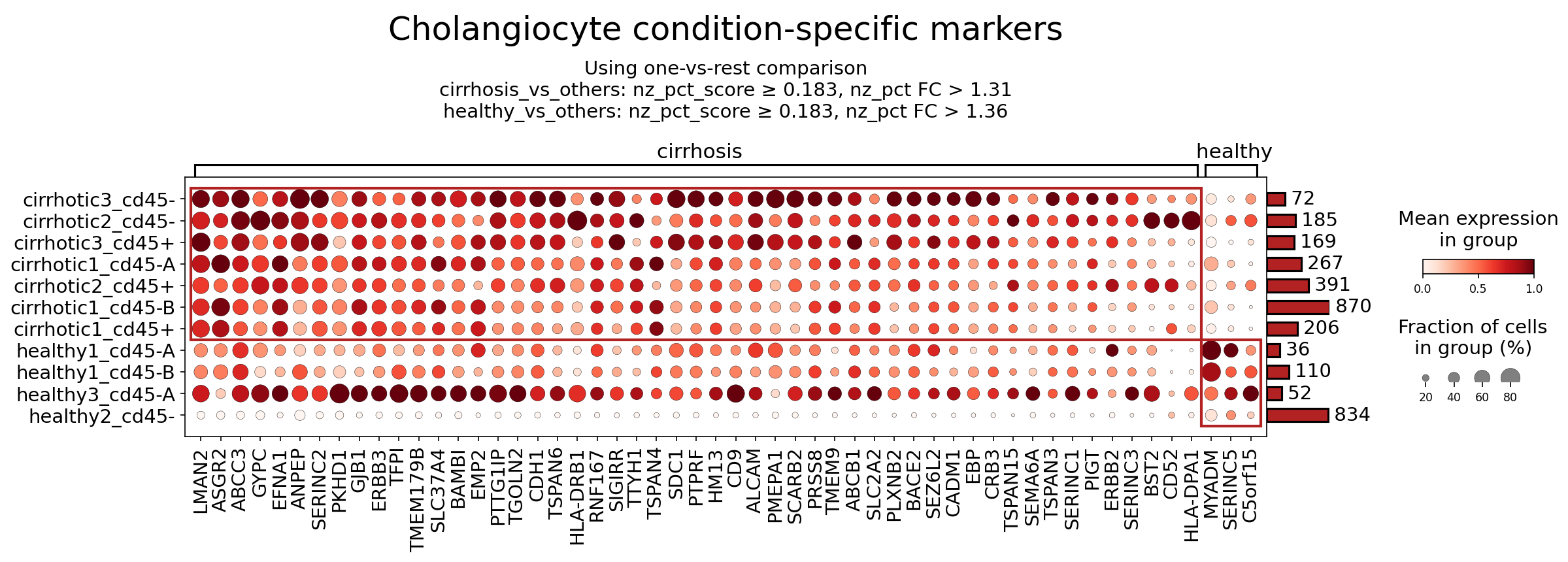
This analysis identifies surfaceome markers specifically expressed on cholangiocytes in liver tissue, comparing cirrhotic conditions against healthy controls. The markers were selected using a one-vs-rest comparison approach, with specified cutoffs for expression percentage score (nz_pct_score) and fold change (nz_pct FC). The dot plot visualizes the mean expression level (color intensity) and the fraction of cells expressing the gene (dot size) for each marker across different samples clustered by condition (cirrhosis vs. healthy). This focused approach aims to pinpoint surface proteins that characterize cholangiocyte states in cirrhosis, offering insights into disease mechanisms and potential therapeutic avenues.
Visual Summary
The dot plot prominently displays surfaceome markers for Cholangiocytes, grouped by individual samples under "cirrhosis" (top) and "healthy" (bottom) conditions.
- Cirrhosis-Associated Markers: A clear pattern of increased expression (darker red dots) and higher prevalence (larger dot size) is observed for numerous markers across the cirrhotic samples compared to healthy ones. Genes such as LMAN2, ASGR2, ABCC3, GYPC, EFNA1, ANPEP, SERINC2, PKHD1, GJB1, ERBB3, TFPI, SLC37A4, EMP2, CDH1, TSPAN4, HLA-DRB1, SDC1, PTPRF, CD9, ALCAM, PMEPA1, SCARB2, ERBB2, and BST2 show high expression and cell prevalence in most cirrhotic samples. This indicates a significant alteration of the cholangiocyte surface proteome during cirrhosis.
- Heterogeneity within Cirrhosis: There is some variability in marker expression across different cirrhotic samples. For example, "cirrhotic3_cd45-" and "cirrhotic2_cd45-" samples generally exhibit higher expression for many markers than "cirrhotic1_cd45-B" or "cirrhotic1_cd45+". This heterogeneity might reflect different stages, immune infiltration (CD45+/-), or etiologies of cirrhosis among patients.
- Healthy Condition Markers: In contrast, cholangiocytes from healthy samples generally show very low or absent expression for most of the markers highly elevated in cirrhosis. A few markers, such as MYADM, SERINC5, and C5orf15, appear to have relatively higher expression in some healthy samples (e.g., healthy1_cd45-A, healthy3_cd45-A) compared to cirrhotic samples. However, the overall visual weight of the plot, likely due to marker selection criteria, emphasizes genes upregulated in the cirrhotic state.
- Cell Count per Sample: The right-hand bar plots indicate the number of cholangiocytes contributing to each sample group (e.g., 72 cells for cirrhotic3_cd45-, 834 for healthy2_cd45-), providing context for the robustness of the observed expression patterns.
Biological Interpretation
Cholangiocytes are crucial epithelial cells lining the bile ducts in the liver. In cirrhosis, these cells undergo significant pathological changes, contributing to disease progression through altered proliferation, inflammation, and fibrosis. The observed changes in surfaceome markers reflect these dynamic cellular responses.
Markers Upregulated in Cirrhosis-Associated Cholangiocytes:
Several surface markers show prominent upregulation in cirrhotic cholangiocytes, suggesting their involvement in disease pathogenesis:
Cell Adhesion and Matrix Remodeling
- SDC1 (Syndecan-1): A transmembrane heparan sulfate proteoglycan involved in cell adhesion, growth factor binding, and extracellular matrix (ECM) interactions. Upregulation of SDC1 is frequently observed in inflammatory conditions and fibrosis, where it can modulate inflammatory signaling and cell proliferation, potentially contributing to cholangiocyte activation and fibrogenesis in cirrhosis GeneCards: SDC1.
- ALCAM (CD166): An adhesion molecule mediating homotypic and heterotypic cell interactions. ALCAM plays roles in immune cell-epithelial interactions, cell migration, and differentiation. Its upregulation in cirrhotic cholangiocytes could indicate increased interaction with immune cells or altered cell migratory properties, critical for tissue remodeling GeneCards: ALCAM.
- TSPANs (e.g., TSPAN4, TSPAN6, TSPAN3, TSPAN15): Tetraspanins are membrane proteins that organize cell surface complexes, influencing cell adhesion, motility, and signaling. Their widespread upregulation suggests a global reorganization of the cholangiocyte membrane, affecting cell-cell communication and responsiveness to the microenvironment in cirrhosis PubMed search: Tetraspanins liver disease cholangiocytes.
- CDH1 (E-cadherin): While often downregulated in epithelial-mesenchymal transition (EMT) during fibrosis, sustained or altered expression in some cholangiocyte populations might reflect compensatory proliferation, attempts at repair, or specific states of cholangiocyte plasticity in cirrhosis.
Growth Factor Signaling and Proliferation
- ERBB2 (HER2), ERBB3 (HER3): These are receptor tyrosine kinases belonging to the epidermal growth factor receptor (EGFR) family. Activation of ErbB signaling pathways promotes cell proliferation, survival, and differentiation. Their significant upregulation suggests enhanced growth factor responsiveness and proliferation of cholangiocytes, contributing to bile duct proliferation (ductular reaction) characteristic of chronic liver injury and cirrhosis GeneCards: ERBB2, GeneCards: ERBB3.
- EFNA1 (Ephrin-A1): A ligand for Eph receptors, involved in contact-dependent cell-cell communication that guides cell migration, axon guidance, and angiogenesis. EFNA1 signaling can influence cell proliferation and contribute to pathological processes like fibrosis GeneCards: EFNA1.
Immune Modulation and Inflammation
- HLA-DRB1, HLA-DPA1 (MHC Class II molecules): The expression of MHC Class II molecules on cholangiocytes indicates an acquired capacity for antigen presentation. This suggests that cirrhotic cholangiocytes may actively participate in immune responses, potentially presenting antigens to T cells and contributing to chronic inflammation and immune-mediated injury in the liver GeneCards: HLA-DRB1, GeneCards: HLA-DPA1.
- BST2 (CD317/Tetherin): A type I transmembrane protein initially identified for its role in antiviral defense, but also implicated in immune cell activation and inflammation. Its upregulation may reflect generalized immune activation or cellular stress within the cirrhotic microenvironment.
Transport and Metabolism
- ABCC3 (MRP3): An ATP-binding cassette (ABC) transporter. ABCC3 is highly induced in cholestatic liver diseases, including cirrhosis, where it serves as an alternative bile acid efflux pump, protecting cholangiocytes from cytotoxic bile acid accumulation and contributing to hepatoprotection GeneCards: ABCC3.
- SLC37A4: Glucose-6-phosphate transporter. Altered expression might reflect modified glucose metabolism in cholangiocytes under disease stress.
Other Notable Markers
- ANPEP (CD13): Aminopeptidase N, a metalloprotease involved in peptide hydrolysis, cell invasion, and angiogenesis. It can act as a marker for epithelial cells and myeloid cells, and its presence on cholangiocytes might signify roles in local peptide metabolism or tissue remodeling.
- PKHD1 (Polycystic Kidney and Hepatic Disease 1): Mutations in PKHD1 cause autosomal recessive polycystic kidney disease (ARPKD) which affects bile duct development. Its altered expression in cirrhosis could be related to dysregulated cholangiocyte morphology or repair processes.
Markers Relatively Higher in Healthy-Associated Cholangiocytes:
The visualization suggests a minor set of genes that show relatively higher expression in some healthy samples. For instance, MYADM (Myeloid Associated Differentiation Marker), SERINC5 (Serine Incorporator 5), and C5orf15 show some presence in healthy cholangiocytes. While less prominent in the plot, these could represent markers of quiescent or non-activated cholangiocyte states, or genes involved in normal, homeostatic functions that are suppressed or diluted in the diseased context. Further analysis with an unbiased selection focused on healthy-specific markers would be beneficial to fully characterize their roles.
Clinical or Translational Implications
The identification of these cholangiocyte-specific surfaceome markers provides valuable insights for diagnostic, prognostic, and therapeutic strategies in liver cirrhosis.
- Diagnostic and Prognostic Biomarkers: Upregulated surface markers like SDC1, ERBB2, ERBB3, ABCC3, and HLA-DRB1/DPA1 could serve as novel biomarkers for cirrhosis progression or severity. These could potentially be detected through non-invasive means (e.g., in circulating extracellular vesicles from bile or blood) or via advanced imaging techniques targeting specific cell surface proteins on cholangiocytes.
Therapeutic Targets
- Anti-proliferative/Anti-fibrotic Strategies: The consistent upregulation of ERBB2 and ERBB3 suggests that targeting ErbB signaling could attenuate pathological cholangiocyte proliferation (ductular reaction) and associated fibrogenesis in cirrhosis. Therapeutic agents such as HER2 inhibitors (e.g., Trastuzumab, Lapatinib), which are established in oncology, could be investigated for their potential to modulate cholangiocyte behavior in liver disease. Similarly, interventions targeting SDC1, for instance, through antagonists or modulators, might reduce inflammatory and fibrotic signaling in the liver.
- Modulation of Cholestasis: The increased expression of ABCC3 highlights its compensatory role in bile acid detoxification during cholestasis in cirrhosis. While protective, understanding its precise regulation could lead to strategies that optimize bile acid transport to alleviate liver injury.
- Immune Modulators: The heightened expression of HLA-DRB1 and HLA-DPA1 on cholangiocytes implies their active participation in presenting antigens and driving chronic inflammation. Targeted modulation of this antigen presentation capacity, perhaps through specific small molecules or antibodies, could mitigate immune-mediated cholangiocyte injury and inflammation without broadly immunosuppressing the patient.
- Cellular Remodeling: The widespread changes in Tetraspanins (TSPANs) and adhesion molecules like ALCAM indicate significant cholangiocyte remodeling. Understanding the specific roles of these molecules could open avenues for therapies that normalize cholangiocyte architecture and function in cirrhosis.
Further experimental validation in relevant models of liver disease and patient samples would be essential to confirm the functional significance of these markers and explore their therapeutic potential.
12. Macrophage Condition-Specific Surfaceome Markers in Liver Cirrhosis
[Analysis Visualization Results]...
Analysis Overview
This analysis identifies surfaceome markers differentially expressed in macrophages isolated from liver samples, comparing 'cirrhosis' and 'healthy' conditions. A dot plot visualization displays the mean expression level (color intensity) and the fraction of cells expressing each marker (dot size) for individual samples within each condition. The markers were selected using a one-vs-rest comparison strategy, identifying genes with significant differential expression and prevalence. This approach aims to pinpoint specific surface proteins that characterize macrophage phenotypes associated with either healthy liver homeostasis or the pathological state of cirrhosis.
Visual Summary
The dot plot clearly segregates samples into 'cirrhosis' and 'healthy' groups, highlighting distinct patterns of surfaceome marker expression.
- Cirrhosis-associated markers: The upper block, representing 'cirrhosis' samples, shows a high prevalence and intense expression (dark red, large dots) for numerous markers including *JAML*, *GPR65*, *FCGR2A*, *BSG*, *FPR1*, *C3AR1*, *IL13RA1*, *CD33*, *ENTPD1*, and *AXL*. These markers are largely absent or expressed at very low levels in 'healthy' samples.
- Healthy-associated markers: Conversely, the lower block for 'healthy' samples exhibits higher expression and prevalence for a different set of markers such as *PILRA*, *LAIR1*, *SIGIRR*, and *SORL1*. These markers show minimal expression in 'cirrhosis' samples.
- Overall Pattern: The visualization effectively illustrates a dramatic shift in macrophage surface proteome composition in the context of cirrhosis, indicative of distinct activation states and functional roles. The consistency across individual samples within each condition underscores the robustness of these differential markers.
Biological Interpretation
The distinct surfaceome marker profiles observed in macrophages from cirrhotic versus healthy livers indicate a profound shift in macrophage identity and function during disease progression. These markers are crucial for cell-cell interactions, immune signaling, and cellular metabolism, all of which are dysregulated in liver cirrhosis.
Markers Upregulated in Cirrhosis Macrophages:
These markers often signify pro-inflammatory, pro-fibrotic, or altered immune-regulatory functions, consistent with the pathological environment of cirrhosis.
- BSG (CD147): Basigin, or CD147, is a heavily glycosylated transmembrane protein that acts as an inducer of matrix metalloproteinases (MMPs). In liver disease, macrophages expressing CD147 can promote fibrosis by stimulating myofibroblast activation and extracellular matrix remodeling. It also plays a role in inflammation and tumor progression, which are relevant in advanced cirrhosis [1].
- Signaling/Mechanism: CD147 interaction with cyclophilins, particularly EMMPRIN (Extracellular Matrix Metalloproteinase Inducer), activates downstream signaling leading to MMP production.
- FCGR2A (CD32a): This activating Fc-gamma receptor is involved in the clearance of immune complexes and the activation of phagocytic cells. Its upregulation suggests enhanced phagocytic activity and pro-inflammatory responses of macrophages in cirrhosis, potentially contributing to chronic inflammation and tissue damage [2].
- Signaling/Mechanism: Binds to the Fc region of IgG, triggering downstream signaling cascades involving ITAM motifs, leading to cell activation, cytokine release, and phagocytosis.
- C3AR1: The receptor for the anaphylatoxin C3a, a potent pro-inflammatory mediator of the complement system. Upregulation of C3AR1 on macrophages in cirrhosis points to a heightened sensitivity to complement activation products, driving inflammatory responses that contribute to liver injury and fibrosis [3].
- Signaling/Mechanism: G-protein coupled receptor signaling, leading to intracellular calcium mobilization, mast cell degranulation, and inflammatory mediator release.
- AXL: AXL is a receptor tyrosine kinase that plays a complex role in chronic inflammation and fibrosis. While known for its role in efferocytosis (clearance of apoptotic cells), AXL signaling can also promote survival and proliferation of cells, and contribute to fibrosis and inflammation in the context of liver disease [4].
- Signaling/Mechanism: Activated by its ligand Gas6, leading to autophosphorylation and activation of downstream pathways like PI3K/Akt and MAPK/ERK, influencing cell survival, proliferation, and anti-inflammatory or pro-fibrotic responses depending on context.
- GPR65 (TDAG8): As a proton-sensing G protein-coupled receptor, GPR65 is activated by acidic pH, a common feature of inflammatory and hypoxic microenvironments, such as those found in cirrhotic livers. Its expression suggests that macrophages in cirrhosis are primed to respond to metabolic stress, potentially promoting inflammatory responses [5].
- Signaling/Mechanism: G-protein coupled receptor, couples to Gs and Gi/o, affecting cAMP levels and downstream signaling.
- ENTPD1 (CD39): CD39 is an ectonucleotidase that hydrolyzes ATP and ADP to AMP, initiating the purinergic signaling cascade that regulates inflammation, thrombosis, and tissue repair. Its high expression on macrophages in cirrhosis suggests an active role in modulating the extracellular nucleotide environment, potentially influencing immunosuppression or pro-fibrotic processes [6].
- Signaling/Mechanism: Converts pro-inflammatory ATP/ADP into AMP, which can then be further metabolized to adenosine by CD73, activating adenosine receptors (e.g., A2AR) that are often immunosuppressive or anti-inflammatory, but can also promote fibrosis depending on the cellular context.
Markers Upregulated in Healthy Macrophages:
These markers typically reflect homeostatic functions, anti-inflammatory, or tolerogenic roles of resident macrophages.
- PILRA: Paired immunoglobin-like type 2 receptor alpha is an inhibitory receptor expressed on myeloid cells. Its presence in healthy macrophages suggests a role in maintaining immune quiescence and preventing excessive inflammatory responses in the steady-state liver [7].
- Signaling/Mechanism: Contains ITIM motifs in its cytoplasmic tail, which, upon phosphorylation, recruit phosphatases (e.g., SHP-1, SHP-2) to dampen activating signals.
- LAIR1: Leukocyte-associated immunoglobulin-like receptor 1 is another inhibitory receptor, activated by collagen. In healthy tissues, LAIR1 acts as an immune checkpoint, suppressing myeloid cell activation upon engagement with collagen, thereby contributing to immune tolerance and preventing autoimmune reactions or excessive inflammation [8].
- Signaling/Mechanism: Contains ITIM motifs, leading to recruitment of phosphatases to inhibit activating signals.
- SIGIRR: Single immunoglobulin and IL-1 related receptor is a negative regulator of Toll-like receptor (TLR) and IL-1 receptor signaling pathways. Its expression in healthy macrophages indicates a mechanism to control inflammatory responses induced by pathogen-associated molecular patterns (PAMPs) and danger-associated molecular patterns (DAMPs), thereby maintaining liver immune homeostasis [9].
- Signaling/Mechanism: Inhibits recruitment of adaptor molecules (e.g., MyD88) to TLR/IL-1R complexes, thus blocking downstream NF-κB and MAPK activation.
- SORL1: Sortilin-related receptor 1, also known as LR11, is a multi-ligand endocytic receptor involved in protein trafficking, lipid metabolism, and neuronal signaling. Its expression in healthy liver macrophages might reflect specific homeostatic functions related to lipoprotein metabolism, antigen presentation, or clearance of cellular debris crucial for maintaining liver health [10].
- Signaling/Mechanism: Involved in receptor-mediated endocytosis and intracellular protein sorting, influencing cellular cholesterol homeostasis and lysosomal trafficking.
Clinical or Translational Implications
The identification of these condition-specific surfaceome markers provides valuable insights into the pathophysiology of liver cirrhosis and offers potential avenues for therapeutic intervention and biomarker discovery.
Therapeutic Targets for Cirrhosis:
- BSG (CD147): Given its role in MMP induction and fibrosis, blocking CD147 with antibodies or small molecules could reduce extracellular matrix deposition and liver stiffness in cirrhosis.
- C3AR1: Antagonists targeting C3AR1 could dampen the complement-driven inflammatory cascade, mitigating liver damage and inflammation in cirrhosis.
- AXL: Inhibitors of AXL are being explored in cancer therapies and could potentially be repurposed to modulate macrophage function and reduce fibrosis and inflammation in cirrhosis.
- GPR65: Modulators of GPR65 activity, especially antagonists, could be beneficial in reducing inflammation driven by the acidic microenvironment in cirrhotic livers.
- FCGR2A: Targeting FCGR2A could modulate immune complex-mediated inflammation.
Biomarkers for Disease Progression or Response to Therapy:
- The distinct expression patterns of markers like *BSG*, *C3AR1*, *AXL*, *PILRA*, and *LAIR1* could serve as novel biomarkers for diagnosing cirrhosis, staging disease severity, or monitoring therapeutic efficacy. Changes in their expression on circulating or liver-resident macrophages could indicate disease progression or response to treatment.
Diagnostic and Stratification Tools:
- The panel of cirrhosis-associated markers could be used to differentiate between healthy and cirrhotic macrophage populations using techniques like flow cytometry or spatial transcriptomics, aiding in diagnosis and patient stratification for clinical trials.
Immunomodulatory Strategies:
- Enhancing the function of healthy macrophage markers (e.g., by activating PILRA, LAIR1, or SIGIRR signaling) could represent a strategy to restore immune homeostasis and resolve inflammation in the early stages of liver injury or prevent progression to cirrhosis.
Potential Therapeutic Agents:
- Monoclonal Antibodies: Targeting surface receptors like CD147, C3AR1, or AXL. (e.g., specific anti-AXL antibodies are in clinical development for cancer).
- Small Molecule Inhibitors: For receptor tyrosine kinases (e.g., AXL inhibitors) or G protein-coupled receptors (e.g., GPR65 antagonists).
- Immune Modulators: Agents that enhance inhibitory signaling pathways or restore homeostatic macrophage phenotypes.
References:
[1] Basigin (CD147) in Liver Disease: GeneCards entry https://www.genecards.org/cgi-bin/carddisp.pl?gene=BSG
[2] FCGR2A (CD32a) function: UniProt entry https://www.uniprot.org/uniprot/P12318
[3] Role of C3a and C3aR1 in inflammation: PubMed search "C3AR1 inflammation liver cirrhosis" https://pubmed.ncbi.nlm.nih.gov/?term=C3AR1+inflammation+liver+cirrhosis
[4] AXL in liver fibrosis and inflammation: PubMed search "AXL receptor liver fibrosis" https://pubmed.ncbi.nlm.nih.gov/?term=AXL+receptor+liver+fibrosis
[5] GPR65 (TDAG8) and pH sensing in immunity: GeneCards entry https://www.genecards.org/cgi-bin/carddisp.pl?gene=GPR65
[6] CD39 in purinergic signaling and inflammation: PubMed search "CD39 ENTPD1 liver inflammation" https://pubmed.ncbi.nlm.nih.gov/?term=CD39+ENTPD1+liver+inflammation
[7] PILRA function: UniProt entry https://www.uniprot.org/uniprot/Q9NVE5
[8] LAIR1 in immune regulation: PubMed search "LAIR1 immune tolerance" https://pubmed.ncbi.nlm.nih.gov/?term=LAIR1+immune+tolerance
[9] SIGIRR as a negative regulator of TLR signaling: GeneCards entry https://www.genecards.org/cgi-bin/carddisp.pl?gene=SIGIRR
[10] SORL1 function: UniProt entry https://www.uniprot.org/uniprot/Q92625
13. Hepatic Stellate Cell Condition-Specific Surfaceome Markers in Cirrhosis
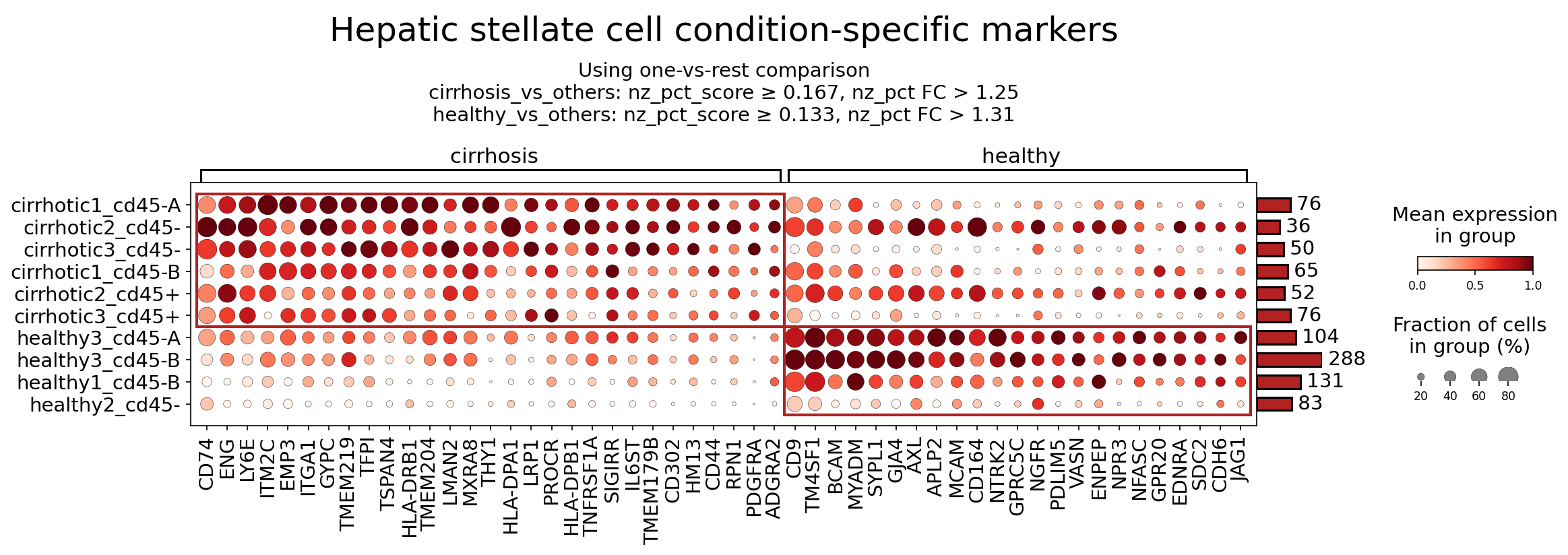
[Analysis Visualization Results]...
Analysis Overview
This analysis identifies condition-specific surfaceome markers in Hepatic stellate cells (HSCs) from single-cell RNA sequencing data comparing cirrhotic and healthy liver tissues. Surfaceome markers are particularly relevant for understanding cell-surface interactions and for their potential as therapeutic targets due to their accessibility. The dot plot visualizes the mean expression level (color intensity) and the fraction of cells expressing each marker (dot size) across individual samples, grouped by cirrhosis and healthy conditions. The analysis utilized a one-vs-rest comparison strategy to identify markers specifically enriched in each condition, with specific nz_pct_score and nz_pct_FC cutoffs for marker selection.
Visual Summary
The dot plot clearly delineates two distinct sets of surfaceome markers: those predominantly expressed in HSCs from cirrhotic samples (left panel) and those enriched in HSCs from healthy samples (right panel).
- Cirrhosis-enriched markers (left panel): A substantial number of genes show high mean expression (dark red dots) and a high fraction of expressing cells (large dots) across all cirrhotic samples (e.g., CD74, ENG, ITGA1, HLA-DRB1, THY1, CD44, PDGFRA). These markers exhibit very low or no expression in healthy samples, indicating a strong upregulation in the disease state. The top red box highlights this clear differential expression pattern.
- Healthy-enriched markers (right panel): Conversely, genes like CD9, TM4SF1, BCAM, AXL, MCAM, NTRK2, and EDNRA display high expression and prevalence in healthy HSCs but are largely absent or expressed at very low levels in cirrhotic samples. The bottom red box highlights this set of markers.
- Sample Variability: Within each condition, there is some variability in expression and fraction of positive cells across individual samples, but the overall trend of differential expression between conditions is robust for the identified markers.
Biological Interpretation
Hepatic stellate cells (HSCs) are the primary collagen-producing cells in liver fibrosis, which is a key component of cirrhosis. In healthy liver, HSCs are quiescent and store vitamin A. Upon liver injury, they activate, proliferate, migrate, and transdifferentiate into myofibroblast-like cells, driving extracellular matrix (ECM) deposition and leading to fibrosis. The identified surfaceome markers reflect distinct activation states and functional roles of HSCs in health versus cirrhosis.
Markers Upregulated in Cirrhotic Hepatic Stellate Cells
- PDGFRA (Platelet-Derived Growth Factor Receptor Alpha):
- Signaling/Mechanism: PDGFRA is a receptor tyrosine kinase whose activation by PDGF ligands is a critical driver of HSC proliferation, migration, and activation. PDGF-PDGFRA signaling is a central pathway in fibrogenesis.
- Relevance to Cirrhosis: Its strong upregulation on cirrhotic HSCs highlights the activated, proliferative, and migratory phenotype characteristic of fibrotic HSCs.
- Reference: GeneCards: PDGFRA
- ENG (Endoglin, CD105):
- Signaling/Mechanism: A co-receptor for the transforming growth factor-beta (TGF-β) superfamily. Endoglin modulates TGF-β signaling, often enhancing its profibrotic effects. It is highly expressed on activated endothelial cells and mesenchymal cells.
- Relevance to Cirrhosis: Elevated ENG expression on activated HSCs indicates increased sensitivity to TGF-β, a master regulator of fibrosis, promoting ECM production and HSC activation. It's also associated with angiogenesis.
- Reference: PubMed search: Endoglin liver fibrosis
- THY1 (Thy-1 cell surface antigen, CD90):
- Signaling/Mechanism: A GPI-anchored glycoprotein involved in cell adhesion, signal transduction, and cell-matrix interactions. It is frequently associated with mesenchymal stem cells and activated fibroblasts.
- Relevance to Cirrhosis: THY1 is a well-established marker of activated HSCs and myofibroblasts in various fibrotic diseases, including liver fibrosis. Its expression is correlated with fibrosis progression and promotes pro-fibrogenic pathways.
- Reference: GeneCards: THY1
- CD44:
- Signaling/Mechanism: A transmembrane glycoprotein and a primary receptor for hyaluronan (HA), a major component of the ECM. CD44 mediates cell adhesion, migration, proliferation, and survival, and is involved in various signaling pathways.
- Relevance to Cirrhosis: Upregulation of CD44 on activated HSCs facilitates their adhesion to the remodeled fibrotic matrix and promotes migration. CD44-HA interactions can activate downstream signaling leading to HSC activation and fibrogenesis.
- Reference: PubMed search: CD44 hepatic stellate cell fibrosis
- MHC Class II molecules (HLA-DRB1, HLA-DRA, HLA-DPA1, HLA-DPB1):
- Signaling/Mechanism: These proteins form major histocompatibility complex (MHC) class II, essential for presenting peptide antigens to CD4+ T lymphocytes, thus initiating adaptive immune responses. They are typically expressed on professional antigen-presenting cells.
- Relevance to Cirrhosis: Their robust expression on cirrhotic HSCs suggests that activated HSCs gain or upregulate antigen-presenting capabilities. This indicates an active role for HSCs in local immune modulation and perpetuating chronic inflammation within the fibrotic liver, potentially presenting autoantigens or pathogen-derived antigens to T cells.
- CD74 (MHC Class II invariant chain):
- Signaling/Mechanism: Acts as a chaperone for MHC class II molecules and also functions as a cell surface receptor for macrophage migration inhibitory factor (MIF), a pro-inflammatory cytokine.
- Relevance to Cirrhosis: Co-expression with MHC Class II on HSCs supports their role in antigen presentation. The CD74-MIF axis is known to promote inflammation and cell proliferation, which are critical components of liver fibrosis.
- ITGA1 (Integrin Alpha-1):
- Signaling/Mechanism: Forms an integrin heterodimer (VLA-1, with ITGB1) that acts as a receptor for collagen and laminin, major components of the ECM. Integrins are crucial for cell-matrix adhesion and transmit mechanical and biochemical signals.
- Relevance to Cirrhosis: Its upregulation indicates increased adhesion of activated HSCs to the fibrotic ECM, facilitating their migration and activation, and contributing to fibrotic remodeling.
- Reference: GeneCards: ITGA1
Markers Upregulated in Healthy Hepatic Stellate Cells
- CD9:
- Signaling/Mechanism: A member of the tetraspanin family, involved in cell adhesion, migration, and membrane organization. Tetraspanins form microdomains with other proteins, influencing their function.
- Relevance to Cirrhosis: High expression in healthy HSCs suggests a role in maintaining HSC quiescence or normal cellular interactions. Its downregulation in cirrhosis could signify a loss of quiescent-associated functions and a transition to an activated state.
- EDNRA (Endothelin Receptor Type A):
- Signaling/Mechanism: A G-protein coupled receptor for endothelin-1 (ET-1), a potent vasoconstrictor and mitogen.
- Relevance to Cirrhosis: While ET-1 signaling is generally profibrotic in the liver, the *downregulation* of EDNRA on cirrhotic HSCs compared to healthy is an interesting observation. This might imply a shift in the receptor profile of activated HSCs (perhaps favoring EDNRB, if expressed), or that EDNRA on quiescent HSCs plays a different homeostatic role that is lost upon activation.
- NTRK2 (Neurotrophic Receptor Tyrosine Kinase 2, TRKB):
- Signaling/Mechanism: The high-affinity receptor for brain-derived neurotrophic factor (BDNF), mediating neuronal survival and plasticity. Found also in non-neuronal cells.
- Relevance to Cirrhosis: Its expression in healthy HSCs suggests potential neurotrophic signaling pathways that may contribute to maintaining HSC quiescence or their normal physiological functions. Its loss in cirrhosis might indicate a disruption of these protective pathways during disease progression.
Clinical or Translational Implications
The identification of surfaceome markers differentially expressed on Hepatic stellate cells in cirrhosis offers significant clinical and translational opportunities, particularly for therapeutic targeting and diagnostic purposes.
Therapeutic Targets for Cirrhosis
- PDGFRA: Given its central role in HSC activation and proliferation, PDGFRA is a prime therapeutic target. Tyrosine kinase inhibitors that target PDGFRA (e.g., imatinib, nintedanib) are already approved for other fibrotic conditions and could be repurposed or developed specifically for liver fibrosis.
- Related Agents: Imatinib, Nintedanib.
- Reference: PubMed search: Imatinib liver fibrosis
- ENG (Endoglin): As a key modulator of TGF-β signaling, ENG presents a promising target. Antibodies that block ENG (e.g., TRC105) have been explored in various fibrotic diseases and cancer, aiming to attenuate TGF-β-driven fibrogenesis.
- Related Agents: Anti-Endoglin antibodies.
- THY1 (CD90): The strong upregulation of THY1 on activated HSCs makes it a potential target. Strategies to inhibit THY1 signaling or deplete THY1-positive activated HSCs could be explored, potentially through antibody-drug conjugates or small molecule inhibitors.
- CD44: Targeting CD44 or its interaction with hyaluronan could disrupt HSC adhesion, migration, and activation. Anti-CD44 antibodies or agents that interfere with HA binding have been investigated in preclinical models.
- ITGA1: Inhibiting ITGA1-mediated adhesion to the ECM could reduce HSC activation and fibrotic progression. Integrin inhibitors are a class of drugs, and specific blockade of VLA-1 could be considered.
- MHC Class II / CD74: Modulating the antigen-presenting function of HSCs could be a novel approach to dampen chronic inflammation in cirrhosis. Therapies targeting specific immune checkpoints or antigen presentation pathways on HSCs might be investigated.
Diagnostic Biomarkers and Monitoring
The differentially expressed surfaceome markers, particularly those highly expressed in cirrhotic HSCs, could serve as non-invasive biomarkers for diagnosing cirrhosis, staging fibrosis severity, or monitoring therapeutic responses. Their cell-surface location makes them accessible for detection through techniques like flow cytometry (if cells are isolated) or potentially through imaging modalities targeting these surface proteins in vivo.
Considerations for Drug Development
Targeting surfaceome markers offers advantages for drug development, as antibodies or small molecules can directly interact with these accessible proteins. However, specificity to activated HSCs (over healthy cells or other cell types) will be crucial to minimize off-target effects. Further validation in preclinical models and clinical studies is essential to confirm efficacy and safety.
14. T cell CD4+ Condition-Specific Surfaceome Markers in Liver Cirrhosis
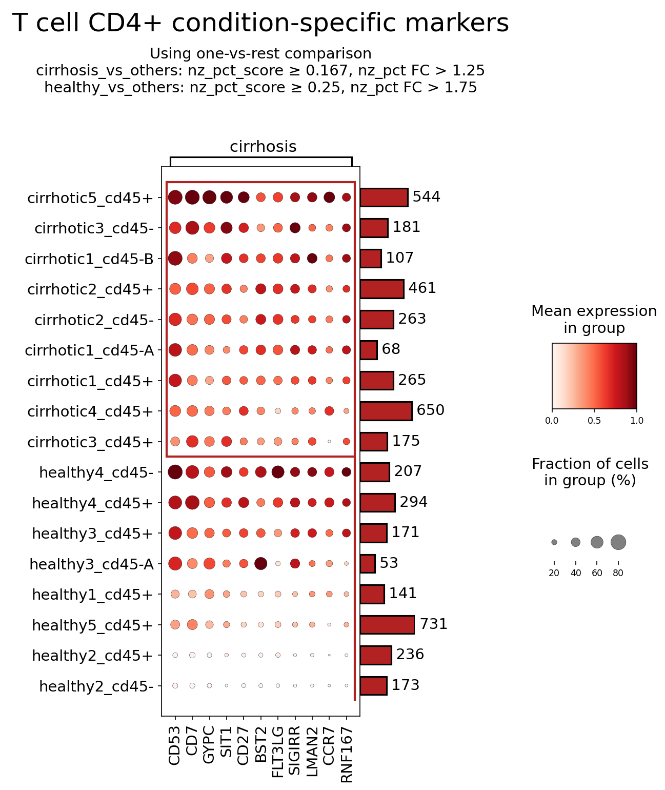
[Analysis Visualization Results]...
Analysis Overview
This analysis investigates the expression of surfaceome markers on CD4+ T cells in liver tissue from individuals with cirrhosis compared to healthy controls. A dot plot visualization was generated using a one-vs-rest comparison, identifying markers that show differential expression patterns between the two conditions. The analysis focused on surfaceome markers to highlight cell-surface proteins that are readily accessible and often implicated in cell-cell interactions, signaling, and as potential therapeutic targets.
Visual Summary
The dot plot displays the mean expression level (color intensity, ranging from white for low to dark red for high) and the fraction of cells expressing the marker (dot size, from small for low to large for high) for eleven surfaceome markers (CD53, CD7, GYPC, SIT1, CD27, BST2, FLT3LG, SIGIRR, LMAN2, CCR7, RNF167) across multiple healthy and cirrhotic liver samples.
Key observations include:
- Elevated Markers in Cirrhosis: Several markers, including CD53, CD7, SIT1, CD27, BST2, FLT3LG, and SIGIRR, show generally higher mean expression and/or a larger fraction of expressing cells across most cirrhotic samples compared to many healthy samples. This suggests a consistent upregulation of these markers on CD4+ T cells in the context of liver cirrhosis.
- Reduced Marker in Cirrhosis: CCR7 exhibits notably lower mean expression and a smaller fraction of expressing cells in most cirrhotic samples compared to several healthy samples (e.g., healthy4_cd45- and healthy3_cd45+), indicating its potential downregulation in disease.
- Variable/Low Expression: Markers such as GYPC, LMAN2, and RNF167 show less consistent patterns or generally low expression across both conditions, making their differential role less clear from this plot alone.
- Sample Heterogeneity: While general trends are observed, there is notable sample-to-sample variability within both the healthy and cirrhotic groups, particularly visible for markers like CCR7 and the general level of expression for most markers. For example, healthy4_cd45- displays high expression of many markers similar to cirrhotic samples, suggesting potential sub-phenotypes or inflammatory states even within the 'healthy' designation, or individual variation.
Biological Interpretation
The observed differential expression of surfaceome markers on CD4+ T cells provides insights into their altered state and function within the cirrhotic liver.
Indicators of T cell Activation and Memory (CD53, CD7, CD27):
- CD53: A tetraspanin, often associated with immune cell activation, adhesion, and trafficking. Its upregulation in cirrhotic CD4+ T cells could reflect increased activation and involvement in cell-cell interactions within the inflamed liver microenvironment. GeneCards: CD53
- CD7: An early T cell differentiation marker, also implicated in T cell activation and adhesion. Elevated CD7 suggests an expanded population of activated T cells or T cells undergoing chronic stimulation in cirrhosis. UniProt: CD7
- CD27: A costimulatory molecule vital for T cell activation, differentiation into memory cells, and survival. Its increased expression on CD4+ T cells in cirrhosis could indicate an accumulation of memory or effector T cell subsets that are chronically engaged in immune responses against liver damage or associated antigens. Loss of CD27 is sometimes seen on highly differentiated or exhausted T cells, so its upregulation here suggests a more active, possibly memory-like, phenotype. GeneCards: CD27
Modulators of Immune Responses (SIT1, BST2, SIGIRR):
- SIT1 (SHP2-interacting transmembrane adaptor protein 1): Known to negatively regulate T cell activation by recruiting phosphatases like SHP2 to the TCR signaling complex. Its upregulation could represent a compensatory mechanism to dampen excessive T cell activation and inflammation in the chronically inflamed cirrhotic liver, or contribute to T cell dysfunction/exhaustion. GeneCards: SIT1
- BST2 (Bone Marrow Stromal Antigen 2 / Tetherin): An interferon-inducible protein with antiviral activity, but also involved in immune regulation. Its elevated expression suggests an ongoing inflammatory or antiviral milieu in the cirrhotic liver, possibly linked to chronic viral hepatitis (if applicable to cirrhosis etiology) or sterile inflammation. BST2 can impact T cell activation and antigen presentation. UniProt: BST2
- SIGIRR (Single Ig and TIR domain containing): A negative regulator of Toll-like receptor (TLR) and IL-1 receptor (IL-1R) signaling, which are critical pathways for innate and adaptive immune responses. Upregulation of SIGIRR on CD4+ T cells might reflect an attempt to control exaggerated inflammatory responses driven by pathogen-associated molecular patterns (PAMPs) or danger-associated molecular patterns (DAMPs) abundant in chronic liver disease. This could lead to a dampened, potentially ineffective, immune response if overexpressed. GeneCards: SIGIRR
T Cell Homing and Trafficking (CCR7):
- CCR7: A chemokine receptor essential for the homing of naive and central memory T cells to secondary lymphoid organs. Its reduced expression on CD4+ T cells in cirrhosis is consistent with a shift from naive/central memory phenotypes to effector or tissue-resident memory T cells that have exited lymphoid organs and accumulated in the chronically inflamed liver. This indicates T cells are likely retaining or re-entering the liver, rather than recirculating to lymphoid tissues. PubMed: CCR7 T cell trafficking
Cellular Communication (FLT3LG):
- FLT3LG (Fms-like tyrosine kinase 3 ligand): Typically produced by stromal cells, T cells, monocytes, and B cells to stimulate FLT3-expressing hematopoietic progenitors (e.g., dendritic cells). Its surface expression on CD4+ T cells, as observed, is noteworthy. It suggests that these T cells might actively contribute to the local immune microenvironment by influencing dendritic cell development or function, potentially sustaining immune activation and inflammation in the cirrhotic liver. GeneCards: FLT3LG
Clinical or Translational Implications
The differential expression of these surfaceome markers identifies potential candidates for therapeutic intervention and biomarkers for disease progression in liver cirrhosis.
Therapeutic Targets for Immune Modulation:
- SIT1 and SIGIRR: As negative regulators of T cell activation and inflammatory signaling, targeting these molecules could offer a strategy to fine-tune immune responses in cirrhosis. Inhibiting their function could potentially enhance beneficial anti-fibrotic or immune-clearance pathways if T cell activity is inappropriately suppressed, or conversely, agonists could dampen detrimental chronic inflammation.
- CD27/CD70 Axis: Modulating the CD27 signaling pathway could be explored. Strategies to enhance or suppress this axis exist in other diseases (e.g., cancer, autoimmunity). For example, blocking CD70 (ligand for CD27) or CD27 could potentially reduce sustained T cell activation and inflammation contributing to cirrhosis.
- FLT3LG: If T cell-derived FLT3LG significantly contributes to local DC activation and subsequent T cell-mediated inflammation, targeting FLT3LG or its receptor (FLT3, typically on DCs) could disrupt this feedback loop and ameliorate liver inflammation.
Biomarkers for Disease Activity/Prognosis:
- The consistent upregulation of markers like CD53, CD7, BST2, FLT3LG, SIT1, and SIGIRR, or the downregulation of CCR7, on CD4+ T cells in the cirrhotic liver could serve as indicators of disease activity, specific immune dysregulation, or potentially predict progression. Monitoring these markers, perhaps via flow cytometry on liver biopsy samples or even circulating T cells (if similar patterns are observed), could offer prognostic value.
Considerations for Cell Trafficking:
- The reduced CCR7 expression suggests a diminished capacity for cirrhotic CD4+ T cells to home to lymphoid organs, indicating their preferential retention or infiltration within the inflamed liver. Therapeutic approaches aimed at modifying T cell trafficking to and from the liver could potentially impact disease progression.
15. Gene Ontology (GSA) Analysis of Cholangiocytes in Liver Cirrhosis
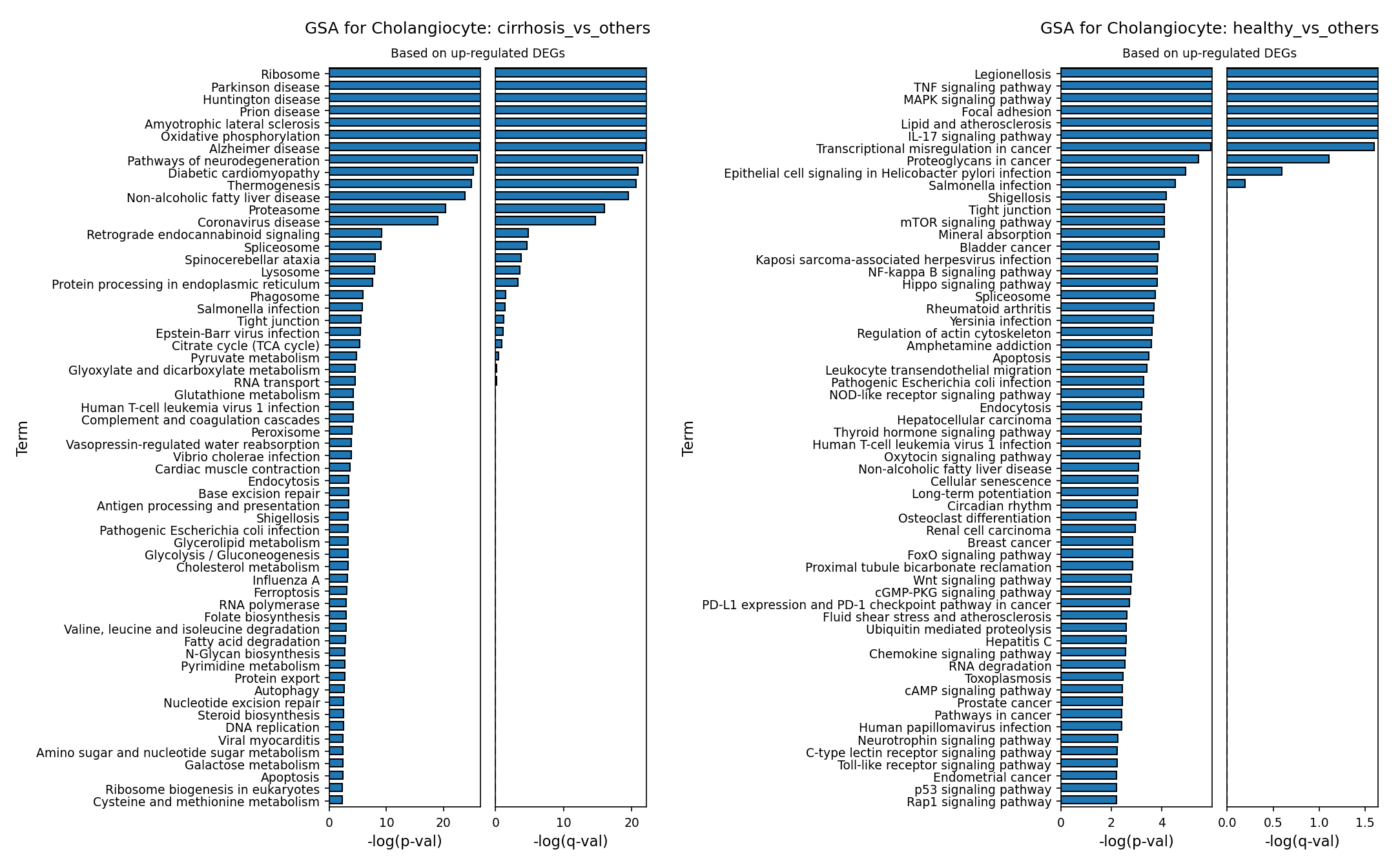
[Analysis Visualization Results]...
Analysis Overview
This analysis investigates Gene Ontology (GO) enrichment in cholangiocytes from human liver tissue, comparing conditions of cirrhosis versus healthy, and healthy versus cirrhosis. The results are based on genes found to be significantly upregulated in each comparison, providing insights into the biological processes and pathways characteristic of cholangiocytes in different disease states. The plots display the top enriched GO terms ranked by their statistical significance (-log(p-val) and -log(q-val)).
Visual Summary
The provided bar plots illustrate the top enriched Gene Ontology (GO) terms for cholangiocytes.
The first plot, "GSA for Cholangiocyte: cirrhosis_vs_others," shows pathways upregulated in cholangiocytes from cirrhotic livers compared to other conditions (primarily healthy). Key observations include:
- High Significance for Protein Synthesis and Degradation: Terms like "Ribosome," "Protein processing in endoplasmic reticulum," "Proteasome," and "Lysosome" are among the most significantly enriched, indicating heightened protein turnover and cellular stress responses.
- Metabolic Reprogramming: Pathways such as "Oxidative phosphorylation," "Citrate cycle (TCA cycle)," "Pyruvate metabolism," and "Thermogenesis" are also highly ranked, suggesting altered energy metabolism.
- Cellular Stress and Disease Associations: Notably, several neurodegenerative disease pathways (e.g., "Parkinson disease," "Huntington disease," "Alzheimer disease") and "Non-alcoholic fatty liver disease" are enriched, pointing to common cellular stress mechanisms.
- Barrier Function: "Tight junction" pathway also appears with notable significance.
The second plot, "GSA for Cholangiocyte: healthy_vs_others," highlights pathways upregulated in cholangiocytes from healthy livers compared to those in cirrhosis. Key observations include:
- Immune and Inflammatory Signaling: "TNF signaling pathway," "MAPK signaling pathway," "IL-17 signaling pathway," and "NF-kappa B signaling pathway" are highly enriched, reflecting active immune surveillance and response.
- Cell Adhesion and Structure: "Focal adhesion" and "Tight junction" pathways are prominent, indicating robust maintenance of cell-matrix interactions and intercellular junctions.
- Metabolic and Growth Regulation: "mTOR signaling pathway" and "Lipid and atherosclerosis" are observed, suggesting active basal metabolic and growth regulatory processes.
- Infection and Cancer-Related Pathways: A diverse set of infection-related and cancer-associated pathways are also significantly enriched.
Biological Interpretation
Cholangiocytes in Cirrhosis: Responding to Chronic Stress
Cholangiocytes in cirrhotic livers exhibit a robust upregulation of pathways related to protein synthesis and degradation machinery (Ribosome, Protein processing in endoplasmic reticulum, Proteasome, Lysosome). This suggests a significant cellular response to chronic injury and stress, possibly involving increased protein turnover for repair, adaptation, or coping with misfolded proteins, a common feature in chronic liver diseases. The enrichment of Oxidative phosphorylation and other metabolic pathways (Citrate cycle, Pyruvate metabolism) indicates a profound shift in energy metabolism, potentially reflecting increased metabolic demand or mitochondrial dysfunction in response to the cirrhotic microenvironment. The appearance of "Non-alcoholic fatty liver disease" is directly relevant to liver pathology, suggesting shared molecular underpinnings or consequences. The frequent enrichment of pathways associated with neurodegenerative diseases (e.g., Parkinson, Huntington, Alzheimer) is intriguing; these diseases often involve protein aggregation, mitochondrial dysfunction, and cellular stress, suggesting that cholangiocytes in cirrhosis may be undergoing similar fundamental stress responses, rather than directly implying neurological pathology within the cholangiocytes themselves. Furthermore, the presence of "Tight junction" pathways suggests altered regulation of barrier function, which is critical for bile duct integrity.
Cholangiocytes in Healthy Liver: Maintaining Homeostasis and Responsiveness
In contrast, cholangiocytes from healthy livers show a significant upregulation of immune and inflammatory signaling pathways (TNF, MAPK, IL-17, NF-kappa B signaling pathways). This indicates that healthy cholangiocytes are actively involved in immune surveillance and maintaining a finely tuned inflammatory balance within the liver, essential for protecting against pathogens and tissue damage. The strong enrichment of Focal adhesion and Tight junction pathways underscores their critical role in maintaining structural integrity, cell-cell communication, and mechanical sensing, which are fundamental for normal bile duct function. Pathways like mTOR signaling highlight active metabolic regulation and cellular growth processes in a healthy, homeostatic state. The presence of infection-related pathways may reflect the cholangiocytes' role as a first line of defense against ascending infections from the gut or as part of the liver's broader immune ecosystem.
Comparison and Context
The stark differences in enriched pathways between cirrhotic and healthy cholangiocytes highlight a clear shift in cellular priorities and function. In cirrhosis, cholangiocytes appear to be in a state of stress response, attempting to manage protein homeostasis and adapt their metabolism to a hostile environment. In health, they are primarily engaged in maintaining structural integrity, active immune signaling, and metabolic regulation. This shift underscores the pathological changes occurring in cholangiocytes during cirrhosis, moving from homeostatic maintenance to stress adaptation.
Clinical or Translational Implications
- Biomarker Discovery: Upregulated genes contributing to pathways like "Ribosome," "Oxidative phosphorylation," and "Protein processing in endoplasmic reticulum" in cirrhotic cholangiocytes could represent novel biomarkers for disease progression or severity in cirrhosis, particularly reflecting cholangiocyte stress or dysfunction. These could potentially be detected in bile or tissue biopsies.
- Therapeutic Targets: Targeting specific pathways found to be dysregulated in cirrhotic cholangiocytes, such as those involved in protein quality control (e.g., ER stress, proteasomal activity) or mitochondrial function, could offer therapeutic avenues to mitigate cholangiocyte injury, improve bile duct integrity, and slow the progression of liver fibrosis in cirrhosis. For example, modulating ER stress responses has shown promise in various fibrotic diseases PubMed Search: ER stress fibrosis.
- Understanding Pathogenesis: A deeper understanding of these pathway shifts provides mechanistic insights into how cholangiocytes contribute to the pathogenesis of cirrhosis, including their role in cholestasis and fibrosis. Identifying factors that drive the altered metabolic state or protein handling could reveal key drivers of disease.
- Drug Repurposing: The enrichment of pathways associated with neurodegenerative diseases, which share common cellular stress mechanisms, might suggest potential for repurposing drugs that target these fundamental cellular processes to treat cholangiocyte pathology in cirrhosis.
16. Gene Set Enrichment Analysis of Cell Type-Specific Pathways in Cirrhosis
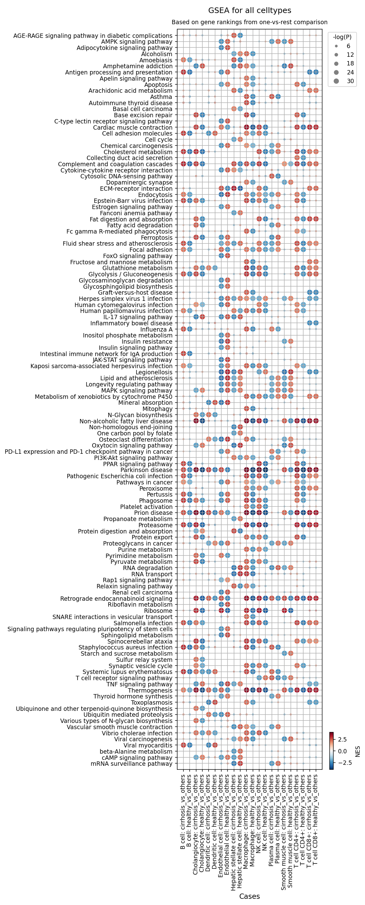
[Analysis Visualization Results]...
Analysis Overview
This analysis presents Gene Set Enrichment Analysis (GSEA) results for various cell types in human liver single-cell RNA-seq data, comparing two conditions: healthy and cirrhosis. The dot plot visualizes the enrichment of 120 selected gene sets (pathways) across different cell types. For each cell type, GSEA was performed to identify pathways enriched or depleted in that specific cell type relative to all other cell types *within the same condition* (i.e., celltype: cirrhosis_vs_others compares B cells in cirrhosis against all other cell types in cirrhosis). The size of the dot corresponds to the statistical significance (-log(p-value)), and the color indicates the Normalized Enrichment Score (NES), with red for positive enrichment (upregulation) and blue for negative enrichment (downregulation).
Visual Summary
The dot plot reveals widespread and complex alterations in pathway enrichment across different cell types in the cirrhotic liver compared to the healthy liver.
- Immune Activation: Many immune-related pathways, such as "Cytokine-cytokine receptor interaction", "Chemokine signaling pathway", "TNF signaling pathway", "JAK-STAT signaling pathway", "Antigen processing and presentation", and "Phagosome", show strong positive enrichment (red, larger dots) across various immune cell types (Macrophages, Dendritic cells, T cells, B cells, Plasma cells) in the cirrhosis_vs_others columns. This indicates a heightened and distinct inflammatory and immune response in these cell types during cirrhosis.
- Fibrosis and ECM Remodeling: Pathways like "ECM-receptor interaction", "Focal adhesion", and "Rap1 signaling pathway" are significantly enriched (red, large dots) in Hepatic stellate cells, Endothelial cells, and Macrophages in the cirrhosis_vs_others context. This highlights their active role in extracellular matrix remodeling and fibrogenesis, a hallmark of cirrhosis.
- Metabolic Dysregulation: Metabolic pathways exhibit mixed and cell-type-specific patterns. For example, "Non-alcoholic fatty liver disease" pathways show negative enrichment (blue, large dots) in Macrophages, Hepatic stellate cells, Endothelial cells, and T cells in cirrhosis_vs_others, suggesting a shift in metabolic distinctions beyond the fatty liver stage. Conversely, "AMPK signaling pathway" often shows positive enrichment in several cell types in cirrhosis.
- Cell Proliferation and Cancer Risk: "Cell cycle", "PI3K-Akt signaling pathway", and "Pathways in cancer" show prominent positive enrichment (red, large dots) in Hepatic stellate cells, Cholangiocytes, Endothelial cells, and some immune cells in the cirrhosis_vs_others condition, suggesting increased proliferative activity and a propensity towards malignancy.
- Immune Checkpoint Pathway: "PD-L1 expression and PD-1 checkpoint pathway in cancer" is positively enriched in Macrophages, Dendritic cells, and T cells (particularly CD8+) in the cirrhotic environment, indicative of immune exhaustion or tolerance mechanisms.
- Viral Pathways: Several viral infection-related pathways (e.g., "Human cytomegalovirus infection", "Herpes simplex virus 1 infection") show enrichment in multiple cell types in cirrhosis_vs_others, potentially pointing to viral etiology, reactivation, or altered antiviral responses.
- Cell Type-Specific Shifts: Comparing a cell type's healthy_vs_others profile to its cirrhosis_vs_others profile reveals shifts. For instance, Macrophages in the healthy liver ("Macrophage: healthy_vs_others") show more distinct metabolic pathway enrichments (e.g., "Fatty acid degradation") that are attenuated or absent in cirrhotic macrophages, which instead show a strong inflammatory and antigen-presenting profile.
Biological Interpretation
The GSEA results provide a detailed molecular landscape of cellular dysfunction and adaptation in the cirrhotic liver.
- Exacerbated Inflammation and Immune Dysfunction: The pervasive upregulation of immune signaling pathways across various immune cells signifies a state of chronic inflammation in cirrhosis. Macrophages, Dendritic cells, and T cells adopt a highly activated and inflammatory phenotype, characterized by enhanced cytokine production, antigen presentation, and phagocytic activity. This sustained inflammatory environment contributes to liver damage and fibrosis progression [PubMed search: chronic inflammation liver cirrhosis].
- Fibrogenesis driven by HSC Activation: Hepatic stellate cells (HSCs) are central to liver fibrosis. Their strong enrichment in "ECM-receptor interaction", "Focal adhesion", "TGF-beta signaling pathway", and "PI3K-Akt signaling pathway" in cirrhosis reflects their activation and transition into myofibroblast-like cells, leading to excessive extracellular matrix deposition and scarring [GeneCards: HSC].
- Metabolic Reprogramming: The observed shifts in metabolic pathways across cell types suggest a profound metabolic reprogramming in cirrhosis. The negative enrichment for NAFLD pathways might indicate that cells in advanced cirrhosis have transitioned beyond the specific metabolic dysregulations associated with early fatty liver disease, or that these pathways are no longer uniquely distinctive compared to other cell types in the cirrhotic milieu.
- Oncogenic Potential: The consistent enrichment of "Cell cycle" and "Pathways in cancer" across multiple cell types, including Hepatic stellate cells, Cholangiocytes, and Endothelial cells, underscores the high risk of developing hepatocellular carcinoma (HCC) or cholangiocarcinoma (CCA) in cirrhotic patients. These pathways indicate increased cellular proliferation and potentially dysregulated growth control.
- Immune Evasion and Exhaustion: The activation of "PD-L1 expression and PD-1 checkpoint pathway" in immune cells (Macrophages, T cells) suggests a mechanism of immune exhaustion or suppression. This could contribute to the liver's inability to clear chronic infections or mount effective anti-tumor responses, perpetuating inflammation and promoting disease progression [UniProt: PD-1, PD-L1].
- Vascular Remodeling and Cholangiopathy: Endothelial cells show enrichment in pathways related to vascular integrity and remodeling, consistent with the development of portal hypertension. Cholangiocytes exhibit enrichment in pathways related to bile duct biology and cancer, indicating their active involvement in cholestasis and potential malignant transformation.
Clinical or Translational Implications
The comprehensive GSEA results offer several insights with clinical relevance:
- Identification of Therapeutic Targets: The consistently enriched inflammatory (e.g., TNF, JAK-STAT) and fibrogenic (e.g., ECM-receptor interaction, PI3K-Akt) pathways across multiple cell types provide promising targets for anti-inflammatory and anti-fibrotic therapies. Modulating these pathways could slow disease progression or reverse damage [PubMed search: liver fibrosis treatment].
- Biomarker Discovery: Cell-type- specific pathway alterations could serve as novel biomarkers for diagnosing cirrhosis, staging disease severity, predicting prognosis, or monitoring treatment response. For instance, distinct gene expression profiles associated with HSC activation could be detectable in peripheral blood or liver biopsies.
- Understanding Cancer Risk: The widespread activation of oncogenic pathways in various cell types highlights the molecular basis for increased cancer risk in cirrhosis. Interventions targeting these pathways could be explored for chemoprevention strategies.
- Immune Modulation Strategies: The presence of immune checkpoint activation suggests that immunomodulatory therapies, similar to those used in cancer, might be considered in cirrhosis. However, careful patient stratification and understanding of the complex immune landscape are crucial to avoid exacerbating inflammation or precipitating autoimmune complications.
- Personalized Medicine: The cell-type-specific nature of pathway dysregulation underscores the importance of a precision medicine approach, where therapies could be tailored based on the dominant cell type involvement and pathway activation profile in individual patients.
17. Discussion
The comprehensive single-cell analysis of human liver tissue provides a high-resolution view of the profound cellular and molecular changes occurring in cirrhosis. Initial UMAP visualizations clearly demonstrate a global transcriptomic separation between healthy and cirrhotic samples, indicative of widespread cellular and molecular reprogramming. This separation is supported by robust cell type annotations across major, minor, and subset levels, ensuring the reliability of downstream analyses.
Population analyses reveal a hallmark of cirrhosis: a drastic reduction in functional hepatocytes, replaced by an expansion of fibrogenic cells such as Hepatic stellate cells and Fibroblasts, alongside increased Cholangiocytes, signifying ongoing liver damage, repair, and ductular reaction. The immune compartment undergoes a significant remodeling, with a prominent increase in macrophages, T cells, and NK cells. Specifically, macrophages show a statistically significant shift towards a pro-inflammatory M1 phenotype, with a concomitant decrease in the reparative M2A subset. Within T cell populations, there is a complex dysregulation characterized by an increase in CD4 Memory T cells, Th22, Regulatory T cells (Tregs), and CD4 Naive T cells, while crucial Cytotoxic T cells are significantly reduced. This suggests a chronic inflammatory milieu with concurrent attempts at immune suppression and an impaired capacity for effector functions, potentially contributing to immune evasion and increased risk of hepatocellular carcinoma (HCC).
Cell-cell interaction (CCI) analyses underscore a highly active and pathological microenvironment in cirrhosis. The cirrhotic liver exhibits a dramatic upregulation of interactions involving fibrogenic pathways, such as various integrins binding to extracellular matrix components like SPP1 (Osteopontin), Fibronectin (FN1), and Collagen (COL). Pro-inflammatory axes like TNF-TNFRSF and CXCL12-CXCR4 are also highly active, largely mediated by macrophages, endothelial cells, and hepatic stellate cells. Increased VEGFA-VEGFR interactions point to active angiogenesis, contributing to portal hypertension. In contrast, healthy liver tissue shows a more quiescent communication network, with some homeostatic interactions (e.g., ANXA1-FPR, ProstaglandinD2) appearing suppressed in cirrhosis, indicating a loss of protective regulatory mechanisms. The involvement of HLA-G-LILRB2 in cirrhosis-specific interactions further suggests an active role of immune suppression, potentially by hepatic stellate cells on T cells.
Surfaceome marker expression analyses provide molecular details for specific cell types. Cirrhotic cholangiocytes upregulate markers like Syndecan-1 (SDC1), ALCAM, various Tetraspanins (TSPANs), and strikingly, the growth factor receptors ERBB2 and ERBB3, along with MHC Class II molecules (HLA-DRB1), indicative of increased proliferation, altered adhesion, and antigen-presenting capabilities. Macrophages in cirrhosis show elevated expression of pro-inflammatory markers such as Basigin (BSG/CD147), FCGR2A, C3AR1, and AXL, while homeostatic markers like PILRA, LAIR1, and SIGIRR are relatively higher in healthy macrophages. Activated Hepatic stellate cells in cirrhosis upregulate key fibrogenic receptors including PDGFRA, Endoglin (ENG/CD105), THY1 (CD90), CD44, and Integrin Alpha-1 (ITGA1), and also express MHC Class II molecules, confirming their activated, profibrotic, and immune-modulatory phenotype. CD4+ T cells in cirrhosis show increased expression of activation/memory markers (CD53, CD7, CD27) and immune modulators (SIT1, BST2, SIGIRR), coupled with reduced CCR7 expression, suggesting their retention and chronic activation within the inflamed liver.
Gene Ontology (GSA) analysis of cholangiocytes in cirrhosis highlights pathways related to protein synthesis, processing, and degradation (e.g., Ribosome, ER stress, Proteasome, Lysosome), alongside metabolic reprogramming (Oxidative phosphorylation, TCA cycle), indicating a cellular stress response. In contrast, healthy cholangiocytes are enriched in immune signaling (TNF, MAPK, NF-kappa B) and structural maintenance (Focal adhesion, Tight junction) pathways. The broader Gene Set Enrichment Analysis (GSEA) reinforces these findings, showing pervasive immune activation, fibrosis, ECM remodeling, and cell proliferation pathways across multiple cell types in cirrhosis. Notably, the 'PD-L1 expression and PD-1 checkpoint pathway' is enriched in macrophages, dendritic cells, and T cells in cirrhosis, further supporting immune exhaustion/tolerance. The presence of 'Pathways in cancer' across several cell types underscores the oncogenic risk in the cirrhotic microenvironment. Overall, the analyses reveal a highly pathogenic cellular ecosystem characterized by chronic inflammation, active fibrogenesis, immune dysregulation, and a heightened proliferative drive, all contributing to the progressive nature of liver cirrhosis and its complications.
Hypotheses:
- The sustained M1 macrophage polarization and decreased M2A macrophage population directly contribute to chronic inflammation and hinder tissue repair in cirrhotic livers.
- Dysregulated cell-cell interactions, particularly the upregulation of SPP1-integrin and PDGFRA signaling, are central drivers of hepatic stellate cell activation and subsequent liver fibrosis in cirrhosis.
- The observed expansion of regulatory T cells (Tregs) and reduction of cytotoxic T cells contribute to an immune-tolerant microenvironment, impairing anti-viral and anti-tumor immunity in cirrhosis, thereby increasing the risk of HCC.
- Cholangiocyte proliferation, driven by ERBB2/ERBB3 signaling, contributes to ductular reaction and biliary fibrosis, further exacerbating liver injury in cirrhosis.
- The loss of homeostatic macrophage surface markers like PILRA and LAIR1 in cirrhosis reflects a compromised ability of macrophages to maintain immune quiescence, contributing to persistent inflammation.
Potential therapeutic targets:
- PDGFRA (Platelet-Derived Growth Factor Receptor Alpha): PDGFRA is strongly upregulated on activated Hepatic stellate cells (HSCs) in cirrhosis. Its activation by PDGF ligands is a critical driver of HSC proliferation, migration, and activation, making it a central pathway in fibrogenesis. Evidence: Analysis shows PDGFRA is significantly upregulated in cirrhotic HSCs compared to healthy. CCI analysis also highlights pro-fibrotic signaling involving HSCs. Literature confirms PDGF-PDGFRA signaling's role in liver fibrosis. Validation: Preclinical models of liver fibrosis can be treated with PDGFRA inhibitors (e.g., imatinib, nintedanib) to assess reductions in HSC activation, ECM deposition, and overall fibrosis. In vitro studies with primary HSCs can confirm proliferation and activation inhibition.
- SPP1 (Osteopontin) and its Integrin Receptors (e.g., αVβ3, αVβ5): SPP1 is a pleiotropic cytokine upregulated in liver injury and cirrhosis, interacting with various integrins on macrophages and HSCs. This interaction promotes inflammation, immune cell recruitment, cell survival, and fibrosis by activating pro-inflammatory and pro-fibrotic phenotypes. Evidence: CCI analysis shows prominent SPP1-integrin interactions in cirrhosis, involving macrophages and HSCs, and are largely absent in healthy samples. Literature supports SPP1's role in inflammation and fibrosis. Validation: Blocking SPP1 or its cognate integrin receptors using neutralizing antibodies or small molecule inhibitors in cirrhotic animal models could reduce inflammation, HSC activation, and fibrosis. In vitro, perturbing this axis can evaluate macrophage polarization and fibrotic gene expression.
- ERBB2 (HER2) and ERBB3 (HER3) on Cholangiocytes: ERBB2 and ERBB3 are receptor tyrosine kinases involved in cell proliferation, survival, and differentiation. Their significant upregulation in cirrhotic cholangiocytes suggests enhanced growth factor responsiveness and pathological proliferation (ductular reaction), contributing to biliary fibrosis. Evidence: Cholangiocyte surfaceome marker analysis shows significant upregulation of ERBB2 and ERBB3 in cirrhotic samples. GSEA also indicates enrichment of 'Cell cycle' and 'Pathways in cancer' in cholangiocytes in cirrhosis. Validation: Investigate the effect of HER2/HER3 inhibitors (e.g., trastuzumab, lapatinib) on cholangiocyte proliferation and fibrogenic activity in vitro and in relevant in vivo models of cholestatic liver injury leading to cirrhosis.
- BSG (CD147) on Macrophages: CD147 is a transmembrane protein that induces matrix metalloproteinases (MMPs), promoting fibrosis by stimulating myofibroblast activation and extracellular matrix remodeling. Its upregulation in cirrhotic macrophages suggests a role in disease progression. Evidence: Macrophage surfaceome marker analysis identifies BSG as significantly upregulated in cirrhotic macrophages. Validation: Employ neutralizing antibodies or small molecule inhibitors against CD147 in macrophage cultures and liver fibrosis models to assess reductions in MMP production, HSC activation, and fibrosis severity.
- C3AR1 on Macrophages: C3AR1 is the receptor for the pro-inflammatory complement component C3a. Its upregulation on macrophages in cirrhosis indicates heightened sensitivity to complement activation products, driving inflammatory responses that contribute to liver injury and fibrosis. Evidence: Macrophage surfaceome marker analysis shows C3AR1 is significantly upregulated in cirrhotic macrophages. GSEA indicates 'Complement and coagulation cascades' enrichment in cirrhosis. Validation: Evaluate the impact of C3AR1 antagonists in animal models of cirrhosis on macrophage activation, inflammatory cytokine production, and fibrosis progression.
- TNF-TNFRSF1B signaling pathway: TNF is a potent pro-inflammatory cytokine that, via TNFRSF1B (TNF-R2), activates pathways leading to inflammation, cell survival, and fibrosis. This axis promotes hepatic stellate cell activation and proliferation in cirrhosis. Evidence: CCI analysis shows significant upregulation of TNF-TNFRSF1B interactions, particularly between T cells and Hepatic stellate cells, in cirrhosis. GSEA also highlights 'TNF signaling pathway' enrichment in multiple immune cells and HSCs in cirrhosis. Validation: Investigate selective TNFRSF1B inhibitors (if available, distinct from broad anti-TNF agents) in localized liver fibrosis models to reduce inflammation and HSC activation without systemic immunosuppression. Assess the impact on liver injury and fibrosis.
- HLA-G-LILRB2 (ILT4) pathway: The HLA-G-LILRB2 axis promotes immune suppression. Its upregulation in cirrhosis, involving interactions between hepatic stellate cells and T cells, could dampen effective anti-fibrotic or anti-tumor immune responses, contributing to immune evasion and fibrosis progression or HCC risk. Evidence: CCI analysis identifies HLA-G-LILRB2 as a significantly upregulated interaction in cirrhosis, involving HSCs and T cells. Validation: Blocking LILRB2 or inhibiting HLA-G expression in in vitro co-culture systems (HSCs with T cells) can assess restoration of T cell effector function or anti-fibrotic immunity. In vivo models with targeted modulators could evaluate effects on fibrosis and immune surveillance.
Follow-up validation ideas:
- Perform flow cytometry or mass cytometry on liver biopsies from healthy and cirrhotic patients to validate the altered proportions of T cell and macrophage subsets, specifically M1/M2A macrophages, Tregs, and Cytotoxic T cells, and their surface marker expression (e.g., CD147, C3AR1, PILRA, LAIR1).
- Conduct ex vivo co-culture experiments using primary human hepatic stellate cells, macrophages, and cholangiocytes. Perturb key interaction pathways identified (e.g., blocking SPP1, PDGFRA, ERBB2/3, or integrins with neutralizing antibodies or small molecule inhibitors) and assess their effects on cell activation, proliferation, migration, and ECM production.
- Utilize spatial transcriptomics or multiplexed immunostaining on liver sections to localize and quantify cells expressing key surface markers (e.g., PDGFRA, ENG, CD147, ERBB2, HLA-DRB1) and specific ligand-receptor interactions identified in CCI analysis within the fibrotic lesion versus non-fibrotic areas.
- Generate in vivo models of liver cirrhosis (e.g., CCl4, bile duct ligation) and test the therapeutic efficacy of targeted interventions (e.g., anti-PDGFRA antibodies, integrin inhibitors, AXL inhibitors, anti-C3AR1) on fibrosis progression, inflammation, and cellular composition using histological, biochemical, and single-cell RNA-seq endpoints.
- Investigate the functional consequences of altered T cell subset proportions by performing T cell functional assays (e.g., cytotoxicity assays, cytokine production) on sorted T cells from cirrhotic livers, and evaluate the impact of modulating SIT1 or SIGIRR expression on T cell activation and exhaustion.
- Perform proteomics on circulating extracellular vesicles or liver biopsies to assess the translational potential of identified surfaceome markers (e.g., SDC1, ERBB2, CD147, PDGFRA) as non-invasive biomarkers for disease severity or treatment response.
Limitations:
This report is based on single-cell RNA sequencing data, which provides a snapshot of gene expression and does not directly measure protein levels or cellular function. Inferences regarding cell-cell interactions and pathway activities are based on transcriptional data and require further experimental validation. The specific etiology and stage of cirrhosis were not provided, which could introduce heterogeneity in findings. Causality cannot be directly inferred from observational transcriptomic changes. While the data suggests potential therapeutic targets, their efficacy and safety in a clinical setting remain to be rigorously tested.
18. Query List
- Show and save UMAPs for condition, sample, major cell type, minor cell type, and celltype_subset in two columns.
- Show major cell type scores on UMAP and save the result.
- Show and save a marker expression dot plot for celltype_subset. SET target_cell = None and var_group_rotation = 45. Leave all other arguments at their default values.
- Show and save a population bar plot of minor cell types.
- Show and save a population bar plot of T cell subsets.
- Show and save box plots of T cell subset populations with statistically significant differences between conditions, if any. Choose ncols based on the total number of panels.
- Show and save a population bar plot of macrophage subsets.
- Show and save box plots of macrophage subset populations with statistically significant differences between conditions, if any. Choose ncols based on the total number of panels.
- Show and save cell-cell interaction patterns. Select at most 80 cell-cell interactions per group.
- Find cell-cell interactions involving major tissue cells, immune cells and stromal cells that differ significantly between conditions, show them as a dot plot, and save the result. Set max_n_items_per_group to 60. For each interaction identified under each condition, using the provided dataset context, describe the associated signaling pathways and mechanisms and, where relevant, its potential as a therapeutic target and related therapeutic agents.
- Show marker expressions for Cholangiocyte in a dot plot, and save the result. Use only surfaceome markers, with at most 50 markers per condition. For each marker identified under each condition, using the provided dataset context, describe the associated signaling pathways and biological mechanisms within the tissue and, where relevant, its potential as a therapeutic target for Cirrhosis and related therapeutic agents.
- Show marker expressions for Macrophage in a dot plot, and save the result. Use only surfaceome markers, with at most 40 markers per condition. For each marker identified under each condition, using the provided dataset context, describe the associated signaling pathways and biological mechanisms within the tissue and, where relevant, its potential as a therapeutic target for Cirrhosis and related therapeutic agents.
- Show marker expressions for Hepatic stellate cell in a dot plot, and save the result. Use only surfaceome markers, with at most 40 markers per condition. For each marker identified under each condition, using the provided dataset context, describe the associated signaling pathways and biological mechanisms within the tissue and, where relevant, its potential as a therapeutic target for Cirrhosis and related therapeutic agents.
- Show marker expressions for T cell CD4+ in a dot plot, and save the result. Use only surfaceome markers, with at most 40 markers per condition. For each marker identified under each condition, using the provided dataset context, describe the associated signaling pathways and biological mechanisms within the tissue and, where relevant, its potential as a therapeutic target for Cirrhosis and related therapeutic agents.
- Show Gene Ontology (GSA) analysis results for Cholangiocyte as a bar plot, and save the result.
- Show Gene Set Enrichment Analysis results as a dot plot, and save it. Set the color map to RdBu_r, n_pws_to_show to 120, and target_cells to None.