Single-Cell Landscape of Colorectal Cancer: Cellular Heterogeneity, Dysregulated Signaling, and Therapeutic Vulnerabilities
This report details a comprehensive single-cell analysis of human colon tissue, elucidating the complex cellular and molecular landscape of Colorectal Cancer. We identified Intestinal Epithelial cells as the tumor origin, characterized by significant aneuploidy, hyperactive cell cycle, and distinct surface marker expression. The tumor microenvironment exhibits profound immune cell remodeling, including an increase in exhausted T cells, regulatory T cells, and pro-tumorigenic macrophages, alongside activated cancer-associated fibroblasts. This analysis highlights key oncogenic pathways and identifies specific cell-cell interactions and surfaceome markers as potential therapeutic targets.
Contents
- Dataset overview
- UMAP Visualization of Cell Populations by Condition, Sample, Cell Type, and Ploidy in Colon Tissue
- Single-Cell UMAP Visualization of Major Cell Type Scores and Ploidy Status
- Marker Expression Dot Plot for Celltype Subsets
- Copy Number Variation Analysis of Intestinal Epithelial Cells in Colon Tissue
- UMAP Visualization of CNV Patterns by Cell Type, Ploidy, Condition, and Sample
- Cell Type Population Analysis in Normal vs. Tumor Colon Tissue
- T Cell Subset Population Analysis in Colorectal Cancer
- Changes in T cell and Innate Lymphoid Cell Subset Proportions in Colorectal Cancer
- Macrophage Subset Population Analysis in Colorectal Cancer
- Macrophage Subset Population Shifts in Colorectal Cancer
- Ploidy Status of Intestinal Epithelial Cells in Normal and Tumor Colon Tissue
- Cell-Cell Interaction Analysis in Colorectal Cancer: Normal vs. Tumor Microenvironment
- Condition-Specific Cell-Cell Interaction Patterns in Colorectal Cancer
- Condition-Specific Cell-Cell Interactions in Colorectal Cancer
- Surfaceome Marker Expression in Intestinal Epithelial Cells: Normal vs. Tumor Conditions
- Fibroblast Condition-Specific Surfaceome Markers in Colorectal Cancer
- T cell CD4+ Condition-Specific Surfaceome Markers in Colorectal Cancer
- Cell Cycle Pathway Gene Expression in Colorectal Cancer Intestinal Epithelial Cells
- Gene Ontology (GSA) Analysis for Intestinal Epithelial Cells in Colon Tissue
- Gene Set Enrichment Analysis (GSEA) of Colon Tissue: Uncovering Pathway Dynamics in Colorectal Cancer
- Discussion
- Query List
0. Dataset overview
Dataset Summary
- Type: Single-cell RNA-seq (AnnData format).
- Dimensions: Contains 48,033 cells and 20,683 genes.
- Species & Tissue: Human, Colon tissue.
- Conditions: Data includes 'normal' and 'tumor' conditions, with 'normal' serving as the reference for comparative analyses.
- Observed Ploidy: The dataset includes ploidy labels: Diploid (93.4%), Aneuploid (6.4%), and Unclear (0.1%).
- CNV Data: Copy Number Variation (CNV) estimates are available.
- Tumor Origin: 'Intestinal Epithelial cell' has been identified as the resolved tumor-origin cell type.
- Major Cell Types: T cell, B cell, Intestinal Epithelial cell, Plasma cell, unassigned, Stromal cell, Myeloid cell, Mast cell.
- Minor Cell Types: T cell CD4+, B cell, Intestinal Epithelial cell, Plasma cell, T cell CD8+, NK cell, unassigned, ILC, Fibroblast, Macrophage, Smooth muscle cell, Mast cell, Dendritic cell.
- Subset Cell Types: Over 40 distinct cell subsets, including various T cell, B cell, and epithelial cell types, as well as myeloid and stromal cells.
- Precomputed Results: The dataset includes precomputed results for Cell-Cell Interactions (CCI), Differential Gene Expression (DEG), Gene Set Enrichment Analysis (GSEA), and Gene Ontology (GSA) for various cell types and conditions.
- Inferred Context: The data is inferred to be related to Colorectal Cancer, given the colon tissue origin, 'tumor' condition, and tumor-originating 'Intestinal Epithelial cells'.
1. UMAP Visualization of Cell Populations by Condition, Sample, Cell Type, and Ploidy in Colon Tissue
[Analysis Visualization Results]...
Analysis Overview
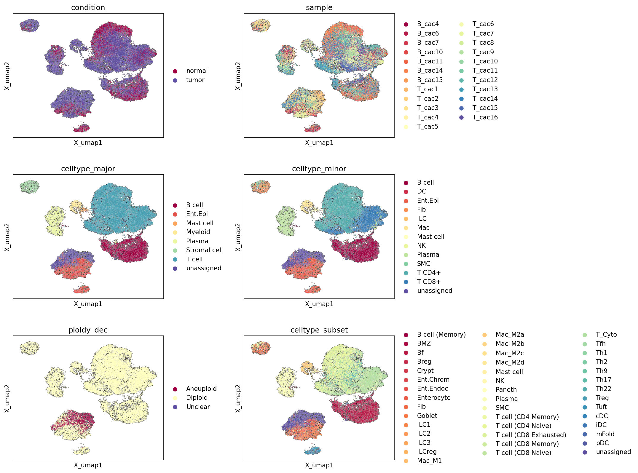
This analysis presents UMAP (Uniform Manifold Approximation and Projection) plots visualizing the relationships between single cells based on their gene expression profiles. The cells are colored by various metadata annotations: condition (normal vs. tumor), sample, celltype_major, celltype_minor, ploidy_dec (diploid vs. aneuploid), and celltype_subset. These visualizations are crucial for assessing the overall data structure, identifying distinct cell populations, evaluating annotation quality, and understanding the distribution of specific biological characteristics, such as tumor presence and ploidy status, across the cellular landscape.
Visual Summary
Condition UMAP
The UMAP colored by condition shows a clear separation between cells originating from 'normal' and 'tumor' tissues. While there is an overlap, particularly in the central dense clusters, distinct regions appear predominantly enriched for either normal or tumor cells. Specifically, several smaller, more peripheral clusters seem to be almost exclusively composed of 'normal' cells, whereas the largest central and lower-left clusters contain a substantial mixture, with some areas more enriched for 'tumor' cells. This indicates that while some cell types might be shared or exhibit similar gene expression states across conditions, others undergo significant transcriptional changes or are unique to either normal or tumor microenvironments.
Sample UMAP
The UMAP colored by sample shows a generally good mixing of cells from different samples across the major clusters, suggesting that batch effects are not strongly driving the primary separation of cell populations. Different samples contribute to various clusters, indicating a diverse representation. However, some smaller, more isolated clusters might be driven by specific samples, which could indicate unique populations or slight sample-specific biases, though overall the embedding appears robust to strong sample-specific artifacts.
Celltype Major UMAP
The celltype_major UMAP reveals well-defined, distinct clusters corresponding to different major cell types.
- Intestinal Epithelial cells (Ent.Epi) form a large, central, and somewhat diffused cluster, consistent with their role as the primary structural and functional cells of the colon, and as the inferred tumor-origin cell type.
- T cells form a prominent, dense cluster, often overlapping with or adjacent to B cells and Plasma cells, reflecting the immune component.
- B cells and Plasma cells form distinct clusters, often in proximity to T cells.
- Myeloid cells, Stromal cells, and Mast cells also occupy relatively distinct regions, suggesting unique transcriptional profiles for these major lineages.
- unassigned cells are minimal and scattered, indicating a high quality of broad cell type annotation.
Celltype Minor UMAP
The celltype_minor UMAP provides more granular detail, showing further subdivision within major cell type clusters.
- T cells CD4+ and T cells CD8+ form separate, yet often adjacent, sub-clusters within the broader T cell major cluster.
- Fibroblasts, Macrophage (Mac), Dendritic cells (DC), and Smooth muscle cells (SMC) show distinct clustering, refining the Stromal cell and Myeloid cell major categories.
- Intestinal Epithelial cells remain a large and somewhat heterogeneous group, which is expected given their diverse functions and the potential for malignant transformation.
Ploidy_dec UMAP
The ploidy_dec UMAP highlights the distribution of aneuploid cells.
- The vast majority of cells are Diploid (light yellow), forming the bulk of most clusters.
- Aneuploid cells (dark red) are predominantly localized to a specific cluster, largely overlapping with a subset of the Intestinal Epithelial cell population. There are also smaller groups of aneuploid cells in other regions, but the main aneuploid cluster is quite distinct.
- Unclear cells are present in very small numbers and scattered.
This distinct clustering of aneuploid cells strongly supports their identification and points to their origin from a specific cellular lineage, consistent with the inferred tumor-origin cell type.
Celltype Subset UMAP
The celltype_subset UMAP offers the highest resolution of cell type annotation.
- Within the Intestinal Epithelial cell population, subsets like Enterocyte, Goblet cell, Paneth cell, Tuft cell, Enterochromaffin cell, and Enteroendocrine cell show distinct spatial arrangements, reflecting their specialized functions and potentially different states within the tumor.
- Immune cell subsets such as various T cell subtypes (e.g., T cell (CD4 Memory), T cell (CD8 Exhausted), T cell (Treg), T cell (Th17)), B cell subtypes (e.g., B cell (Follicular), B cell (Memory)), and Macrophage subtypes (e.g., Macrophage (M1), Macrophage (M2A)) form highly resolved and distinct clusters. This indicates robust sub-clustering and annotation.
Biological Interpretation
The UMAP visualizations provide critical insights into the cellular landscape of colorectal cancer. The clear separation of cells by condition (normal vs. tumor) suggests significant transcriptional reprogramming associated with tumorigenesis, affecting multiple cell types within the tumor microenvironment.
The distinct clustering of Intestinal Epithelial cells and their partial overlap with tumor cells and aneuploid cells strongly supports the inference that Intestinal Epithelial cells are the Resolved tumor-origin cell type. The clustering of Aneuploid cells into specific regions, primarily within the Intestinal Epithelial cell clusters, further confirms these cells likely represent the cancerous epithelial population undergoing genomic instability. This is a hallmark of many solid tumors, including colorectal cancer. GeneCards: Colorectal Neoplasms
The intricate immune cell landscape revealed by the celltype_major, celltype_minor, and celltype_subset UMAPs is highly relevant to colorectal cancer. The presence of diverse T cell, B cell, Plasma cell, and Myeloid cell populations and their specific spatial relationships with epithelial cells suggest complex immune responses within the tumor microenvironment. For instance, specific T cell subsets (e.g., T cell (CD8 Exhausted), T cell (Treg)) or Macrophage phenotypes (e.g., M1, M2A) are known to play distinct roles in anti-tumor immunity or tumor promotion, respectively. Their clustering patterns indicate a good resolution of these states, which would be foundational for subsequent differential expression or cell-cell interaction analyses to understand their contributions to disease progression. PubMed: Tumor microenvironment in colorectal cancer
The relatively good mixing of sample across major clusters indicates that general cell type differences, rather than patient-specific variations or technical artifacts, primarily drive the observed cell population structure. This strengthens confidence in cross-sample comparisons and the generalizability of findings regarding cell type composition and state changes.
Annotation Notes
The UMAPs demonstrate a high quality of cell type annotation across all levels of granularity (celltype_major, celltype_minor, celltype_subset). Each annotated group forms largely distinct and coherent clusters, indicating that the assigned labels accurately reflect underlying biological distinctions in gene expression. The low number of unassigned cells further supports the comprehensiveness of the current annotation scheme. The clear separation of diploid and aneuploid cells, and the localization of aneuploidy to the inferred tumor-origin cells, also validates the ploidy estimation. These robust annotations provide a strong foundation for downstream analyses of differential gene expression, pathway enrichment, and cell-cell interactions within the context of colorectal cancer.
2. Single-Cell UMAP Visualization of Major Cell Type Scores and Ploidy Status
[Analysis Visualization Results]...
Analysis Overview
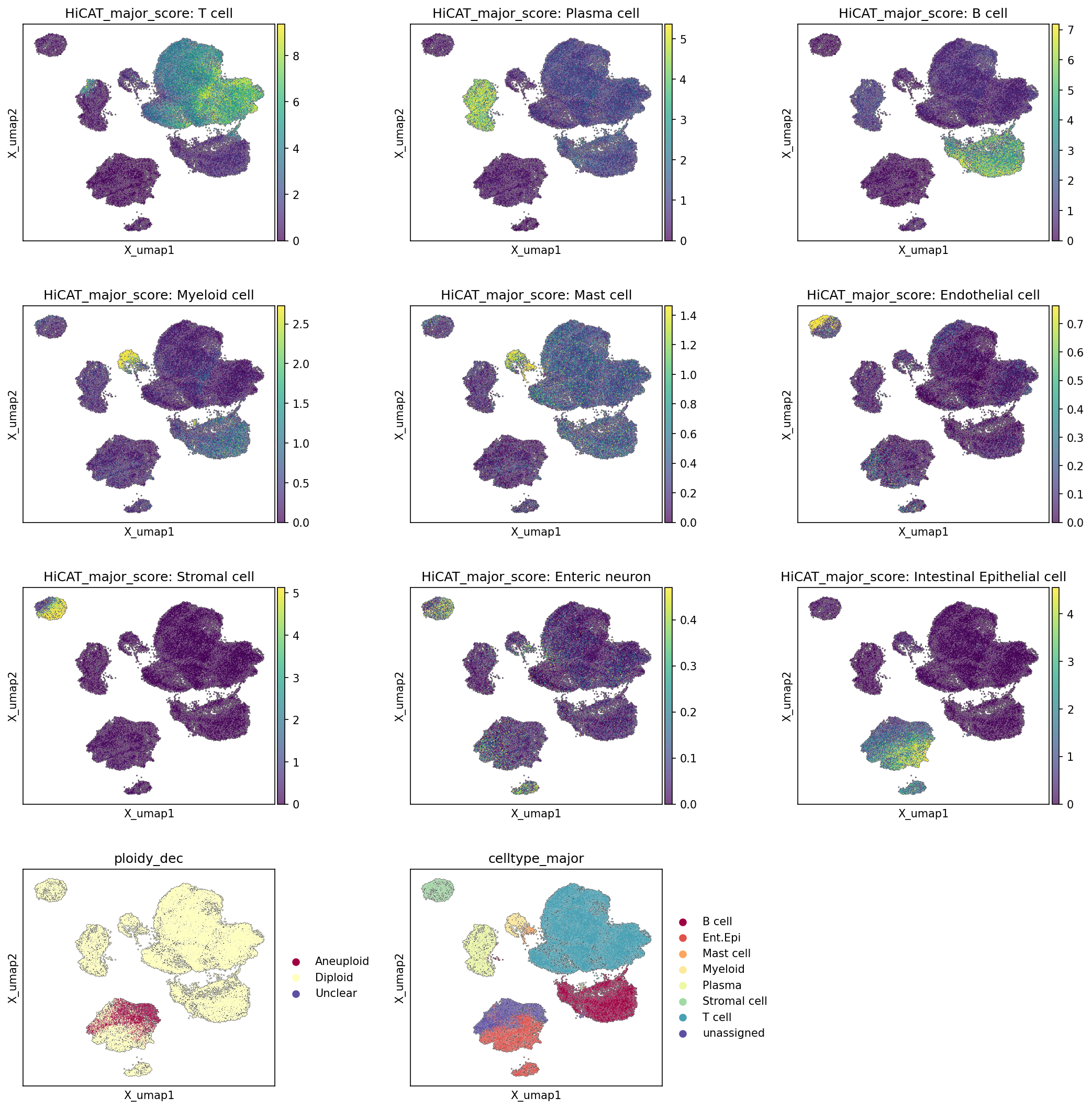
This analysis provides a Uniform Manifold Approximation and Projection (UMAP) visualization of single-cell RNA-sequencing data, displaying the distribution of major cell type scores, inferred ploidy status, and final major cell type annotations across the dataset. The primary goal is to assess the quality of cell type annotation and to visually identify cell populations, particularly the tumor-originating cells, within the embedding space. The data is derived from human Colon tissue with an inferred context of Colorectal Cancer, where Intestinal Epithelial cells are identified as the likely tumor-origin cell type.
Visual Summary
The UMAP plots effectively delineate distinct cellular populations based on gene expression profiles.
Major Cell Type Scores:
- Separate UMAPs, each colored by the "HiCAT_major_score" for a specific cell type, show that each major cell type largely occupies a well-defined and distinct region within the UMAP embedding. For instance, T cell scores are highest in the top-right cluster, Plasma cell scores in a distinct top-middle cluster, and Intestinal Epithelial cell scores are concentrated in a large cluster on the bottom-left. This indicates robust and specific assignment of cells to their respective major cell types.
- The visualization of scores rather than discrete labels highlights the confidence of assignment, with brighter yellow regions indicating higher confidence/score for that cell type.
- Minor cell types like Enteric neuron and Endothelial cell show very small, distinct clusters, reflecting their lower abundance or more specialized transcriptional profiles compared to more numerous immune or epithelial populations.
Ploidy Status:
- The ploidy_dec UMAP clearly separates cells based on their inferred ploidy. A prominent cluster on the bottom-left is predominantly colored in red, indicating Aneuploid cells. The vast majority of the remaining cells throughout the UMAP are colored in light yellow, representing Diploid cells. A very small fraction of cells are marked Unclear (purple), suggesting some ambiguity in their ploidy determination.
Major Cell Type Annotation:
- The celltype_major UMAP provides the overall categorical assignment of cells to their major types, serving as a comprehensive overview and visual reference. This plot confirms the distinct clustering observed in the individual major cell type score plots.
- Comparing the ploidy_dec plot with the celltype_major plot reveals a striking overlap: the large Aneuploid cluster (red in ploidy_dec) precisely corresponds to the Intestinal Epithelial cell cluster (light blue in celltype_major).
Biological Interpretation
The UMAP visualizations provide critical insights into the cellular landscape of the Colon tissue, particularly in the context of inferred Colorectal Cancer.
- Tumor Cell Identification: The strong co-localization of Intestinal Epithelial cell scores with the Aneuploid cell population is a key finding. Given that Intestinal Epithelial cells are the 'Resolved tumor-origin cell type' and aneuploidy is a hallmark of cancer, this cluster most likely represents the malignant epithelial cells [1]. This visual confirmation strengthens the inference of Colorectal Cancer within this dataset.
- Tumor Microenvironment: The remaining major cell types, including T cell, B cell, Plasma cell, Myeloid cell, Stromal cell, and Mast cell, form distinct clusters that are predominantly diploid. These populations collectively represent the tumor microenvironment (TME) and normal tissue components surrounding the malignant epithelial cells. Their spatial separation in the UMAP suggests distinct transcriptional states and functions, which is typical for immune and stromal cells interacting with or infiltrating a tumor.
- Cell Type Annotation Quality: The clear segregation of cells by their major cell type scores and the consistent clustering in the celltype_major plot indicate high quality and reliability of the cell type annotations. Each major cell type forms a distinct and well-defined cluster, suggesting that the clustering algorithms and annotation pipeline have successfully identified biologically meaningful cell populations.
- Cellular Diversity: The presence of various immune cells (T, B, Plasma, Myeloid, Mast), stromal cells, and other specialized cell types (Endothelial, Enteric neuron) highlights the complex cellular heterogeneity of the colon tissue. This diversity is crucial for understanding tissue function in normal conditions and its dysregulation in disease states like cancer.
Annotation Notes
The UMAP visualizations effectively demonstrate the clustering of single cells based on their transcriptional profiles and the consistency of major cell type assignments. The clear separation of aneuploid, epithelial tumor cells from diploid, non-malignant cells provides robust evidence for the initial cell type annotations and validates the identification of the tumor compartment. This robust annotation forms a strong foundation for subsequent, more detailed differential expression or cell-cell interaction analyses.
---
References:
- Aneuploidy in Cancer: [PubMed Search: "aneuploidy cancer hallmark" - link to search results] (PubMed Search)
3. Marker Expression Dot Plot for Celltype Subsets
[Analysis Visualization Results]...
Analysis Overview
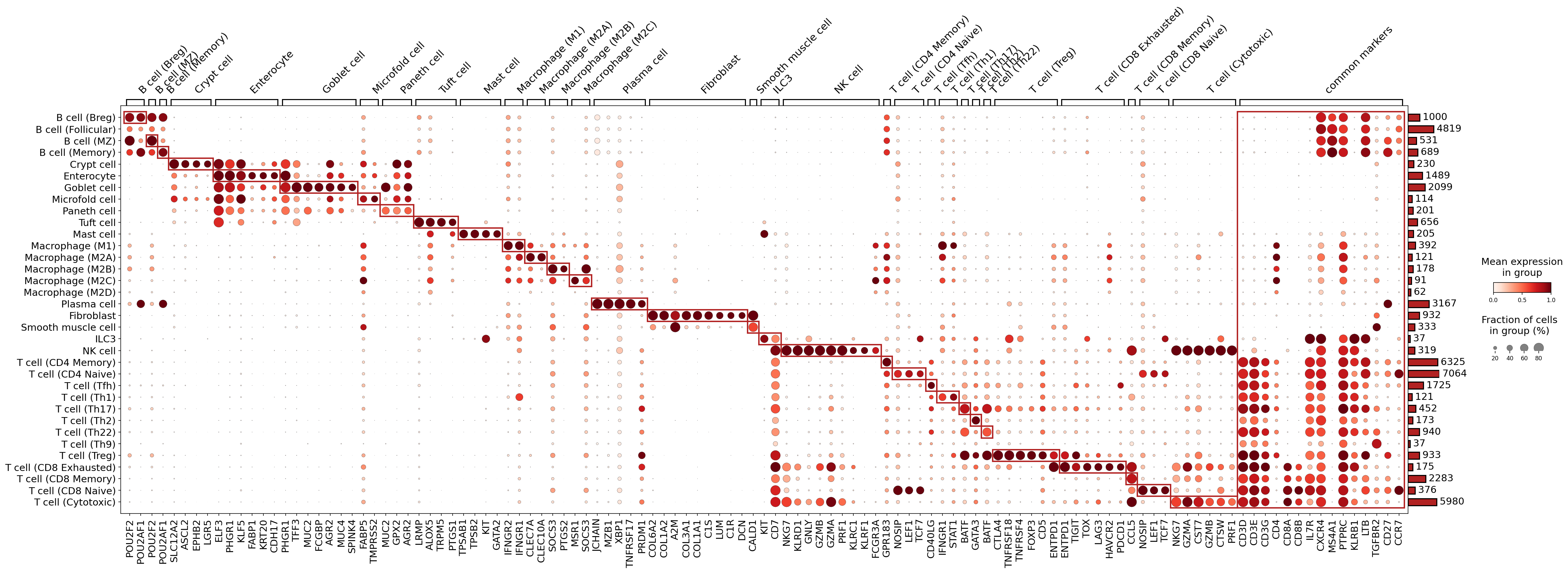
This analysis presents a dot plot illustrating the expression of selected marker genes across various celltype_subset populations. The primary objective is to visually confirm the distinctiveness and biological coherence of these granular cell type annotations based on their gene expression profiles. Each dot's size represents the fraction of cells within a given celltype_subset that express a particular gene, while the color intensity reflects the mean expression level of that gene within the expressing cells of that subset. This plot serves as a critical quality control step for single-cell RNA-seq data annotation.
Visual Summary
The dot plot displays a comprehensive overview of marker gene expression across 36 distinct celltype_subset groups.
- Clustered Patterns: Cell type subsets and marker genes are organized, likely by hierarchical clustering, revealing distinct blocks of high and specific expression. This structure aids in identifying cell lineage-specific and subset-specific markers.
- Specificity and Expression Levels: Many cell types exhibit highly specific marker gene expression, characterized by large, dark red dots (high fraction of expressing cells and high mean expression). For example, B cell (Breg) shows strong expression of *POU2F2* and *POU2AF1*, while Enterocyte expresses *KRT20* and *CDH17*.
- Common Markers: A column labeled "common markers" on the far right indicates genes expressed broadly across multiple cell types. The accompanying bar chart quantifies the number of cells in each celltype_subset, providing context for the observed expression patterns.
- Variability within Lineages: Within major lineages (e.g., T cells, Macrophages), distinct marker profiles delineate different subsets, supporting the granularity of the celltype_subset annotations. For instance, different T cell subsets express distinct combinations of genes such as *LEF1*, *TCF7* for naive T cells, *FOXP3* for T cell (Treg), and *GZMA*, *GZMB*, *PRF1* for cytotoxic T cell populations.
- Red Boxes: Red rectangular outlines highlight blocks of genes that are co-expressed and define specific cell populations or closely related cell clusters, further emphasizing the distinct marker profiles.
Biological Interpretation
The marker expression patterns observed in the dot plot largely support the biological identity and distinctiveness of the annotated celltype_subset populations, aligning well with known biological roles in the colon tissue context.
B Cell Lineage:
- B cell (Breg), B cell (Follicular), B cell (MZ), and B cell (Memory) display shared B cell markers such as *POU2F2* and *POU2AF1*.
- *MZB1* is specifically enriched in B cell (MZ), consistent with its role in marginal zone B cells.
- *MS4A1* (CD20) is a pan-B cell marker evident across these subsets.
- *JCHAIN* and *PRDM1* are highly expressed in Plasma cell, confirming their differentiation from B cells and their role in antibody production PubMed: 35058694.
Intestinal Epithelial Cell Lineage:
- Crypt cell shows strong expression of *LGR5*, a well-known stem cell marker in the intestinal crypts GeneCards: LGR5.
- Enterocyte is characterized by *KRT20* and *CDH17* expression, typical markers of mature enterocytes.
- Goblet cell shows high expression of *MUC2*, the primary mucin produced by goblet cells for mucus secretion GeneCards: MUC2.
- Paneth cell expresses *LYZ*, indicative of its role in secreting antimicrobial peptides.
- Tuft cell expresses *TRPM5* and *POU2F3*, which are characteristic markers for this chemosensory cell type.
Myeloid Lineage:
- Macrophage (M1), Macrophage (M2A), Macrophage (M2B), Macrophage (M2C) show common macrophage markers like *C1QA*, *C1QB*, *C1QC*, *MRC1* (CD206), and *MSR1*.
- Distinct polarization states are suggested by the differential expression of markers within these subsets (e.g., *CCL5* in Macrophage (M1)).
- *CLEC10A* and *CLEC9A* are expressed in certain macrophage/DC subsets, consistent with antigen-presenting cell functions.
Stromal Cells:
- Fibroblast populations are identified by expression of collagen genes (*COL1A1*, *COL1A2*, *COL3A1*) and proteoglycans (*DCN*, *LUM*), which are central to extracellular matrix production GeneCards: COL1A1.
- Smooth muscle cell shows expression of *ACTA2*, a marker for alpha-smooth muscle actin.
T Cell Lineage:
- Pan-T cell markers *CD3D*, *CD3E*, *CD3G* are highly expressed across all T cell subsets.
- T cell (CD4 Memory) and T cell (CD4 Naive) are distinguished by *CD4* expression, along with genes like *IL7R* and *CCR7* for naive cells, and memory-associated genes.
- T cell (Treg) is strongly characterized by *FOXP3* expression, the master regulator of regulatory T cells GeneCards: FOXP3.
- T cell (Th1), T cell (Th17), T cell (Th2), T cell (Th22), T cell (Th9) show distinct, albeit sometimes overlapping, profiles. *STAT1* is observed in T cell (Th1), *GATA3* in T cell (Th2).
- T cell (CD8 Exhausted), T cell (CD8 Memory), T cell (CD8 Naive), and T cell (Cytotoxic) are distinguished by *CD8A* and *CD8B* expression.
- T cell (Cytotoxic) and T cell (CD8 Exhausted) show high expression of effector molecules like *GZMA*, *GZMB*, *PRF1*, and *NKG7*. Exhausted T cells specifically express immune checkpoint markers such as *LAG3*, *TIGIT*, and *PDCD1*.
Other Immune Cells:
- NK cell displays classical NK cell markers like *KLRD1* (CD94), *NKG7*, *GZMB*, and *PRF1*.
- Mast cell is identified by *KIT* (CD117) and *TPSAB1*, consistent with mast cell identity GeneCards: KIT.
Annotation Notes
The celltype_subset annotations appear to be well-supported by the specific expression patterns of known marker genes. The distinct blocks of marker expression for each subset, along with the differentiation of related subsets (e.g., within T cells and B cells), provide confidence in the quality and resolution of the cell type assignments. The presence of expected canonical markers across diverse cell lineages in a human colon context further validates the annotation strategy. No major discrepancies or highly ambiguous populations are immediately apparent, suggesting robust cell type identification at this granular level.
4. Copy Number Variation Analysis of Intestinal Epithelial Cells in Colon Tissue
[Analysis Visualization Results]...
Analysis Overview
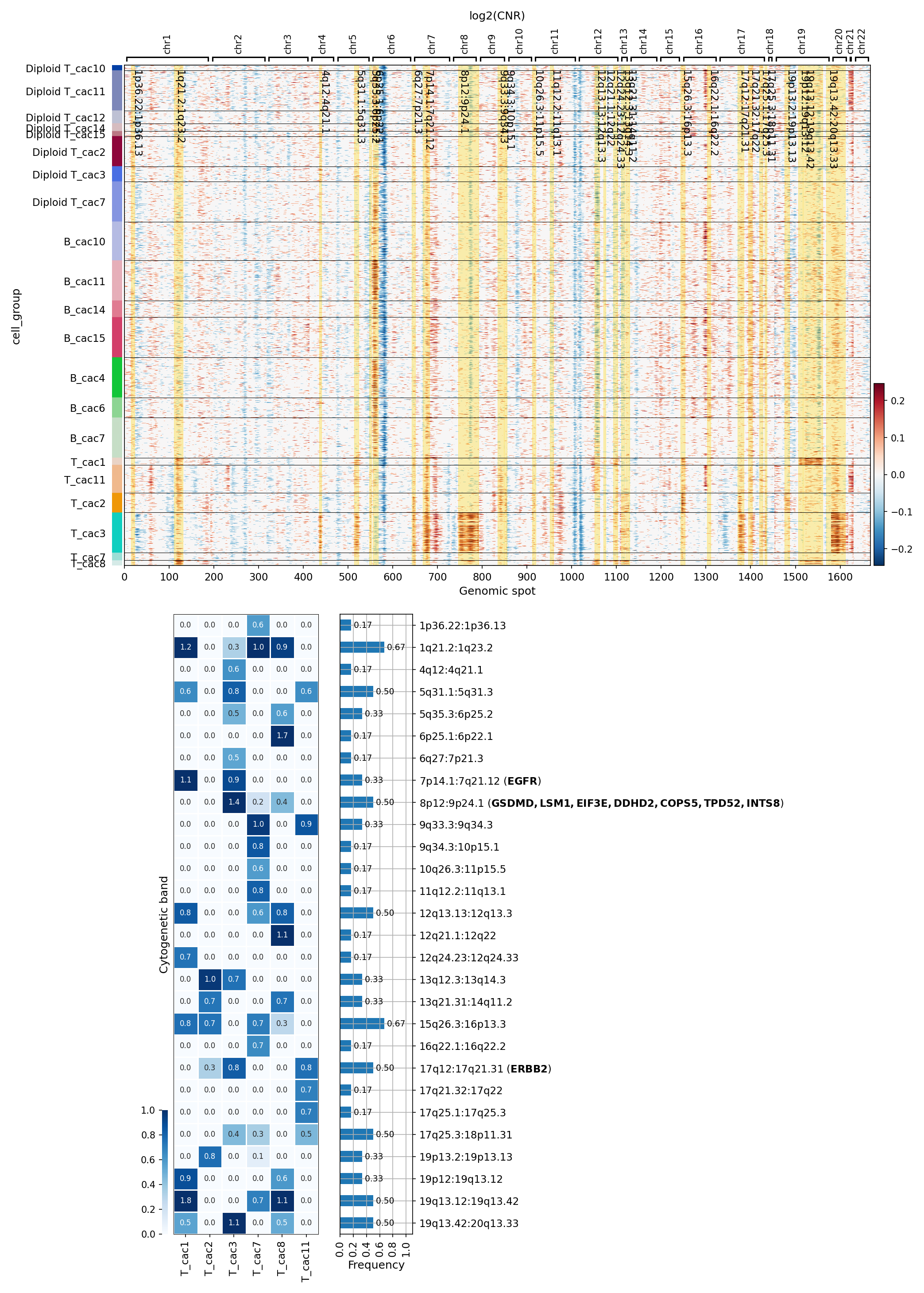
This analysis presents a copy number variation (CNV) heatmap for Intestinal Epithelial cells, identified as the tumor-origin cell type, across various samples from human colon tissue. The heatmap visualizes the log2(Copy Number Ratio) (CNR) across genomic spots for each sample, grouped by sample. Alongside the heatmap, a summary of significantly amplified cytogenetic regions is provided, including their frequency across samples and associated genes. This investigation aims to identify recurrent genomic alterations characteristic of colorectal cancer within the epithelial cell compartment.
Visual Summary
The heatmap displays log2(CNR) values for Intestinal Epithelial cells from different samples, ordered along chromosomes 1 to 22. Red color indicates amplification (positive log2(CNR)), blue indicates deletion (negative log2(CNR)), and yellow/white represents neutral or diploid regions.
- Sample-Specific CNV Patterns: A clear distinction is observed between samples. Several samples (e.g., B_cac10, B_cac11, B_cac14, B_cac15, B_cac4, B_cac6, B_cac7) exhibit widespread and prominent amplifications (red regions) and some deletions (blue regions), indicative of significant genomic instability. In contrast, samples prefixed with "Diploid T_cac" (e.g., Diploid T_cac10 through Diploid T_cac3) show markedly less CNV activity, aligning with their "Diploid" designation and suggesting lower tumor burden or a less aggressive tumor phenotype in the Intestinal Epithelial cells within these samples.
- Recurrent Amplifications: Notable regions of amplification frequently observed across the cac samples include segments on chromosomes 7, 8, 13, and 20. These appear as distinct red bands spanning multiple samples.
- Recurrent Deletions: Some regions, such as parts of chromosome 4, 6, and 18, show recurrent deletions (blue bands), though less pronounced or frequent than amplifications in this specific visualization.
- Summary of Amplified Regions: The bar chart on the right quantifies the frequency of significantly amplified cytogenetic bands. Top frequently amplified regions include:
1q21.2:1q23.2 (Frequency 0.67)
4q12:4q21.1 (Frequency 0.67)
7p14.1:7q21.12 (Frequency 0.33, encompassing EGFR)
- 8p12:9p24.1 (Frequency 0.50, encompassing GSDMD, LSM1, EIF3E, DDHD2, COP5, TPD52, INTS8)
15q26.3:16p13.3 (Frequency 0.67)
17q12:17q21.31 (Frequency 0.50, encompassing ERBB2)
19p13.2:19p13.13 (Frequency 0.50)
19q13.42:20q13.33 (Frequency 0.50)
- CNA Score Matrix: The smaller heatmap on the left displays a CNA score for selected samples (T_cac1, T_cac2, T_cac3, T_cac8, T_cac11) across the cytogenetic bands, providing a sample-specific view of amplification intensity.
Biological Interpretation
The observed CNV patterns in Intestinal Epithelial cells are highly consistent with the genomic instability characteristic of Colorectal Cancer, the inferred disease context. The distinction between samples with high and low CNV burdens underscores the heterogeneity often seen in tumor samples, possibly reflecting varying stages of tumor progression or different cellular compositions within the original sample (even when focusing on IECs).
Oncogenic Amplifications:
- EGFR (Epidermal Growth Factor Receptor) amplification on 7p14.1:7q21.12 is a well-established driver event in various cancers, including colorectal cancer. EGFR signaling promotes cell proliferation, survival, and metastasis. GeneCards: EGFR
- ERBB2 (HER2, Epidermal Growth Factor Receptor 2) amplification on 17q12:17q21.31 is another critical oncogenic event. While more famously associated with breast and gastric cancers, ERBB2 amplification is found in a subset of colorectal cancers, promoting aggressive tumor growth. GeneCards: ERBB2
- The region 8p12:9p24.1 containing genes like GSDMD (Gasdermin D) and EIF3E (Eukaryotic Translation Initiation Factor 3 Subunit E) is also frequently amplified. GSDMD plays a role in pyroptosis, a form of programmed cell death, and its dysregulation can impact tumor immunity and progression. EIF3E is involved in protein synthesis, and its amplification can contribute to increased protein production favoring cancer cell growth.
- Chromosomal Aneuploidy: The frequent amplifications on chromosomes 1, 4, 7, 8, 15, 17, 19, and 20 highlight recurrent chromosomal alterations that likely confer a selective advantage to the tumor cells, enabling their survival, proliferation, and malignant progression. These regions may harbor other oncogenes or regulatory elements critical for cancer development.
Clinical or Translational Implications
The identification of specific and recurrent CNVs, particularly those involving known oncogenes, holds significant clinical relevance for colorectal cancer.
- Biomarker Identification: Amplifications of genes like EGFR and ERBB2 can serve as crucial diagnostic or prognostic biomarkers. Their presence may indicate a more aggressive tumor phenotype or predict response to specific therapies.
- Therapeutic Targeting: The observed amplifications of EGFR and ERBB2 point towards potential therapeutic strategies. Patients with EGFR amplification might benefit from anti-EGFR monoclonal antibodies (e.g., cetuximab, panitumumab). Similarly, anti-ERBB2 (HER2) targeted therapies, such as trastuzumab or pertuzumab, are increasingly being explored and used in ERBB2-amplified colorectal cancer.
- Genomic Heterogeneity and Treatment: The variability in CNV patterns across different tumor samples underscores the importance of genomic profiling for personalized medicine approaches. Recognizing this heterogeneity can guide treatment selection and help anticipate potential resistance mechanisms.
- Disease Monitoring: Monitoring the evolution of these CNVs, potentially through liquid biopsies or serial single-cell analyses, could provide insights into disease progression or recurrence.
5. UMAP Visualization of CNV Patterns by Cell Type, Ploidy, Condition, and Sample
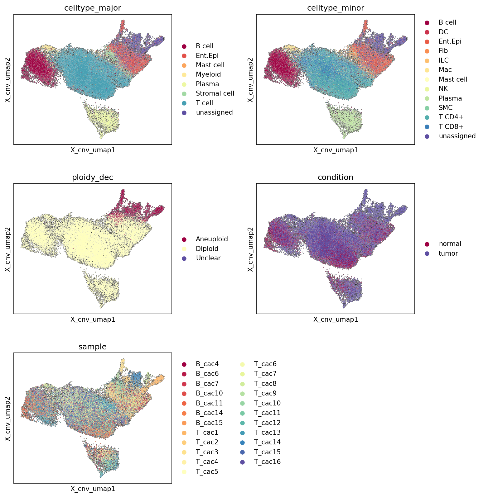
[Analysis Visualization Results]...
Analysis Overview
This analysis presents UMAP visualizations of single-cell RNA-seq data, specifically leveraging copy number variation (CNV) estimates to generate the embedding. The goal is to visualize the distribution of cells based on their CNV patterns, colored by major cell type, minor cell type, ploidy status, experimental condition (normal vs. tumor), and individual sample. This provides a comprehensive overview of cellular heterogeneity, identifying populations with distinct CNV profiles and linking them to biological annotations. The inferred context is Colorectal Cancer, with Intestinal Epithelial cells identified as the tumor-origin cell type.
Visual Summary
Cell Type Distribution (Major and Minor)
The UMAPs colored by celltype_major and celltype_minor reveal a structured organization of cell populations based on their CNV profiles.
- Major Cell Types: Intestinal Epithelial cells (Ent.Epi) form a distinct cluster, predominantly located towards the upper right region of the UMAP. T cells, Myeloid cells, Stromal cells, B cells, Plasma cells, and Mast cells form other distinct clusters or are more broadly distributed, primarily in the central and left regions.
- Minor Cell Types: A similar pattern is observed at the minor cell type resolution. Intestinal Epithelial cells remain a prominent cluster. Immune cell subtypes (e.g., T CD4+, T CD8+, B cell, Plasma cell, Macrophage, DC) and Stromal cells (Fibroblast, Smooth muscle cell) are generally well-separated, suggesting unique CNV landscapes or localization patterns.
Ploidy and Condition Association
The UMAPs colored by ploidy_dec and condition provide crucial insights into the malignant nature of the tissue.
- Ploidy Status: A distinct cluster of 'Aneuploid' cells (red) is evident in the upper right region of the UMAP. The vast majority of cells elsewhere are 'Diploid' (yellow), forming a large, diffuse cluster spanning the rest of the embedding. This clear separation indicates that aneuploidy is a strong driver of the CNV-based UMAP embedding.
- Condition: Cells from the 'tumor' condition (blue/purple) largely co-localize with the 'Aneuploid' cluster. Conversely, cells from the 'normal' condition (red) are almost entirely restricted to the 'Diploid' regions of the UMAP. This strong correlation between 'tumor' condition and 'Aneuploid' status is a key finding.
Sample Distribution
The UMAP colored by sample shows that cells from different samples are generally well-integrated within their respective biological clusters (e.g., aneuploid/tumor vs. diploid/normal), rather than forming sample-specific clusters.
- Intermixing: Multiple samples (e.g., B_cac4, B_cac6, T_cac10, T_cac11) contribute to the 'Aneuploid'/'tumor' cluster, indicating that the observed CNV patterns are not driven by a single sample but are a consistent feature across several tumor samples. Similarly, various samples contribute to the 'Diploid'/'normal' cell populations. This suggests that batch effects are minimal, and the UMAP effectively captures biological variation.
Biological Interpretation
The CNV-based UMAP clearly delineates distinct cell populations, primarily separating malignant cells from healthy cells and immune/stromal components based on their genomic integrity.
- Identification of Malignant Cells: The most striking observation is the strong co-localization of 'Aneuploid' cells with the 'tumor' condition. Furthermore, comparing this region with the celltype_major plot, the 'Aneuploid' and 'tumor' cluster predominantly consists of 'Intestinal Epithelial cells'. Given that 'Intestinal Epithelial cell' is the resolved tumor-origin cell type in this dataset and the inferred context is Colorectal Cancer, this cluster represents the malignant, cancerous epithelial cells. Their distinct positioning in the UMAP, driven by CNVs, confirms their abnormal genomic landscape characteristic of cancer.
- Tumor Microenvironment Components: The 'Diploid' region of the UMAP, largely corresponding to the 'normal' condition but also present within the 'tumor' samples, comprises a diverse array of cell types, including T cells, B cells, Plasma cells, Myeloid cells, and Stromal cells. These populations likely represent the immune and stromal components of the tumor microenvironment (TME) in tumor samples, and the cellular composition of normal colon tissue in control samples. Their diploid status is expected, as these are typically non-malignant cells, even if they are interacting with the tumor.
- CNV as a Principal Driver of Heterogeneity: The effectiveness of CNV patterns in separating tumor cells from normal cells and other stromal/immune cells highlights CNVs as a fundamental biological feature distinguishing cancer cells. This UMAP projection based on CNVs provides strong evidence for the presence and genomic abnormality of malignant epithelial cells.
- Data Quality and Integration: The good intermixing of cells from different samples within their respective biological clusters (e.g., tumor/aneuploid vs. normal/diploid) suggests successful integration of data from various samples without significant batch effects confounding the biological signal related to CNVs and cell identity.
Annotation Notes
- The clear separation between aneuploid/tumor cells and diploid/normal cells strongly supports the accuracy of the ploidy_dec and condition annotations, and their biological relevance.
- The clustering of Intestinal Epithelial cells within the aneuploid/tumor region is consistent with their identification as the tumor-origin cell type, bolstering the confidence in cell type annotations for these critical populations.
- The UMAP visualization provides a robust validation of the presence of distinct malignant cell populations based on genomic features (CNVs) and their association with the 'tumor' condition and specific cell types.
6. Cell Type Population Analysis in Normal vs. Tumor Colon Tissue
[Analysis Visualization Results]...
Analysis Overview
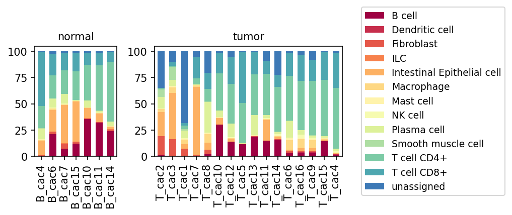
This analysis presents a stacked bar plot illustrating the relative proportions of minor cell types across individual samples from both normal and tumor colon tissue. The visualization provides a high-level overview of the cellular composition, highlighting shifts in cell type abundances associated with the disease state (Colorectal Cancer, as inferred from the data context). Each bar represents a distinct sample, with segments showing the percentage of different minor cell populations within that sample.
Visual Summary
The bar plot effectively contrasts the cellular landscape between normal and tumor colon tissues.
Normal Samples:
- Normal samples (B_cac4 to B_cac14) show a relatively consistent cellular composition.
- Intestinal Epithelial cells (light orange) constitute a substantial and consistent portion of the normal tissue.
- T cell CD4+ (light green) and T cell CD8+ (teal) populations are present, along with Smooth muscle cells (light green, similar hue to T cell CD4+ but distinct segment), B cells (maroon), Plasma cells (light yellow-green), and Macrophages (light yellow).
Tumor Samples:
- Tumor samples (T_cac2 to T_cac4) exhibit a much more heterogeneous and profoundly altered cellular composition compared to normal samples.
- Intestinal Epithelial cells are highly variable. While some tumor samples (e.g., T_cac7) show a high proportion, many others show significantly reduced proportions, indicating either high immune/stromal infiltration or lower tumor purity in those specific biopsies.
- Immune cell infiltration is a striking feature. T cell CD4+ and T cell CD8+ populations appear markedly increased in many tumor samples, often dominating the cellular landscape where epithelial cells are less abundant.
- Fibroblasts (orange-red) show a noticeable increase in many tumor samples, suggesting an expanded stromal compartment.
- "unassigned" cells (dark blue) are notably more abundant in a significant number of tumor samples compared to normal tissue. In some tumor samples (e.g., T_cac1, T_cac8, T_cac10, T_cac12, T_cac5, T_cac13, T_cac11, T_cac14, T_cac16, T_cac9, T_cac15, T_cac4), this unassigned population represents a substantial fraction (up to 25-50% in some cases), indicating challenges in cell type annotation within the tumor context or the presence of novel cell states.
- Other immune cells like B cells, Plasma cells, Macrophages, and Dendritic cells show variable proportions across tumor samples.
Biological Interpretation
The observed shifts in cell populations are highly consistent with the known biology of Colorectal Cancer, which is inferred as the disease context.
- Dysregulation of Intestinal Epithelial Cells: As 'Intestinal Epithelial cell' is identified as the 'Resolved tumor-origin cell type,' the variability and often reduced relative proportion of these cells in tumor samples suggest a complex tumor microenvironment. Tumor samples can consist of varying proportions of malignant epithelial cells, stromal cells, and immune infiltrates. The overall decrease in epithelial cells in some tumor samples, contrasted with their dominance in normal tissue, points to a significant stromal and immune cell expansion within the tumor.
Immune Infiltration and Tumor Microenvironment (TME) Remodeling:
- Increased T cells (CD4+ and CD8+): The elevated presence of T cells in tumor samples is a hallmark of immune infiltration into the TME. These T cells represent the host's immune response to the tumor. CD8+ T cells (cytotoxic T lymphocytes, CTLs) are crucial for anti-tumor immunity, while CD4+ T cells play diverse roles, including helper functions (Th1, Th2, Th17) and regulatory functions (Tregs) that can either promote or suppress anti-tumor responses. Their increased presence suggests an active immunological battle within the tumor.
- Expanded Fibroblast Population: The increase in Fibroblasts in tumor tissue is indicative of cancer-associated fibroblasts (CAFs). CAFs are key components of the tumor stroma, known to remodel the extracellular matrix, promote tumor growth, angiogenesis, and suppress anti-tumor immunity, thereby playing a critical role in disease progression in colorectal cancer.
- Other Immune Cells: The variable presence of B cells, Plasma cells, Macrophages, Dendritic cells, and NK cells further highlights the complexity of the immune response within the TME. Tumor-associated macrophages (TAMs), for instance, can differentiate into pro-tumorigenic M2-like phenotypes that support tumor growth and metastasis [NCBI].
- Significance of "unassigned" cells: The substantial increase in "unassigned" cell populations in tumor samples is a critical observation. This could signify:
- Tumor-specific cell states: Malignant epithelial cells undergoing de-differentiation or acquiring novel states, or highly plastic stromal/immune cells adopting tumor-specific phenotypes not well captured by existing annotations.
- Technical challenges: Limitations of current annotation strategies in capturing the full spectrum of cellular heterogeneity, especially in complex disease environments. Further investigation is warranted to characterize these populations.
Clinical or Translational Implications
The distinct differences in cellular composition between normal and tumor tissues have several important clinical and translational implications for Colorectal Cancer:
- Prognostic and Predictive Biomarkers: The proportion and composition of immune infiltrates, particularly T cells and CAFs, are known to correlate with patient prognosis and response to therapy in CRC [NCBI]. For example, a higher density of CD8+ T cells within the tumor is often associated with a better prognosis. These cellular signatures could serve as biomarkers for patient stratification.
- Immunotherapy Targets: The significant immune infiltration, particularly of T cells, in the tumor microenvironment suggests that many colorectal cancers might be amenable to immunotherapy. The presence of these immune cells indicates that the immune system is recognizing the tumor, and strategies like immune checkpoint blockade (e.g., targeting PD-1/PD-L1 or CTLA-4) could enhance this anti-tumor response [NCBI]. The composition of the TME, including the balance of effector T cells, regulatory T cells, and myeloid-derived suppressor cells, can determine response to such therapies.
- Stromal Targeting: The increased fibroblast population highlights the tumor stroma as a potential therapeutic target. Strategies aimed at depleting CAFs or reprogramming their pro-tumorigenic functions could improve treatment outcomes by reducing immunosuppression and promoting drug delivery.
- Understanding Tumor Heterogeneity: The considerable inter-sample variability in cell type proportions within the tumor group underscores the high cellular heterogeneity of colorectal cancer. This emphasizes the need for personalized therapeutic approaches that consider the unique cellular composition of each patient's tumor.
- Future Research into "unassigned" populations: The large "unassigned" population in tumor samples represents an opportunity for discovery. Characterizing these cells could unveil novel tumor-specific cell states, new drivers of cancer progression, or previously unknown immune evasion mechanisms, potentially leading to the identification of new therapeutic targets.
7. T Cell Subset Population Analysis in Colorectal Cancer
[Analysis Visualization Results]...
Analysis Overview
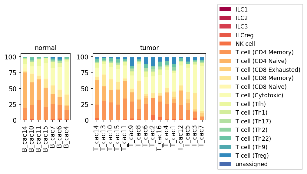
This analysis presents a population bar plot illustrating the relative distribution of various T cell subsets, as well as innate lymphoid cells (ILCs) and NK cells, across individual samples from both normal colon tissue and colorectal tumor tissue. The goal is to identify shifts in immune cell composition within the T cell compartment in the tumor microenvironment compared to normal conditions, providing insights into the immune landscape in colorectal cancer.
Visual Summary
The plot displays the proportions of different T cell subsets and related immune cells (ILCs, NK cells) for each sample, grouped by 'normal' and 'tumor' conditions.
- Normal Tissue Samples: The T cell compartment in normal colon tissue samples (B_cac14, B_cac10, B_cac11, etc.) is predominantly composed of conventional T cells, including T cell (CD4 Memory), T cell (CD4 Naive), T cell (CD8 Memory), and T cell (CD8 Naive). T cell (Cytotoxic) and T cell (Tfh) are also present in smaller proportions. Critically, T cell (CD8 Exhausted) and T cell (Treg) populations appear minimal or absent in these normal samples. ILCs and NK cells constitute a very small fraction.
- Tumor Tissue Samples: In stark contrast, tumor tissue samples (T_cac14, T_cac13, T_cac10, etc.) exhibit marked alterations in the T cell landscape:
- Increased T cell (CD8 Exhausted): A significant and consistent increase in T cell (CD8 Exhausted) is observed across almost all tumor samples. In some samples (e.g., T_cac16, T_cac4), these exhausted cells represent a substantial proportion, indicating a chronic antigen-driven state.
- Increased T cell (Treg): Regulatory T cells (Treg) also show a noticeable increase in most tumor samples compared to normal tissue, appearing as a prominent blue band at the top of the bars.
- Reduced Proportions of Naive/Memory T cells: Concurrently, the relative proportions of T cell (CD4 Memory), T cell (CD4 Naive), T cell (CD8 Memory), and T cell (CD8 Naive) appear generally reduced in tumor samples, likely due to the expansion of exhausted and regulatory T cell populations.
- Variable Cytotoxic T cells: The proportion of T cell (Cytotoxic) appears variable, but in some tumor samples, it is relatively low, possibly reflecting dysfunction or displacement by exhausted phenotypes.
- Helper T cell (Th) subsets, ILCs, and NK cells: While present, these populations generally remain in lower proportions, with no clear overarching pattern of substantial increase or decrease visible at this resolution, though specific shifts might exist.
Biological Interpretation
The observed shifts in T cell subset populations in colorectal tumor tissue compared to normal colon tissue are highly indicative of an altered, often immunosuppressive, tumor microenvironment (TME) characteristic of cancer.
- T cell Exhaustion in the TME: The dramatic increase in T cell (CD8 Exhausted) cells in tumor samples is a critical finding. T cell exhaustion is a state of T cell dysfunction that arises during chronic antigen stimulation, such as in cancer. Exhausted T cells lose their effector functions (e.g., cytokine production, cytolytic activity) and upregulate inhibitory receptors (e.g., PD-1, CTLA-4), contributing significantly to tumor immune evasion [1]. This suggests that while T cells are present and reacting to tumor antigens, their ability to effectively eliminate cancer cells is severely compromised.
- Regulatory T Cells (Tregs) and Immune Suppression: The expansion of T cell (Treg) populations in the tumor microenvironment is a well-documented mechanism of immunosuppression in cancer. Tregs suppress effector T cell responses, thereby promoting tumor growth and metastasis by dampening anti-tumor immunity [2]. Their increased presence suggests an active mechanism by which the tumor is evading immune surveillance.
- Impaired Anti-tumor Immunity: The relative decrease in naive and memory T cells, coupled with the increase in exhausted CD8 T cells and Tregs, paints a picture of an TME where effective anti-tumor immunity is suppressed. The T cells that are present are either functionally impaired (exhausted) or actively suppressing other immune cells (Tregs), hindering a robust immune response against the tumor.
Clinical or Translational Implications
These findings have important implications for understanding colorectal cancer progression and for developing or refining immunotherapeutic strategies:
- Prognostic Biomarkers: A higher proportion of exhausted CD8 T cells and Tregs in the tumor microenvironment could serve as a prognostic indicator for more aggressive disease progression or poorer patient outcomes in colorectal cancer.
- Rationale for Immunotherapy: The prominent presence of exhausted CD8 T cells strongly supports the use of immune checkpoint inhibitors (ICIs) targeting pathways like PD-1/PD-L1 or CTLA-4. By blocking these inhibitory signals, ICIs aim to reinvigorate exhausted T cells and restore their anti-tumor functions [3]. The increased Tregs also suggest potential benefits from therapies that deplete or inhibit Treg function.
- Personalized Medicine: Characterizing the T cell immune landscape in individual patient tumors could help stratify patients who are more likely to respond to specific immunotherapies (e.g., those with high T cell exhaustion might benefit more from checkpoint blockade).
- Monitoring Treatment Efficacy: Changes in the proportions of these T cell subsets (e.g., a decrease in exhausted T cells or Tregs, or an increase in functional cytotoxic T cells) could serve as biomarkers to monitor the efficacy of immunotherapy treatments.
---
References
- T cell exhaustion:
PubMed search: T cell exhaustion cancer
- Regulatory T cells in cancer:
PubMed search: Regulatory T cells colorectal cancer
- Immune checkpoint inhibitors:
PubMed search: immune checkpoint inhibitors cancer
8. Changes in T cell and Innate Lymphoid Cell Subset Proportions in Colorectal Cancer
[Analysis Visualization Results]...
Analysis Overview
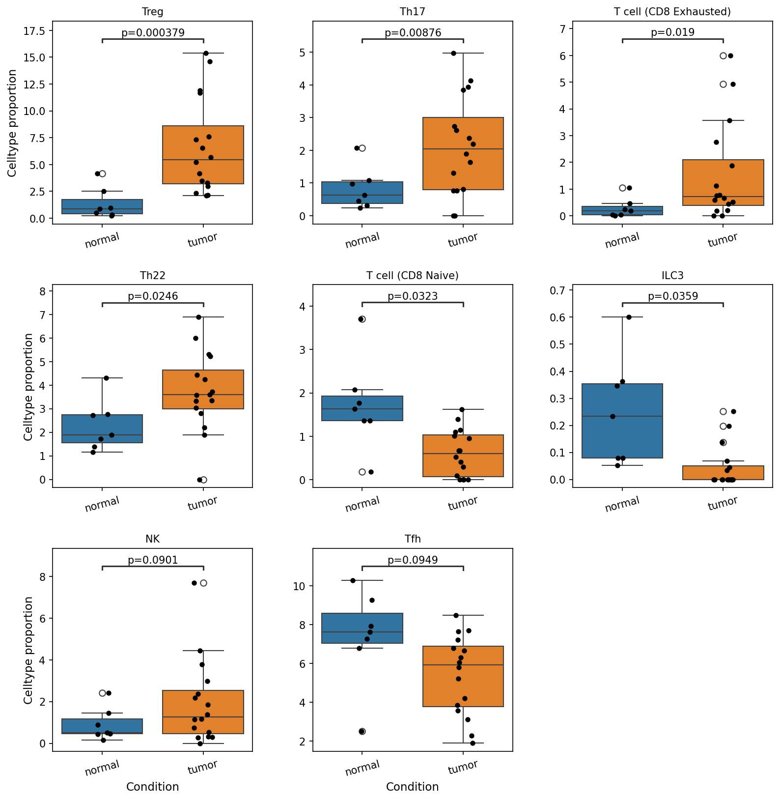
This analysis investigates the differential abundance of T cell and selected innate lymphoid cell (ILC) subset populations between normal colon tissue and colorectal tumor samples. Box plots visualize the proportions of these cell types, with statistical significance determined by a comparison between the 'normal' and 'tumor' conditions. The analysis aimed to identify cell subsets that show statistically significant shifts in their proportions in the tumor microenvironment, which is critical for understanding immune evasion and anti-tumor responses in colorectal cancer.
Visual Summary
The box plots illustrate significant shifts in the proportions of several T cell and innate lymphoid cell subsets when comparing normal colon tissue to tumor samples. A total of eight cell populations exhibited statistically significant differences (p-value < 0.1):
Increased in Tumor:
- Treg cells (Regulatory T cells): Show a substantial and highly significant increase in tumor samples (p=0.000379). The median proportion rises from approximately 1.5% in normal to about 5.5% in tumor.
- Th17 cells (T helper 17 cells): Display a significant increase in tumor tissues (p=0.00876), with the median proportion approximately doubling from around 1% to 2.5%.
- T cell (CD8 Exhausted): Significantly elevated in tumor samples (p=0.019), increasing from very low proportions (<0.5%) in normal tissue to a median of about 0.8% in tumors, with some samples showing much higher proportions.
- Th22 cells (T helper 22 cells): Exhibit a significant increase in tumor samples (p=0.0246), with the median proportion rising from approximately 2% to 3.5%.
- NK cells (Natural Killer cells): Show a marginally significant increase in tumor samples (p=0.0901), with a slight rise in median proportion.
Decreased in Tumor:
- T cell (CD8 Naive): Significantly reduced in tumor samples (p=0.0323), with the median proportion dropping from approximately 1.7% in normal to about 0.7% in tumors.
- ILC3 (Group 3 Innate Lymphoid Cells): Significantly decreased in tumor samples (p=0.0359), with the median proportion falling sharply from around 0.2% in normal to near zero in tumors.
- Tfh cells (T follicular helper cells): Show a marginally significant decrease in tumor samples (p=0.0949), with the median proportion declining from approximately 8% to 6%.
Overall, the tumor microenvironment appears to be characterized by an accumulation of immunosuppressive or pro-tumorigenic T cell subsets (Treg, Th17, Th22, CD8 Exhausted) and a reduction in potentially anti-tumor or homeostatic populations (CD8 Naive, ILC3, Tfh).
Biological Interpretation
These findings provide critical insights into the immune landscape of Colorectal Cancer (CRC) and align with known mechanisms of tumor immune evasion and progression.
- Immunosuppressive Environment: The pronounced increase in Treg cells is a hallmark of many cancers, including CRC. Tregs suppress anti-tumor immune responses by inhibiting effector T cells, thereby promoting immune evasion and tumor growth 1. Similarly, the increase in CD8 Exhausted T cells signifies a dysfunctional anti-tumor response, where cytotoxic T cells lose their effector functions due to chronic antigen exposure and inhibitory signals within the tumor microenvironment 2.
- Pro-tumorigenic Inflammation: The significant increase in Th17 cells and Th22 cells suggests an inflammatory environment that can be hijacked by the tumor. Th17 cells, through cytokines like IL-17, can promote angiogenesis, tumor growth, and metastasis in CRC, although their role can be context-dependent 3. Th22 cells, producing IL-22, can support epithelial cell proliferation and survival, potentially contributing to tumor progression 4.
- Compromised Anti-tumor Immunity and Homeostasis:
- The decrease in CD8 Naive T cells could indicate either reduced infiltration of new naive T cells into the tumor site or their extensive differentiation into other states, possibly exhausted or regulatory phenotypes, or a systemic depletion.
- The reduction in ILC3 is notable. ILC3s are crucial for maintaining gut barrier integrity and promoting tissue homeostasis. Their decrease could reflect a disruption of the normal mucosal immune architecture in the tumor, potentially contributing to dysregulation of the local immune response or impaired barrier function in the context of CRC 5.
- The decrease in Tfh cells may impair humoral immunity against the tumor, as Tfh cells are essential for effective B cell responses and the generation of high-affinity antibodies.
- NK Cell Dynamics: The marginal increase in NK cells might suggest an attempt by the innate immune system to respond to the tumor. However, the functional status of these NK cells in the tumor microenvironment often becomes suppressed, limiting their anti-tumor efficacy despite increased numbers 6.
Collectively, these shifts paint a picture of a tumor microenvironment that actively recruits and expands immunosuppressive and pro-inflammatory T cell subsets while potentially impairing the influx or function of beneficial immune populations, facilitating tumor growth and immune evasion in the colon.
Clinical or Translational Implications
The observed alterations in T cell and ILC subset proportions have several potential clinical and translational implications for colorectal cancer:
- Biomarkers for Prognosis and Response: The elevated proportions of Treg cells, Th17 cells, and CD8 Exhausted T cells could serve as prognostic biomarkers, potentially indicating a more aggressive disease or poorer response to conventional therapies. Conversely, higher proportions of specific anti-tumor immune cells (if functional) or ILC3s in early stages might correlate with better outcomes.
- Immunotherapy Targets: The dominance of immunosuppressive cells (Treg, CD8 Exhausted) highlights the potential for immune checkpoint blockade therapies or strategies aimed at reversing T cell exhaustion. Depleting Tregs or re-invigorating exhausted CD8 T cells could enhance anti-tumor immunity 2.
- Modulating Inflammation: The increased Th17 and Th22 populations suggest that targeting specific inflammatory pathways driven by these cells (e.g., IL-17, IL-22) might be a therapeutic avenue, although the complex context-dependent roles require careful consideration.
- Restoring Homeostasis: Strategies to restore ILC3 populations or their functions might help normalize the gut immune environment and potentially enhance anti-tumor responses or improve barrier function in CRC.
- Patient Stratification: Understanding these cellular shifts could enable better stratification of patients for different immunotherapy approaches or combination therapies, leading to more personalized treatment strategies.
References:
- Treg cells in Cancer: PubMed Search for "regulatory T cells cancer immune evasion" https://pubmed.ncbi.nlm.nih.gov/?term=regulatory+T+cells+cancer+immune+evasion
- CD8 T cell Exhaustion: PubMed Search for "CD8 T cell exhaustion cancer immunotherapy" https://pubmed.ncbi.nlm.nih.gov/?term=CD8+T+cell+exhaustion+cancer+immunotherapy
- Th17 cells in CRC: PubMed Search for "Th17 colorectal cancer prognosis" https://pubmed.ncbi.nlm.nih.gov/?term=Th17+colorectal+cancer+prognosis
- Th22 cells and IL-22 in Cancer: PubMed Search for "Th22 IL-22 cancer proliferation" https://pubmed.ncbi.nlm.nih.gov/?term=Th22+IL-22+cancer+proliferation
- ILC3s in Gut Immunity/Cancer: PubMed Search for "ILC3 gut cancer" https://pubmed.ncbi.nlm.nih.gov/?term=ILC3+gut+cancer
- NK cells in Tumor Microenvironment: PubMed Search for "NK cell tumor microenvironment suppression" https://pubmed.ncbi.nlm.nih.gov/?term=NK+cell+tumor+microenvironment+suppression
9. Macrophage Subset Population Analysis in Colorectal Cancer
[Analysis Visualization Results]...
Analysis Overview
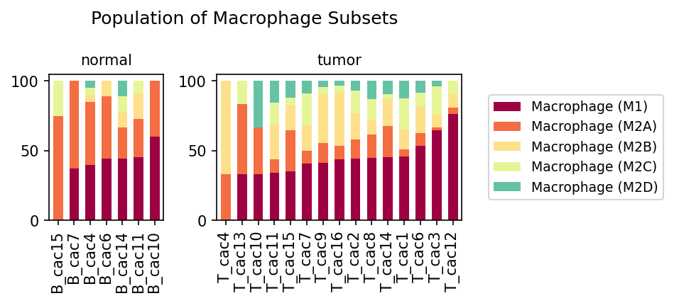
This analysis investigates the proportional distribution of various macrophage subsets (Macrophage (M1), Macrophage (M2A), Macrophage (M2B), Macrophage (M2C), and Macrophage (M2D)) within normal colon tissue and colorectal tumor samples. The goal is to identify shifts in macrophage polarization that may be associated with the disease state, providing insights into the immune microenvironment of Colorectal Cancer.
Visual Summary
The stacked bar plot visualizes the relative proportions of five macrophage subsets across multiple normal (B_cac samples) and tumor (T_cac samples) samples.
- Normal Samples: In normal colon tissue samples, the macrophage population is predominantly composed of Macrophage (M1) and Macrophage (M2A). While there is some variability, Macrophage (M2A) often represents a substantial fraction, sometimes comparable to or slightly exceeding Macrophage (M1). Macrophage (M2B), Macrophage (M2C), and Macrophage (M2D) are consistently present in relatively low proportions across all normal samples.
- Tumor Samples: A notable shift in macrophage subset composition is observed in tumor samples compared to normal tissue.
- Increased Macrophage (M1): Macrophage (M1) proportions show a distinct increase in most tumor samples, often becoming the predominant subset. In some samples (e.g., T_cac3, T_cac12), Macrophage (M1) accounts for approximately 65-75% of the total macrophage population.
- Decreased Macrophage (M2A): Concomitantly, Macrophage (M2A) generally exhibits a reduced presence in tumor samples, often becoming a minor component (typically less than 20%).
- Varied M2B/C/D: Macrophage (M2B) shows a considerable increase in some tumor samples (e.g., T_cac4, T_cac13), where it can constitute up to 50% of the macrophage population. Macrophage (M2C) and Macrophage (M2D) also appear slightly elevated in some tumor samples compared to their levels in normal tissue, though they generally remain minor subsets.
- Inter-sample Heterogeneity: There is significant heterogeneity in macrophage subset distribution among individual tumor samples, indicating diverse immune microenvironments within different tumors.
Biological Interpretation
Macrophages are highly plastic immune cells that polarize into distinct functional phenotypes, traditionally categorized as M1 (pro-inflammatory, anti-tumorigenic) and M2 (anti-inflammatory, pro-tumorigenic, tissue repair). The observed shifts in macrophage populations within the colorectal tumor microenvironment offer critical biological insights.
- Shift Towards Macrophage (M1) Dominance: The most striking finding is the substantial increase in Macrophage (M1) proportions in many tumor samples. M1 macrophages are known for their ability to produce pro-inflammatory cytokines, present antigens, and directly kill tumor cells, thus playing a crucial role in anti-tumor immunity PubMed Search: M1 macrophage anti-tumor function cancer. This M1 prevalence could indicate an active immune response against the tumor in these patients, or it might represent a complex and possibly dysfunctional M1-like state within the highly immunosuppressive tumor microenvironment.
- Decreased Macrophage (M2A) and Increased Macrophage (M2B): The reduction in Macrophage (M2A) suggests a decrease in certain Th2-mediated responses or immune regulatory pathways commonly associated with this subtype. Conversely, the rise in Macrophage (M2B) in some tumors is noteworthy. M2B macrophages are induced by immune complexes and Toll-like receptor agonists, mediating both pro-inflammatory and immunoregulatory functions PubMed Search: M2B macrophage function cancer. Their variable increase suggests a more complex regulatory landscape in specific tumor contexts.
- Colorectal Cancer Context: In colorectal cancer, tumor-associated macrophages (TAMs) are typically considered major contributors to tumor progression, often adopting an M2-like phenotype that promotes angiogenesis, immune suppression, and metastasis GeneCards: CD68 TAM CRC. The observed M1-dominant shift in many of these tumor samples challenges a simplistic view of TAMs as universally M2-polarized in CRC. This suggests a more heterogeneous and dynamic immune landscape, where different polarization states coexist or dominate depending on the individual tumor and its microenvironment. This could reflect varying disease stages, genetic backgrounds, or responses to extrinsic factors in the tumor.
Clinical or Translational Implications
The diverse macrophage polarization patterns observed in colorectal cancer samples have several potential clinical implications:
- Prognostic and Predictive Biomarkers: The specific composition of macrophage subsets, particularly the M1/M2 balance and the prevalence of M2B, could serve as prognostic indicators for disease progression or as predictive biomarkers for response to specific therapies.
- Personalized Immunotherapy Strategies: The significant heterogeneity across tumor samples highlights the need for personalized treatment approaches. For patients with a predominantly M1 macrophage profile, therapies aimed at enhancing M1 activity or overcoming M1 suppression might be effective. Conversely, patients with increased M2B or other M2 subtypes might benefit from strategies designed to reprogram M2 macrophages towards an M1 phenotype or inhibit their pro-tumor functions.
- Drug Development: A deeper understanding of the molecular mechanisms driving these specific macrophage polarization states (e.g., M1 dominance, M2B increase) could lead to the identification of novel therapeutic targets to modulate the immune microenvironment in CRC.
- Further Validation: Correlating these observed macrophage population shifts with clinical outcomes, tumor genetics, and patient responses to different treatments is essential to translate these findings into actionable clinical strategies.
10. Macrophage Subset Population Shifts in Colorectal Cancer
[Analysis Visualization Results]...
Analysis Overview
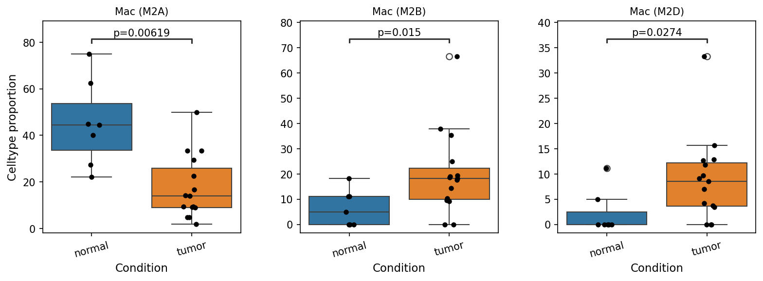
This analysis investigates the proportional representation of different macrophage subsets—Mac (M2A), Mac (M2B), and Mac (M2D)—between normal colon tissue and colorectal tumor samples. The goal is to identify statistically significant differences in these cell populations that may contribute to the understanding of the tumor microenvironment (TME) in colorectal cancer. The analysis utilized single-cell RNA-seq data from human colon tissue, comparing 'tumor' conditions against 'normal' as a reference.
Visual Summary
The box plots display the celltype proportion for three macrophage subsets across normal and tumor conditions, with statistically significant differences highlighted:
- Mac (M2A) Proportions: The proportion of Mac (M2A) cells is significantly *lower* in tumor samples compared to normal samples (p=0.00619). In normal tissue, the median proportion is approximately 45%, while in tumor tissue, it drops to about 15%. This indicates a substantial depletion or shift away from the M2A phenotype in the colorectal tumor microenvironment.
- Mac (M2B) Proportions: Conversely, the proportion of Mac (M2B) cells is significantly *higher* in tumor samples compared to normal samples (p=0.015). The median proportion increases from approximately 5% in normal tissue to around 15-20% in tumor tissue, with some tumor samples showing particularly high proportions (outliers near 70%). This suggests an enrichment of M2B macrophages in the tumor.
- Mac (M2D) Proportions: Similarly, Mac (M2D) cells show a statistically significant *increase* in their proportion in tumor samples compared to normal samples (p=0.0274). The median proportion in normal tissue is very low, around 1-2%, which rises to about 8-9% in tumor tissue, indicating a notable expansion of this macrophage subset within the tumor microenvironment.
Biological Interpretation
Macrophages are highly plastic immune cells that play a crucial role in the tumor microenvironment, often polarizing into distinct phenotypes that can either inhibit (M1-like) or promote (M2-like) tumor growth. The observed shifts in macrophage subset populations in colorectal cancer are highly indicative of an immunosuppressive and pro-tumorigenic environment.
- Decrease in Mac (M2A): M2A macrophages are typically associated with allergic responses and wound healing, often induced by IL-4 and IL-13. Their decreased presence in tumor samples could suggest a divergence from a classical tissue repair-like environment or a specific context where these macrophages are outcompeted or reprogrammed into other M2 subtypes more conducive to tumor progression. While their role in cancer can be complex, a reduction might imply a loss of certain potentially anti-tumorigenic or early pro-resolving functions, or simply that the tumor microenvironment favors other M2 subtypes.
- Increase in Mac (M2B) and Mac (M2D): Both M2B and M2D macrophages are generally considered pro-tumorigenic.
- M2B macrophages, often activated by immune complexes and TLR agonists, are known to produce both pro- and anti-inflammatory cytokines. Their elevated levels in tumor could contribute to a complex inflammatory milieu that ultimately supports tumor growth and immune evasion.
- M2D macrophages (also known as "tumor-associated macrophages" or TAMs in some contexts) are particularly recognized for their strong pro-tumorigenic activities. They are often induced by adenosine and TLR signaling and are implicated in promoting angiogenesis, immunosuppression, and metastasis. An increase in M2D populations is a common feature of many cancers and is associated with poor prognosis. Their enrichment in colorectal tumors strongly points towards an enhanced immunosuppressive state and active tumor progression. [For a general overview of TAMs and their M2 subtypes in cancer, refer to PubMed search on "M2 macrophages cancer" or "tumor associated macrophages colorectal cancer"]
Collectively, these findings demonstrate a clear polarization of macrophages within the colorectal tumor microenvironment towards pro-tumorigenic M2B and M2D phenotypes, while M2A macrophages are diminished. This shift underscores the dynamic re-education of immune cells by the tumor to foster its growth and evade immune surveillance.
Clinical or Translational Implications
The distinct shifts in macrophage subsets between normal and tumor colon tissue have several potential clinical and translational implications for colorectal cancer:
- Biomarkers: The differential proportions of Mac (M2A), Mac (M2B), and Mac (M2D) could serve as potential diagnostic or prognostic biomarkers. For instance, a higher ratio of M2B/M2D to M2A macrophages might indicate a more aggressive tumor phenotype or poorer patient outcome.
- Therapeutic Targets: Given the pro-tumorigenic roles of M2B and M2D macrophages, targeting these specific subsets or the signaling pathways that drive their polarization and function represents a promising therapeutic strategy. This could involve approaches to reprogram these macrophages to an M1-like anti-tumor phenotype, deplete them, or inhibit their immunosuppressive and pro-angiogenic activities. [For therapeutic targeting of TAMs in cancer, see reviews indexed in PubMed on "macrophage reprogramming cancer therapy"]
- Immunotherapy Enhancement: Understanding the precise macrophage landscape in colorectal cancer patients could aid in personalizing immunotherapeutic strategies. Combining existing immunotherapies with agents that modulate macrophage polarization might improve treatment efficacy by converting an immunosuppressive TME into an immune-responsive one.
11. Ploidy Status of Intestinal Epithelial Cells in Normal and Tumor Colon Tissue
[Analysis Visualization Results]...
Analysis Overview
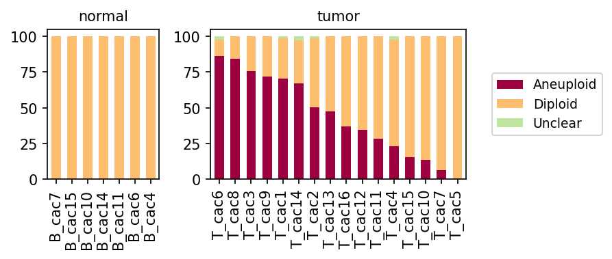
This analysis investigates the ploidy status (diploid, aneuploid, or unclear) of Intestinal Epithelial cells, identified as the tumor-origin cell type, across different samples from both normal and colorectal cancer conditions. The goal is to determine if and how the chromosomal stability of these cells changes in the context of disease, providing insight into the genomic characteristics of the malignant population.
Visual Summary
The stacked bar plots display the proportional distribution of ploidy states (Aneuploid, Diploid, Unclear) within Intestinal Epithelial cells for individual samples, separated by condition.
- Normal Condition: In all normal colon tissue samples (e.g., B_cac7, B_cac15), Intestinal Epithelial cells are almost exclusively Diploid (represented by the orange bars). The proportion of aneuploid or unclear cells is negligible or entirely absent in these healthy samples, indicating chromosomal stability.
- Tumor Condition: In contrast, Intestinal Epithelial cells from tumor samples exhibit a clear and often substantial presence of Aneuploid cells (maroon bars).
- A significant proportion of aneuploid cells is observed in most tumor samples, ranging from approximately 10% to over 85%. For instance, samples like T_cac6, T_cac8, T_cac3, T_cac9, and T_cac1 show a very high percentage of aneuploid Intestinal Epithelial cells (e.g., ~70-85%).
- There is considerable heterogeneity in the degree of aneuploidy across different tumor samples. Some tumor samples (e.g., T_cac15, T_cac10, T_cac7, T_cac5) show a lower proportion of aneuploid cells, with diploid cells remaining dominant, while still presenting a notable aneuploid fraction compared to normal tissue.
- A minor "Unclear" ploidy component (light green) is also visible in a few tumor samples, though it constitutes a very small fraction of the total cells.
Biological Interpretation
The observed ploidy patterns strongly support the inference of Intestinal Epithelial cells as the malignant population in colorectal cancer.
- Aneuploidy as a Hallmark of Cancer: The dramatic shift from a predominantly diploid state in normal Intestinal Epithelial cells to a highly aneuploid state in tumor Intestinal Epithelial cells is a classic hallmark of cancer. Aneuploidy, an abnormal number of chromosomes, contributes to genomic instability, which drives oncogenesis, tumor heterogeneity, and resistance to therapy PubMed Search: aneuploidy cancer hallmark.
- Confirmation of Tumor-Origin Cell Type: The finding that aneuploidy is almost exclusively observed in Intestinal Epithelial cells within the tumor context, and not in the normal colon, directly validates the designation of "Intestinal Epithelial cell" as the tumor-origin cell type. This confirms that these are the cells undergoing malignant transformation and accumulating chromosomal abnormalities.
- Tumor Heterogeneity: The variability in the proportion of aneuploid cells across different tumor samples (e.g., T_cac6 vs. T_cac5) highlights the genomic and cellular heterogeneity common in colorectal cancer. This variation could reflect:
- Differences in tumor purity, where some samples might contain a higher admixture of non-malignant stromal or immune cells, thus diluting the observed aneuploid fraction.
- Intrinsic biological heterogeneity in the tumor clone, with some tumors being more chromosomally unstable or having evolved more aneuploid clones than others.
- Different stages or aggressiveness of the tumors from which the samples were obtained.
Clinical or Translational Implications
The detection of aneuploidy in Intestinal Epithelial cells within tumor samples holds several clinical and translational implications:
- Diagnostic Value: The presence of significant aneuploidy in Intestinal Epithelial cells is a strong indicator of malignancy and can aid in the pathological diagnosis of colorectal cancer. While traditionally assessed by other methods like karyotyping or FISH, single-cell analysis provides cell-type-specific resolution.
- Prognostic Potential: In many cancers, the degree of aneuploidy is correlated with tumor aggressiveness, risk of recurrence, and patient prognosis GeneCards: Aneuploidy & Cancer. Tumors with a higher burden of aneuploidy might indicate more advanced or aggressive disease requiring tailored therapeutic approaches.
- Therapeutic Strategies: Understanding the aneuploid status of tumor cells can inform therapeutic strategies. Tumors with high genomic instability and aneuploidy might respond differently to certain chemotherapies or targeted agents. Furthermore, the observed heterogeneity suggests that a "one-size-fits-all" approach might be suboptimal, emphasizing the need for personalized medicine.
- Monitoring Disease Progression: Tracking changes in aneuploidy levels could potentially be used to monitor disease progression or response to treatment, although this requires longitudinal studies and specific methodologies.
12. Cell-Cell Interaction Analysis in Colorectal Cancer: Normal vs. Tumor Microenvironment
[Analysis Visualization Results]...
Analysis Overview
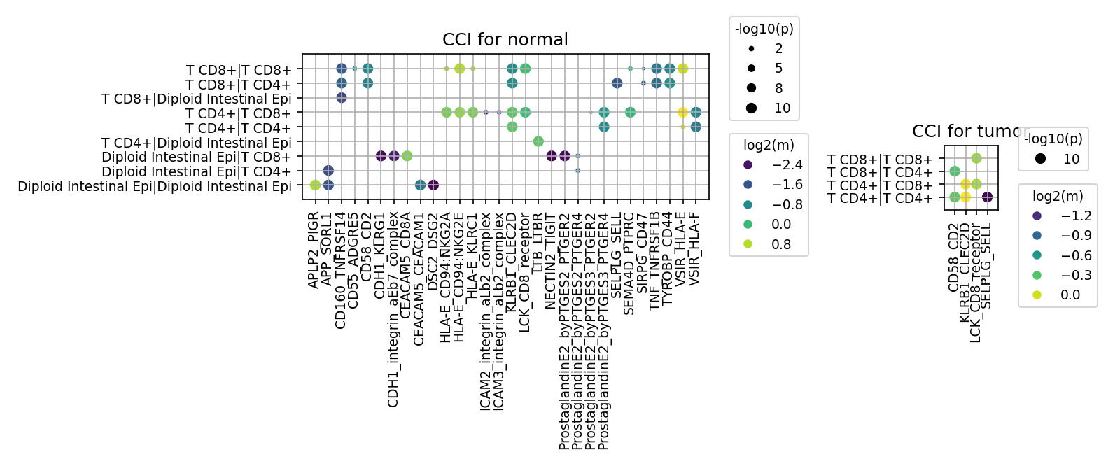
This analysis investigates cell-cell interaction (CCI) patterns in human colon tissue, comparing normal and tumor conditions. The focus is on interactions involving key cell types implicated in colorectal cancer: Intestinal Epithelial cells (identified as the tumor-origin cell type), Fibroblasts, Macrophages, and T cell subsets (CD4+ and CD8+). The CellPhoneDB tool was used to identify ligand-receptor interactions, with results filtered to show at most 80 significant pairs per group (p-value < 0.05, mean expression > 0.01). This analysis aims to highlight how the cellular communication landscape changes in the colorectal tumor microenvironment compared to normal tissue.
Visual Summary
CCI for Normal Condition
The dot plot for the normal condition displays a rich and diverse network of cell-cell interactions.
- Interacting Cell Types: Interactions are observed between various T cell subsets (CD8+ T cells with CD8+ T cells, CD8+ T cells with CD4+ T cells, CD4+ T cells with CD4+ T cells) and, notably, between Diploid Intestinal Epithelial cells and T cells (CD8+ and CD4+). Diploid Intestinal Epithelial cells also show homotypic interactions.
- Ligand-Receptor Pairs: A broad range of ligand-receptor pairs are identified, indicating complex cellular communication. These include:
- Adhesion and Epithelial Integrity: CDH1-CDH1 (E-cadherin homophilic adhesion, crucial for epithelial barrier), ICAM1-CD11A, ICAM2-CD11A.
- Immune Recognition and Regulation: HLA-E-CD94/NKG2A, HLA-E-TCR, CD58-ADGRE5, CD160-HVEM, NECTIN2-TIGIT (an immune checkpoint molecule), KLRB1-CLEC2D.
- Prostaglandin Signaling: Multiple interactions involving Prostaglandin E2 and its receptors (PTGER2, PTGER4), suggesting local inflammatory or immunomodulatory processes.
- Other: APLP2-PIGR, CDX2-CEACAM (adhesion/signaling), DSC2-DSG2A.
- Interaction Strength: Many interactions show high significance (large dot size, indicating low p-value) and moderate to high mean expression (green to yellow dot color). This signifies active and robust communication networks in healthy colon tissue.
CCI for Tumor Condition
The dot plot for the tumor condition presents a strikingly sparse interaction landscape compared to the normal state.
- Interacting Cell Types: A dramatic observation is the complete absence of significant interactions (within the top 80 pairs displayed) involving Intestinal Epithelial cell, Fibroblast, or Macrophage with any of the other target cell types, or among themselves. The only interactions that meet the significance thresholds are exclusively between T cell subsets (CD8+ T cells with CD8+ T cells, CD8+ T cells with CD4+ T cells, CD4+ T cells with CD8+ T cells, CD4+ T cells with CD4+ T cells).
- Ligand-Receptor Pairs: The detected pairs are limited to those relevant for T cell-T cell communication:
- CD58-CD2: A well-known co-stimulatory pathway for T cell activation and adhesion.
- KLRB1-CLEC2D: Interactions involving KLRB1 (CD161), often found on NK cells and some T cells, can mediate inhibitory signals.
- LCK-CD8: While LCK is an intracellular kinase, this pair might represent interactions involving the CD8 co-receptor that influence LCK-mediated signaling pathways crucial for T cell activation.
- T_receptor-SELL: Suggests T cell receptor interactions with L-selectin (SELL), relevant for T cell homing and adhesion.
- Interaction Strength: The few remaining interactions generally show moderate significance and mean expression.
Comparative Summary
There is a profound shift in the cell-cell interaction patterns from normal to tumor conditions. The diverse network seen in normal tissue, which includes significant epithelial-immune and stromal communications, is largely replaced by a highly restricted set of T cell-T cell interactions in the tumor microenvironment. The absence of significant interactions involving the tumor-origin Intestinal Epithelial cell (and Fibroblast, Macrophage) in the tumor context, despite being explicitly requested, is a critical finding, implying a severe disruption or suppression of these communication pathways in cancer.
Biological Interpretation
Normal Colon Homeostasis and Immune Surveillance
In the normal colon, the rich interaction network reflects a healthy tissue microenvironment. The presence of Diploid Intestinal Epithelial cell interactions with T cells, along with homotypic epithelial interactions via CDH1-CDH1 (E-cadherin) and DSC2-DSG2A (desmogleins/desmocollins), underscores robust epithelial barrier function and communication [PubMed: 31213348]. Immune interactions like HLA-E-CD94/NKG2A and HLA-E-TCR suggest active immune surveillance by T cells and NK cells, essential for maintaining gut immune tolerance and defense [PubMed: 25687707]. Prostaglandin signaling pathways highlight dynamic regulation of inflammation and cellular processes within the colon.
Remodeling of the Tumor Microenvironment
The dramatic reduction and re-patterning of cell-cell interactions in the tumor condition point to a significant remodeling of the tumor microenvironment (TME).
- Loss of Epithelial-Immune/Stromal Crosstalk: The most striking observation is the absence of significant interactions involving Intestinal Epithelial cells (the resolved tumor-origin cell type), Fibroblasts, and Macrophages in the tumor environment. This could indicate several biological processes:
- Immune Evasion: Tumor cells (derived from Intestinal Epithelial cells) may downregulate specific surface molecules or modify their secretome to evade immune recognition or interaction, contributing to an immunosuppressive TME.
- Disrupted Homeostasis: The normal intricate crosstalk between epithelial cells, stromal cells (Fibroblasts), and immune cells (Macrophages, T cells) is fundamental for tissue organization and function. Its disruption in tumor suggests a highly dysregulated microenvironment.
- Context of Analysis: It's important to note that the filter (n_pairs_to_show=80, pval_cutoff=0.05, mean_cutoff=0.01) may exclude less significant interactions. However, the complete absence among the top pairs suggests these interactions are either significantly reduced in strength/frequency or overshadowed by other dominant signals.
- Dominance of T Cell-T Cell Interactions: The prominence of T cell-T cell interactions in the tumor microenvironment suggests that while T cells are present, their primary communication might be intra-T-cellular or focused on self-regulation rather than directly interacting with tumor cells or other stromal components.
- CD58-CD2 is an important co-stimulatory pathway, essential for T cell activation and adhesion [GeneCards: CD2]. Its presence could indicate ongoing T cell activation or memory responses, but without interactions with tumor cells, its efficacy might be limited.
- KLRB1-CLEC2D (CD161-CLEC2D) signaling often conveys inhibitory signals, potentially contributing to T cell exhaustion or anergy within the TME, a common feature of advanced cancers [PubMed: 32669485].
- LCK-CD8 and T_receptor-SELL further emphasize intra-T cell signaling and adhesion mechanisms, which might be critical for T cell function (or dysfunction) in the absence of robust tumor-directed interactions.
Clinical or Translational Implications
- Biomarkers of TME Disruption: The dramatic shift in CCI patterns, particularly the loss of epithelial-immune/stromal interactions in the tumor, could serve as a signature of TME dysregulation in colorectal cancer. This altered interaction landscape could be explored as a prognostic or predictive biomarker for disease progression or response to therapy.
- Therapeutic Target Prioritization:
- Restoring Epithelial-Immune Crosstalk: Strategies aimed at re-establishing or enhancing crucial interactions between tumor-origin Intestinal Epithelial cells and immune cells could improve anti-tumor immunity. For instance, modulating surface proteins on tumor cells to promote interaction with T cells or macrophages might be beneficial.
- Modulating T Cell-T Cell Dynamics: The observed T cell-T cell interactions in the tumor suggest opportunities for immunotherapy. Targeting inhibitory pathways like KLRB1-CLEC2D could alleviate T cell exhaustion. Conversely, enhancing co-stimulatory signals such as CD58-CD2 could boost T cell effector functions within the TME [PubMed: 30127599].
- Investigating the Missing Links: Further investigation into *why* epithelial-immune/stromal interactions are absent from the top 80 pairs in the tumor is crucial. Are these interactions completely shut down, or merely downregulated and outcompeted by other signals? Identifying the mechanisms behind this loss could reveal novel therapeutic vulnerabilities for restoring immune recognition and control of tumor cells.
- Experimental Validation: The identified specific ligand-receptor pairs, especially those exclusively present in the tumor or significantly altered, warrant experimental validation. For example, KLRB1-CLEC2D could be investigated for its role in T cell exhaustion in colorectal cancer models, and blocking this interaction could be tested as a therapeutic strategy.
13. Condition-Specific Cell-Cell Interaction Patterns in Colorectal Cancer
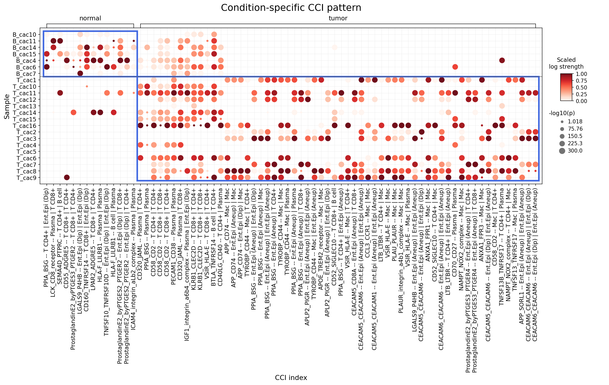
[Analysis Visualization Results]...
Analysis Overview
This analysis identifies cell-cell interactions (CCIs) involving Intestinal Epithelial cells (the resolved tumor-origin cell type), major immune cells (T cells, NK cells, ILC, B cells, Plasma cells, Macrophages, Dendritic cells, Mast cells), and stromal cells (Fibroblasts, Smooth muscle cells) that differ significantly between 'normal' and 'tumor' conditions in human colon tissue. The CellPhoneDB method was used to infer these interactions, with results presented as a dot plot, where dot color indicates the scaled log strength of the interaction and dot size represents the -log10(p-value) for significance. The goal is to highlight key signaling pathways and mechanisms that are perturbed in the tumor microenvironment, identify potential therapeutic targets, and suggest relevant therapeutic agents.
Visual Summary
The dot plot prominently displays condition-specific cell-cell interaction patterns. A clear distinction is observed between 'normal' and 'tumor' samples, with 'B_cac' samples primarily associated with the 'normal' condition and 'T_cac' samples with the 'tumor' condition.
- Differential Interaction Landscape: The 'tumor' condition exhibits a substantially higher number of strong and significant cell-cell interactions compared to the 'normal' condition. Interactions in the 'tumor' context are frequently depicted with darker red colors (higher scaled log strength) and larger dot sizes (higher significance, lower p-value), indicating robust and widespread cellular communication changes.
- Tumor-Specific Interactions: The right half of the plot, corresponding to the 'tumor' condition, is rich with strong interactions. Notably, many of these involve 'Intestinal Epithelial cell (Aneup)', which signifies aneuploid (likely cancerous) intestinal epithelial cells. These cells show extensive interactions with various immune cells, including Macrophages, T CD4+ cells, T CD8+ cells, and Plasma cells.
- Key Interaction Families in Tumor: Several families of interactions are particularly prominent in the tumor samples:
- CEACAMs: Interactions involving CEACAM5 and CEACAM6 are highly frequent and strong between aneuploid Intestinal Epithelial cells and Macrophages, T cells, Plasma cells, and even other aneuploid Intestinal Epithelial cells (homophilic interactions).
- PPIA_BSG (CD147): This interaction is robustly observed between aneuploid Intestinal Epithelial cells and Macrophages or Plasma cells.
- Prostaglandin E2 (PGE2) pathway: Interactions via PTGES3/PTGES and PTGER2 are notable between aneuploid Intestinal Epithelial cells and T CD4+ cells.
- Immune checkpoint/regulatory molecules: Interactions such as VSIR_HLA-E, LGALS9_P4HB, and CD70_CD27 appear in the tumor microenvironment, involving aneuploid Intestinal Epithelial cells and immune cells.
- Normal Condition Interactions: In contrast, the 'normal' condition (left half) shows fewer and generally weaker interactions, predominantly involving 'Intestinal Epithelial cell (Dip)' (diploid, likely healthy) and various immune cells. While some interactions (e.g., certain PGE2-related ones) are present in both conditions, their overall strength and prevalence are markedly higher in the tumor context.
- Ploidy-Specific Differences: The distinction between 'Ent.Epi (Dip)' and 'Ent.Epi (Aneup)' is critical. Most of the highly significant tumor-specific CCIs involve aneuploid Intestinal Epithelial cells, underscoring their unique communicative roles in the neoplastic microenvironment.
Biological Interpretation
The observed shifts in cell-cell interactions from normal to tumor conditions provide critical insights into the pathophysiology of Colorectal Cancer (CRC), especially considering that Intestinal Epithelial cells are the tumor-origin cell type. The prevalence of interactions involving aneuploid Intestinal Epithelial cells highlights their active role in shaping the tumor microenvironment (TME).
- CEACAM-Mediated Pro-Tumorigenic Signaling (CEACAM5, CEACAM6)
- Interactions: CEACAM5_CEACAM6 -- Ent.Epi (Aneup) | Macrophage, CEACAM5_CEACAM6 -- Ent.Epi (Aneup) | T CD4+, CEACAM5_CEACAM6 -- Ent.Epi (Aneup) | Plasma, CEACAM5_CEACAM6 -- Ent.Epi (Aneup) | Ent.Epi (Aneup).
- Pathways and Mechanisms: Carcinoembryonic antigen-related cell adhesion molecules (CEACAMs), particularly CEACAM5 and CEACAM6, are often overexpressed in various cancers, including CRC. They function as both homophilic and heterophilic adhesion molecules. Their interactions promote tumor cell proliferation, survival, invasion, and metastasis. In the TME, CEACAMs can engage with receptors on immune cells (e.g., CEACAM1 on myeloid cells), leading to immune evasion by inhibiting anti-tumor immune responses or inducing immune cell anergy/exhaustion. These molecules are also involved in angiogenesis and resistance to apoptosis.
- Reference: GeneCards: CEACAM5, GeneCards: CEACAM6
- CD147 (PPIA_BSG) in Tumor Progression
- Interactions: PPIA_BSG -- Ent.Epi (Aneup) | Macrophage, PPIA_BSG -- Ent.Epi (Aneup) | Plasma.
- Pathways and Mechanisms: CD147 (also known as Basigin or EMMPRIN), encoded by *BSG*, and its ligand PPIA (Cyclophilin A) form a crucial interaction pair. CD147, highly expressed on tumor cells, stimulates stromal cells (like macrophages and fibroblasts) to produce matrix metalloproteinases (MMPs), which degrade the extracellular matrix, facilitating tumor invasion and metastasis. It also plays roles in glycolysis, angiogenesis, and tumor cell proliferation. Its interaction with immune cells can modulate their function, potentially contributing to an immunosuppressive TME.
- Reference: GeneCards: BSG (CD147)
- Prostaglandin E2 (PGE2) Signaling in Immune Modulation
- Interactions: ProstaglandinE2_byPTGES3_PTGER2 -- Ent.Epi (Aneup) | T CD4+, ProstaglandinE2_byPTGES_PTGER2 -- Ent.Epi (Aneup) | T CD4+.
- Pathways and Mechanisms: Prostaglandin E2 (PGE2), synthesized by cyclooxygenase (COX) enzymes (like COX-2, often elevated in CRC) and prostaglandin E synthase (PTGES, PTGES3), exerts its effects through G-protein coupled receptors (PTGER1-4, EP1-EP4). The interaction with PTGER2 on T CD4+ cells indicates that tumor-derived PGE2 can profoundly influence T cell function. PGE2 is a potent immunosuppressive mediator in the TME, inhibiting T cell proliferation and cytokine production, promoting the differentiation of regulatory T cells (Tregs), and inducing T cell exhaustion. It also contributes to angiogenesis and tumor growth. While present in normal tissue for homeostatic immune regulation, its overexpression and enhanced signaling in tumor lead to pathology.
- Reference: PubMed: Prostaglandin E2 in cancer
- Immune Checkpoint and Regulatory Molecules
Interactions:
- VSIR_HLA-E -- Ent.Epi (Aneup) | Macrophage: VSIR (VISTA) is an immune checkpoint molecule that suppresses T cell responses. Its interaction with HLA-E on macrophages suggests a mechanism for tumor cells to modulate macrophage function and potentially contribute to an immunosuppressive environment.
- LGALS9_P4HB -- Ent.Epi (Aneup) | T CD4+: Galectin-9 (LGALS9) is a secreted lectin that can bind to receptors like TIM-3 on T cells, inducing T cell apoptosis and promoting immune tolerance, thus contributing to immune evasion.
- CD70_CD27 -- Ent.Epi (Aneup) | Plasma / T CD4+: The CD70-CD27 axis is a T cell costimulatory pathway. While typically activating T cells, sustained CD70 signaling (e.g., from tumor cells) can lead to T cell exhaustion in chronic contexts like cancer.
- Pathways and Mechanisms: These interactions represent various strategies employed by tumor cells and associated stromal/immune cells to evade immune surveillance and promote an immunosuppressive TME, a hallmark of cancer.
- References: GeneCards: VSIR (VISTA), GeneCards: LGALS9 (Galectin-9), GeneCards: CD70
- Adhesion, Metabolism, and Inflammation Links
- Interactions: TYROBP_CD44 -- Ent.Epi (Aneup) | Macrophage, NAMPT_NOX2_complex -- Ent.Epi (Aneup) | T CD4+ / Plasma, ANXA1_FPR1 -- Ent.Epi (Aneup) | Macrophage.
Pathways and Mechanisms:
- CD44 is a cell adhesion molecule and a cancer stem cell marker. Its interaction with TYROBP (DAP12, an adaptor protein for activating receptors) in macrophages could modulate macrophage activation and function within the TME, potentially promoting pro-tumorigenic phenotypes.
- NAMPT (Nicotinamide phosphoribosyltransferase), the rate-limiting enzyme in NAD+ biosynthesis, can also act as an extracellular cytokine (eNAMPT). NOX2 (NADPH oxidase 2) generates reactive oxygen species (ROS). This complex interaction suggests cross-talk in NAD+ metabolism and ROS signaling, contributing to inflammation and immunosuppression in the TME.
- ANXA1 (Annexin A1) and FPR1 (Formyl Peptide Receptor 1) typically mediate anti-inflammatory and pro-resolving responses. However, in cancer, ANXA1 can have context-dependent roles, sometimes promoting tumor growth and metastasis, especially through interactions with immune cells.
- References: GeneCards: CD44, GeneCards: NAMPT, GeneCards: ANXA1
Clinical or Translational Implications
The identification of these significantly altered cell-cell interactions in the tumor microenvironment provides a rich set of potential therapeutic targets for Colorectal Cancer. Targeting these interactions could disrupt tumor growth, metastasis, and immune evasion.
- Targeting CEACAMs (CEACAM5, CEACAM6):
- Therapeutic Potential: Given their high expression on CRC cells and their roles in promoting cancer progression and immune evasion, CEACAM5 and CEACAM6 are attractive targets. Disrupting their homophilic and heterophilic interactions could inhibit tumor growth and metastasis.
- Relevant Therapeutic Agents: Monoclonal antibodies targeting CEACAM5 (e.g., Arcitumomab, Tusamitamab ravtansine - a CEACAM5-targeting antibody-drug conjugate) are in various stages of clinical development for CRC and other CEACAM5-expressing cancers. Similar strategies could be developed for CEACAM6.
- Reference: PubMed: CEACAM5 as a therapeutic target
- Modulating CD147 (PPIA_BSG) Activity:
- Therapeutic Potential: Inhibiting CD147/PPIA interactions could reduce MMP production, thereby curbing tumor invasion and metastasis, as well as impacting tumor angiogenesis and metabolism.
- Relevant Therapeutic Agents: Small molecule inhibitors, antibodies, or peptides designed to block CD147 or its interaction with PPIA are under investigation for cancer therapy.
- Reference: PubMed: CD147 in cancer therapy
- Inhibiting PGE2 Signaling:
- Therapeutic Potential: Blocking PGE2 production or its downstream signaling can reverse immunosuppression, reduce inflammation, and inhibit tumor growth. This strategy is particularly relevant given the widespread immunosuppressive effects of PGE2 in the TME.
- Relevant Therapeutic Agents: Non-steroidal anti-inflammatory drugs (NSAIDs) that inhibit COX enzymes (e.g., aspirin, celecoxib) have shown chemopreventive effects in CRC. More specifically, EP2/EP4 receptor antagonists (e.g., TPST-1495, ONO-AE3-208) are being explored to directly block PGE2's pro-tumorigenic and immunosuppressive effects.
- Reference: PubMed: PGE2 inhibitors in CRC
- Targeting Immune Checkpoints and Regulatory Pathways:
- Therapeutic Potential: Manipulating immune checkpoint molecules like VSIR (VISTA) or pathways involving LGALS9 (Galectin-9) and CD70/CD27 could help reactivate anti-tumor immune responses, particularly in T CD4+ cells and macrophages.
- Relevant Therapeutic Agents: Anti-VISTA antibodies are in early clinical development. Galectin-9 inhibitors are also being investigated to restore T cell function. Strategies targeting the CD70-CD27 axis could involve CD70 inhibitors or CD27 agonists, depending on the desired immune modulation.
- Reference: PubMed: VISTA and cancer immunotherapy
- Addressing Metabolic and Adhesion Interactions:
- Therapeutic Potential: Disrupting interactions involving CD44 and NAMPT, or ANXA1 and FPR1, could impact cancer stem cell properties, tumor-associated inflammation, and metabolic reprogramming in the TME.
- Relevant Therapeutic Agents: CD44 inhibitors (e.g., antibodies, small molecules) aim to target cancer stem cells and reduce metastasis. NAMPT inhibitors (e.g., FK866) aim to starve tumor cells of NAD+, thereby impacting their metabolism and survival. FPR1 antagonists or modulators could be investigated to alter the pro-tumorigenic effects of ANXA1.
- Reference: PubMed: NAMPT inhibitors in cancer
These findings emphasize the complex interplay between tumor cells and their microenvironment in CRC and suggest that a multi-pronged therapeutic approach, perhaps combining existing immunotherapies with agents targeting these specific CCI pathways, could offer improved outcomes for patients.
14. Condition-Specific Cell-Cell Interactions in Colorectal Cancer
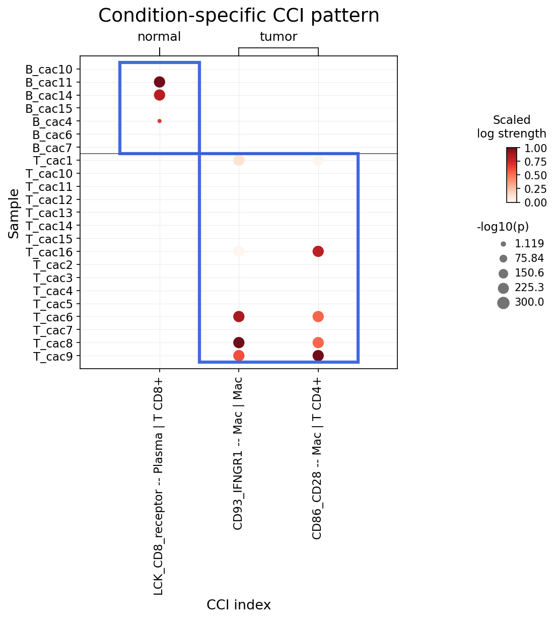
[Analysis Visualization Results]...
Analysis Overview
This analysis investigates condition-specific cell-cell interactions (CCI) involving Intestinal Epithelial cells, major immune cells (T cells, B cells, Plasma cells, Myeloid cells, Mast cells), and Stromal cells in Colon tissue, focusing on genes related to immune checkpoints and the cell cycle. The results are visualized as a dot plot, highlighting significant differences in interaction strength and statistical significance between normal and tumor conditions. The inferred disease context is Colorectal Cancer, given the tissue origin, the presence of tumor samples, and the identification of Intestinal Epithelial cells as the tumor-origin cell type.
Visual Summary
The dot plot displays three distinct cell-cell interaction pairs, showing their strength (scaled log strength, color intensity) and statistical significance (-log10(p), dot size) across different samples and conditions.
- Normal Condition (left panel): A strong and highly significant interaction labeled LCK_CD8_receptor -- Plasma | T CD8+ is predominantly observed in several 'normal' samples (B_cac10, B_cac11, B_cac14). Interactions involving macrophages (CD93_IFNGR1 -- Mac | Mac and CD86_CD28 -- Mac | T CD4+) are largely absent or very weak in normal samples.
- Tumor Condition (right panel): In contrast to the normal condition, the LCK_CD8_receptor -- Plasma | T CD8+ interaction is absent or very weak. Instead, two other interactions are markedly elevated:
- CD93_IFNGR1 -- Mac | Mac: This macrophage-macrophage interaction shows moderate to high strength and significance in several 'tumor' samples (e.g., T_cac6, T_cac7, T_cac8, T_cac9).
- CD86_CD28 -- Mac | T CD4+: This interaction between macrophages and CD4+ T cells also exhibits moderate to high strength and significance across multiple 'tumor' samples (e.g., T_cac16, T_cac6, T_cac7, T_cac8, T_cac9).
Overall, the plot reveals a clear shift in prominent cell-cell communication patterns between normal and tumor colorectal tissue, particularly involving immune cell subsets.
Biological Interpretation
The observed shifts in cell-cell interactions provide insights into the altered immune landscape within the colorectal tumor microenvironment.
1. LCK_CD8_receptor -- Plasma | T CD8+ Interaction (Reduced in Tumor)
- Signaling Pathways and Mechanisms: This interaction involves LCK (Lymphocyte-specific protein tyrosine kinase), a crucial kinase in T cell receptor (TCR) signaling, and the CD8 co-receptor complex, indicating its role in CD8+ T cell activation and function. While the precise ligand on Plasma cells interacting with the LCK/CD8 receptor on CD8+ T cells is not explicitly named, the presence of LCK in the target gene list underscores its importance. LCK is essential for initiating downstream signaling pathways upon TCR engagement, leading to T cell activation, proliferation, and effector functions. Plasma cells, terminally differentiated B cells, are primarily antibody producers but can also present antigens or provide co-stimulatory signals.
- Biological Context: The strong presence of this interaction in normal tissue suggests a robust baseline immune surveillance or an active immune response by CD8+ T cells influenced by Plasma cells in healthy colon. Its marked reduction or absence in tumor samples could indicate a suppression or dysfunction of CD8+ T cell activation and effector mechanisms, which is a common immune evasion strategy employed by tumors. This dampening of cytotoxic T cell activity is detrimental for anti-tumor immunity.
2. CD93_IFNGR1 -- Mac | Mac Interaction (Elevated in Tumor)
- Signaling Pathways and Mechanisms: This interaction involves CD93, a C-type lectin-like receptor expressed on myeloid cells, and IFNGR1, a component of the Interferon Gamma Receptor. IFN-gamma signaling via IFNGR1 is a key pathway for macrophage activation, particularly driving pro-inflammatory M1-like polarization associated with anti-tumor responses. CD93 has been implicated in cell adhesion, phagocytosis, and inflammation, and can modulate macrophage survival and cytokine production. An increased interaction between macrophages themselves, mediated by these molecules, could suggest altered communication or recruitment patterns, potentially influencing their activation state or spatial organization within the tumor microenvironment.
- Biological Context: The elevated CD93_IFNGR1 -- Mac | Mac interaction in tumor samples suggests a heightened or dysregulated macrophage activity. While IFN-gamma can promote anti-tumor immunity, chronic or aberrant signaling in the tumor microenvironment can also contribute to pro-tumor inflammation, immune suppression (e.g., through induction of M2-like macrophages), or T cell exhaustion depending on the context and other co-occurring signals. CD93 itself has been linked to tumor angiogenesis and progression in various cancers [1]. This interaction could be indicative of macrophages adopting pro-tumor phenotypes (e.g., tumor-associated macrophages, TAMs) that facilitate tumor growth and metastasis.
3. CD86_CD28 -- Mac | T CD4+ Interaction (Elevated in Tumor)
- Signaling Pathways and Mechanisms: This is a canonical co-stimulatory interaction crucial for T cell activation. CD86 is a co-stimulatory molecule expressed on antigen-presenting cells (APCs) like macrophages, while CD28 is its receptor on T cells, including CD4+ T cells. Engagement of CD28 by CD86 provides a vital "second signal" necessary for full T cell activation, preventing anergy (unresponsiveness) and promoting proliferation and cytokine production following T cell receptor (TCR) engagement.
- Biological Context: The increased CD86_CD28 -- Mac | T CD4+ interaction in tumor samples suggests enhanced co-stimulatory signaling between macrophages and CD4+ T cells. While essential for effective T cell responses, sustained or excessive co-stimulation within the tumor microenvironment can have complex outcomes. It might initially drive T cell activation, but prolonged stimulation without effective antigen clearance can lead to T cell exhaustion, particularly if inhibitory immune checkpoints (e.g., PD-1/PD-L1) are also active. This interaction can also promote the differentiation of regulatory T cells (Tregs), which suppress anti-tumor immunity. Thus, while seemingly pro-inflammatory, this elevated interaction in the tumor context might ultimately contribute to an immunosuppressive environment or an ineffective anti-tumor response.
Clinical or Translational Implications
The condition-specific cell-cell interactions identified in this analysis offer potential avenues for therapeutic intervention in Colorectal Cancer.
- Targeting the LCK_CD8_receptor -- Plasma | T CD8+ axis (for restoration/enhancement):
- Therapeutic Potential: The diminished presence of this interaction in tumor suggests impaired cytotoxic T cell function. Strategies to restore or enhance this specific signaling pathway could boost anti-tumor CD8+ T cell responses.
- Therapeutic Agents: Approaches could include T cell activators, adoptive T cell therapies, or novel immunotherapies aimed at reversing T cell exhaustion and promoting robust LCK-mediated signaling. While direct LCK modulators might be challenging due to systemic effects, combination therapies that relieve T cell suppression might indirectly reactivate this axis.
- References: LCK's role in T cell activation [2].
Targeting the CD93_IFNGR1 -- Mac | Mac axis (for inhibition/repolarization)
- Therapeutic Potential: The heightened macrophage-macrophage interaction via CD93 and IFNGR1 in tumor tissue suggests a potentially pro-tumorigenic macrophage phenotype or inflammatory environment. Modulating this interaction could repolarize tumor-associated macrophages (TAMs) from an immunosuppressive (M2-like) to an anti-tumor (M1-like) state.
Therapeutic Agents
- CD93 inhibitors: Antibodies or small molecules targeting CD93 could block its pro-tumor functions, potentially inhibiting angiogenesis and macrophage survival within the tumor.
- Macrophage repolarization strategies: Agents like CSF1R inhibitors (e.g., pexidartinib) could deplete or reprogram TAMs, thus indirectly impacting macrophage-macrophage interactions and their functional states [3].
- References: CD93 in cancer [1]. CSF1R inhibition in cancer [3].
Targeting the CD86_CD28 -- Mac | T CD4+ axis (for modulation/rebalancing)
- Therapeutic Potential: The increased CD86-CD28 co-stimulation in tumor, while seemingly activating, might contribute to T cell exhaustion or Treg induction. Therapeutic strategies would aim to re-balance this signaling to favor effective anti-tumor immunity without causing broad immunosuppression.
Therapeutic Agents
- Immune Checkpoint Inhibitors (ICIs): Combining CD28 co-stimulation with checkpoint blockade (e.g., anti-PD-1/PD-L1 antibodies like Pembrolizumab or Nivolumab) can help overcome T cell exhaustion that may arise from chronic CD86-CD28 signaling in the tumor microenvironment, thereby unleashing effective anti-tumor responses [4].
- Treg-depleting agents: If this interaction is found to promote Treg expansion, strategies to deplete Tregs (e.g., anti-CD25 antibodies or low-dose cyclophosphamide) could be explored to enhance overall anti-tumor immunity.
- References: CD28 co-stimulation and T cell exhaustion in cancer [4].
---
References:
[1] For information on CD93, refer to GeneCards: https://www.genecards.org/cgi-bin/carddisp.pl?gene=CD93
[2] For information on LCK's role in T cell activation, refer to PubMed search: "LCK T cell activation cancer" https://pubmed.ncbi.nlm.nih.gov/?term=LCK+T+cell+activation+cancer
[3] For information on CSF1R inhibitors and macrophage repolarization, refer to PubMed search: "CSF1R inhibitor tumor associated macrophages" https://pubmed.ncbi.nlm.nih.gov/?term=CSF1R+inhibitor+tumor+associated+macrophages
[4] For information on CD28 co-stimulation and immune checkpoint inhibitors in cancer, refer to PubMed search: "CD28 immune checkpoint cancer" https://pubmed.ncbi.nlm.nih.gov/?term=CD28+immune+checkpoint+cancer
15. Surfaceome Marker Expression in Intestinal Epithelial Cells: Normal vs. Tumor Conditions
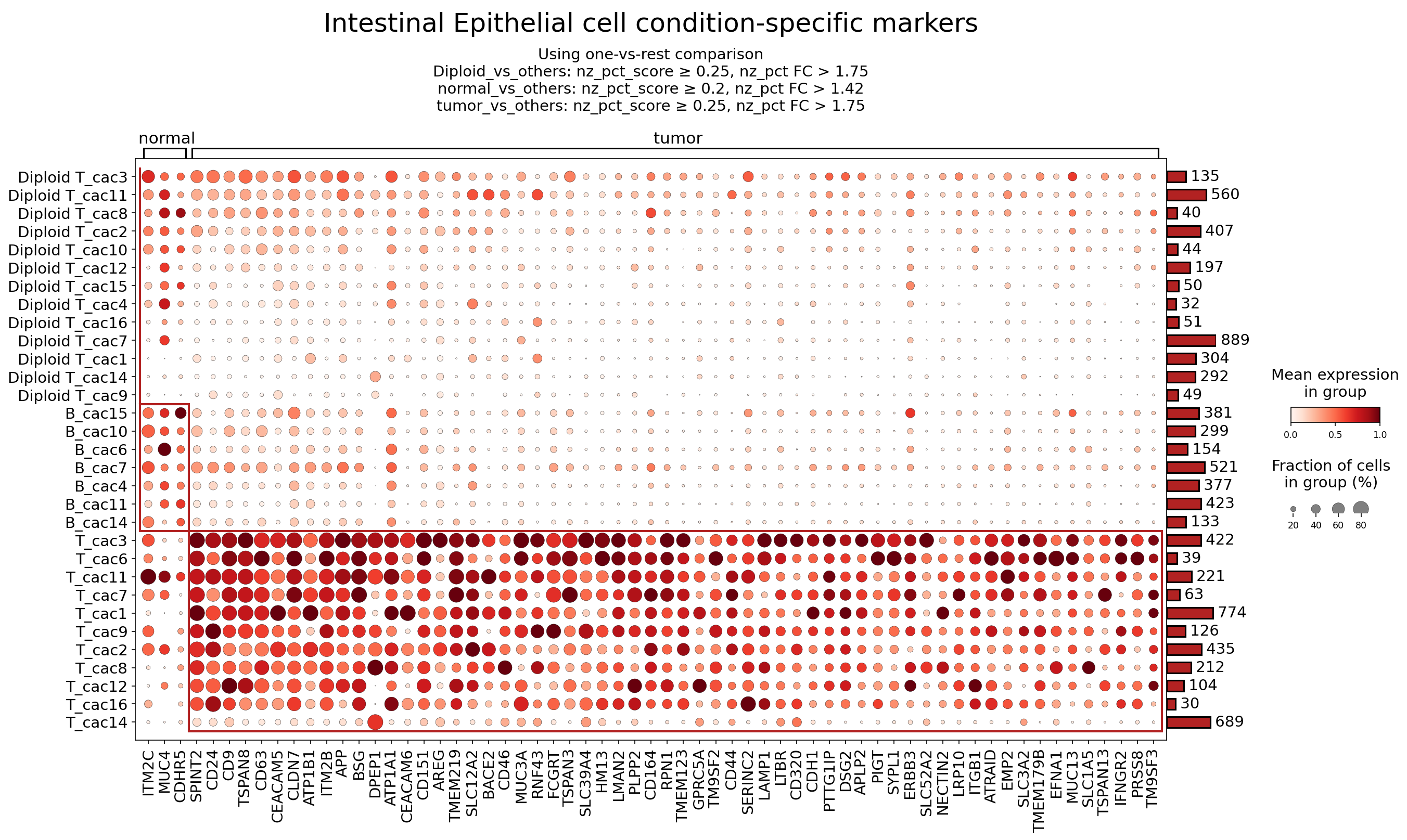
[Analysis Visualization Results]...
Analysis Overview
This analysis presents a dot plot illustrating the expression of surfaceome markers in Intestinal Epithelial cells, comparing normal and tumor conditions. The cells are further stratified by ploidy status and sub-clustering (cac groups), revealing condition-specific marker patterns. The primary goal was to identify surfaceome markers that are differentially expressed, particularly in tumor-derived Intestinal Epithelial cells, to pinpoint potential therapeutic targets or biomarkers for Colorectal Cancer. Markers were selected based on a one-vs-rest comparison with specific expression and fold change cutoffs, focusing on surfaceome-only genes, with up to 60 markers displayed per condition.
Visual Summary
The dot plot categorizes Intestinal Epithelial cell clusters into "normal" (primarily Diploid T_cacX clusters) and "tumor" (primarily T_cacX and some B_cacX clusters). Each row represents a specific cell cluster, while columns correspond to individual surfaceome marker genes.
- Normal Condition Markers: The Diploid T_cacX clusters (top rows) generally show low to moderate expression (lighter red dots) and lower fractions of expressing cells (smaller dot sizes) for most of the displayed markers. Genes like ITM2C, MUC4, and SPINT2 show some presence in these normal clusters, but their overall signal is less intense and less widespread than in tumor clusters.
- Tumor Condition Markers: A striking pattern emerges in the T_cacX clusters (bottom rows), which are associated with the tumor condition. These clusters exhibit high expression (darker red dots) and a high fraction of expressing cells (larger dot sizes) for a substantial number of surfaceome markers. The red boxes highlight regions of particularly strong and widespread expression across these tumor clusters.
- Differential Expression: Many genes, including CEACAM5, CD164, MUC13, ERBB3, CD44, BSG, DPEP1, ATP1A1, CDH1, CD24, and TSPAN8, are highly upregulated and prevalent in the tumor-associated Intestinal Epithelial cells compared to their normal counterparts, suggesting their roles in colorectal cancer progression. The strong signal for these markers across multiple tumor sub-clusters indicates a robust tumor-specific expression signature.
Biological Interpretation
The distinct expression profiles observed in tumor-associated Intestinal Epithelial cells highlight several surfaceome proteins that play crucial roles in cancer biology, particularly in the context of Colorectal Cancer (CRC). These markers can serve as potential diagnostic biomarkers, prognostic indicators, or therapeutic targets.
Here, we discuss several prominent markers highly expressed in tumor Intestinal Epithelial cells:
Established Therapeutic Targets & Key Markers
- CEACAM5 (Carcinoembryonic Antigen-related Cell Adhesion Molecule 5)
- Signaling Pathways/Mechanisms: CEACAM5 is a cell adhesion glycoprotein involved in various cellular processes including cell adhesion, cell signaling, and immune regulation. In cancer, it can promote cell proliferation, survival, and metastasis. It is often aberrantly expressed in various cancers, including CRC, where it acts as an oncofetal antigen.
- Relevance to CRC: CEACAM5 is a well-known biomarker for CRC, with elevated serum levels indicating advanced disease and recurrence. Its overexpression in tumor cells can contribute to tumor growth and invasiveness.
Therapeutic Agents:
- Established: CEACAM5 is an established target for antibody-drug conjugates (ADCs) like Sacituzumab Tirumontecan (a TROP2 ADC, but CEACAM5 is also an ADC target in development) and investigational CEACAM5-targeting ADCs (e.g., tusamitamab ravtansine) in various adenocarcinomas, including CRC PubMed Search: CEACAM5 antibody drug conjugate colorectal cancer.
- Supporting Evidence: High CEACAM5 expression is frequently observed in CRC and is associated with poorer prognosis.
- ERBB3 (Erb-B2 Receptor Tyrosine Kinase 3)
- Signaling Pathways/Mechanisms: ERBB3 is a member of the epidermal growth factor receptor (EGFR) family. It lacks intrinsic kinase activity but heterodimerizes with other EGFR family members (especially ERBB2/HER2) and activates downstream signaling pathways such as the PI3K/Akt/mTOR pathway, promoting cell growth, proliferation, and survival.
- Relevance to CRC: ERBB3 overexpression and activation are implicated in resistance to anti-EGFR therapies in CRC and contribute to tumor progression, metastasis, and recurrence. It is a critical survival signaling node in many cancers.
Therapeutic Agents:
- Established: While not a primary therapeutic target itself like EGFR or HER2, ERBB3 inhibition is a strategy to overcome resistance to other EGFR-targeted therapies. Agents targeting ERBB3 (e.g., anti-ERBB3 antibodies like patritumab deruxtecan in other cancers) are under investigation, often in combination with other agents, for various solid tumors including CRC UniProt: ERBB3.
- Supporting Evidence: ERBB3 signaling is crucial for tumor cell survival and a common mechanism of acquired resistance to targeted therapies in CRC.
- CD44 (CD44 Molecule)
- Signaling Pathways/Mechanisms: CD44 is a transmembrane glycoprotein primarily involved in cell-cell adhesion, cell-matrix interactions, and cell migration, acting as a receptor for hyaluronic acid (HA). It is also a key marker for cancer stem cells (CSCs) and plays roles in tumor initiation, progression, metastasis, and drug resistance.
- Relevance to CRC: High CD44 expression, particularly of specific splice variants (e.g., CD44v6), is associated with increased invasiveness, metastatic potential, and recurrence in CRC. It is often used as a marker to identify CRC stem cell populations.
Therapeutic Agents:
- Potential: CD44-targeted therapies, including antibodies and small molecules that block its interaction with HA or inhibit its signaling, are under preclinical and early clinical investigation for various cancers, including CRC GeneCards: CD44.
- Supporting Evidence: CD44 expression is correlated with CSC properties and poor prognosis in CRC.
Less Well-Characterized Markers with Potential as Therapeutic Targets
- MUC13 (Mucin 13, cell surface associated)
- Signaling Pathways/Mechanisms: MUC13 is a transmembrane mucin that can modulate cell adhesion, proliferation, and apoptosis. It has an intracellular domain capable of interacting with signaling molecules, potentially influencing pathways relevant to cell survival and inflammation.
- Relevance to CRC: Overexpression of MUC13 has been reported in various cancers, including CRC, where it is associated with tumor progression, metastasis, and drug resistance. It may serve as a pro-survival factor for cancer cells.
Therapeutic Potential:
- Potential: As a cell surface protein, MUC13 represents a potential target for antibody-based therapies or ADCs, particularly given its high and selective expression in tumor epithelial cells PubMed Search: MUC13 colorectal cancer therapy.
- Supporting Evidence: MUC13 promotes oncogenic processes in several gastrointestinal cancers.
- CD24 (CD24 Molecule)
- Signaling Pathways/Mechanisms: CD24 is a small, heavily glycosylated, GPI-anchored cell surface protein. It is involved in cell adhesion, migration, and anti-apoptotic signaling. In cancer, CD24 is often expressed on CSCs and can promote tumor growth, metastasis, and immune evasion by interacting with various ligands, including P-selectin and Siglecs.
- Relevance to CRC: CD24 overexpression is associated with tumor aggressiveness, metastatic potential, and poor prognosis in CRC. It is frequently identified as a CSC marker in CRC.
Therapeutic Potential:
- Potential: Given its cell surface location and association with CSCs, CD24 is being investigated as a target for antibody-mediated therapies to eliminate CSC populations in various cancers, including CRC UniProt: CD24.
- Supporting Evidence: CD24 is a prognostic factor and a functional molecule in CRC stemness.
- TSPAN8 (Tetraspanin 8)
- Signaling Pathways/Mechanisms: TSPAN8 belongs to the tetraspanin family, which acts as scaffolding proteins on the cell surface, forming complexes with other proteins (integrins, growth factor receptors) to regulate cell adhesion, migration, proliferation, and invasion.
- Relevance to CRC: TSPAN8 is often highly expressed in CRC and is linked to increased tumor cell motility, invasiveness, and metastatic dissemination. It can facilitate tumor-stromal interactions and angiogenesis.
Therapeutic Potential:
- Potential: Modulating TSPAN8 function could interfere with tumor invasion and metastasis. Antibodies targeting TSPAN8 or inhibitors that disrupt its interactions are potential therapeutic strategies under exploration GeneCards: TSPAN8.
- Supporting Evidence: TSPAN8 plays a significant role in CRC metastasis.
- BSG (Basigin / CD147)
- Signaling Pathways/Mechanisms: BSG (also known as CD147 or EMMPRIN) is a transmembrane glycoprotein that plays a key role in promoting matrix metalloproteinase (MMP) production by stromal cells, thereby facilitating extracellular matrix degradation and tumor invasion. It also influences glycolysis, angiogenesis, and cell signaling.
- Relevance to CRC: High expression of BSG is frequently observed in CRC and is associated with tumor progression, invasiveness, and poor prognosis. It can promote an aggressive tumor phenotype.
Therapeutic Potential:
- Potential: BSG is considered a promising target for anti-cancer therapy, with several anti-CD147 antibodies and small molecule inhibitors being developed to block its pro-invasive and pro-angiogenic activities UniProt: BSG.
- Supporting Evidence: BSG contributes to aggressive tumor behavior in CRC.
- ATP1A1 (ATPase Na+/K+ Transporting Subunit Alpha 1)
- Signaling Pathways/Mechanisms: ATP1A1 is the catalytic subunit of the Na+/K+-ATPase pump, which maintains ion gradients across the cell membrane. Beyond its pumping function, it acts as a signal transducer, interacting with various signaling molecules (e.g., Src, PI3K) to regulate cell proliferation, apoptosis, and differentiation.
- Relevance to CRC: Dysregulation of Na+/K+-ATPase activity and expression, particularly ATP1A1, has been observed in various cancers, including CRC, influencing tumor cell metabolism, survival, and drug resistance.
Therapeutic Potential:
- Potential: Cardiac glycosides (e.g., ouabain, digoxin), which are known inhibitors of Na+/K+-ATPase, have shown anti-cancer activity in preclinical studies and are being investigated as potential repurposed drugs or lead compounds for CRC therapy, potentially by targeting ATP1A1 PubMed Search: ATP1A1 colorectal cancer therapy.
- Supporting Evidence: ATP1A1 signaling axis can drive oncogenic processes in CRC.
Clinical or Translational Implications
The identification of these highly expressed surfaceome markers in tumor-derived Intestinal Epithelial cells has several clinical and translational implications for Colorectal Cancer:
- Diagnostic and Prognostic Biomarkers: Genes like CEACAM5, CD44, MUC13, and CD24 show strong differential expression between normal and tumor tissue, making them excellent candidates for diagnostic markers (e.g., in tissue biopsies or liquid biopsies) and prognostic indicators to predict disease aggressiveness and patient outcomes.
Therapeutic Targets:
- Established Targets: CEACAM5 and ERBB3 are part of signaling pathways already targeted in oncology, suggesting existing therapeutic modalities (e.g., ADCs, receptor tyrosine kinase inhibitors) could be leveraged or adapted for CRC. Their high expression on tumor epithelial cells makes them ideal candidates for targeted drug delivery.
- Novel/Emerging Targets: CD44, MUC13, CD24, TSPAN8, BSG, and ATP1A1 represent promising candidates for the development of novel therapies. Their roles in CSC properties, metastasis, and tumor survival suggest that targeting them could impede critical aspects of cancer progression and potentially overcome drug resistance. Further research, including antibody-drug conjugate development or small molecule inhibitors, could be pursued.
- Patient Stratification: Understanding the expression profiles of these markers could enable more precise patient stratification for clinical trials and treatment decisions. For instance, patients with high ERBB3 expression might benefit from combination therapies that block both EGFR and ERBB3 signaling.
- Resistance Mechanisms: Several of these markers (e.g., ERBB3, CD44) are implicated in mechanisms of therapeutic resistance, highlighting their potential as targets to circumvent resistance to current standard-of-care treatments in CRC.
Further validation through immunohistochemistry, *in situ* hybridization, or functional studies in patient-derived organoids or xenograft models would be essential to confirm their utility as robust biomarkers and therapeutic targets in CRC.
16. Fibroblast Condition-Specific Surfaceome Markers in Colorectal Cancer
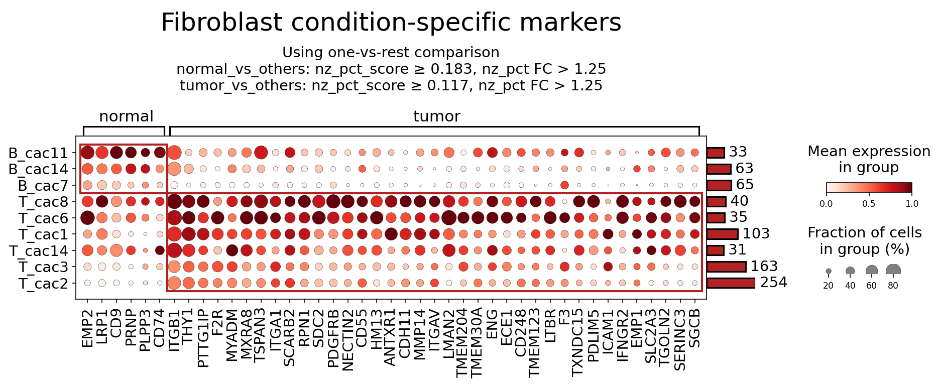
[Analysis Visualization Results]...
Analysis Overview
This analysis aimed to identify surfaceome markers expressed by Fibroblasts, comparing their expression profiles between 'normal' and 'tumor' conditions within the human Colon tissue context, highly relevant to Colorectal Cancer (CRC). Using single-cell RNA-seq data, a one-vs-rest differential expression approach was applied to identify markers specific to fibroblast subpopulations in each condition. The results, visualized as a dot plot, highlight distinct surfaceome signatures that delineate fibroblast states in health and disease, emphasizing their dynamic roles within the tumor microenvironment.
Visual Summary
The dot plot displays the mean expression (color intensity) and fraction of cells expressing (dot size) for a selection of surfaceome markers across different fibroblast clusters under 'normal' and 'tumor' conditions.
- Normal Condition Fibroblasts (B_cac clusters): Fibroblast clusters like B_cac11, B_cac14, and B_cac7 show detectable but generally lower expression levels and smaller fractions of positive cells for markers such as EMP2, LRP1, CD9, PRNP, PLPP3, and CD74. These likely represent quiescent or homeostatic fibroblasts within the normal colon tissue.
- Tumor Condition Fibroblasts (T_cac clusters): In stark contrast, fibroblast clusters T_cac8, T_cac6, T_cac1, T_cac14, T_cac3, and T_cac2, which are associated with the tumor microenvironment, exhibit a dramatic upregulation of a broad panel of surfaceome markers. Many of these markers show both high mean expression (dark red dots) and high fractions of expressing cells (large dot sizes) across multiple tumor-associated fibroblast (CAF) clusters. Prominent examples include PDGFRB, ANTXR1, CDH11, MMP14, ITGAV, ENG, F3, LTBR, ICAM1, and IFNGR2, among others like PTTG1IP, F2R, MYADM, MXRA8, TSPAN3, ITGA1, SCARB2, RPN1, SDC2, NECTIN2, CD55, HM13, LMAN2, TMEM204, TMEM30A, CD248, TMEM123, TXNDC15, PDLIM5, EMP1, SLC2A3, TGOLN2, SERINC3, and SGCB.
- Key Observation: The distinct and significantly enriched expression pattern of numerous surfaceome markers in tumor-associated fibroblasts underscores a profound phenotypic shift and activation of these cells within the colorectal cancer microenvironment compared to normal tissue. The red box visually highlights the highly active and distinct marker profile of tumor-associated fibroblasts.
Biological Interpretation
The observed differential surfaceome marker expression reflects the profound functional changes undergone by fibroblasts in the tumor microenvironment, leading to their transformation into Cancer-Associated Fibroblasts (CAFs). These CAFs are crucial orchestrators of tumor progression in Colorectal Cancer, contributing to extracellular matrix remodeling, angiogenesis, immunosuppression, and metastasis.
Selected Tumor-Associated Fibroblast Markers and their Biological Roles:
PDGFRB (Platelet-Derived Growth Factor Receptor Beta):
- Signaling Pathways: A receptor tyrosine kinase activated by PDGF ligands, which drives cell proliferation, migration, and survival through pathways such as PI3K/Akt and MAPK/ERK 1.
- Role in TME: PDGFRB is a canonical marker for CAFs and a key mediator of their activation and recruitment. Its signaling promotes desmoplasia, angiogenesis, and creates a supportive niche for tumor cells, significantly contributing to CRC progression 2.
ANTXR1 (Anthrax Toxin Receptor 1 / TEM8):
- Signaling Pathways: Functions in cell adhesion and motility, binding to collagen IV. It plays a role in VEGF-independent angiogenesis and is involved in integrin-mediated adhesion 3.
- Role in TME: Highly expressed in tumor vasculature and CAFs, ANTXR1 promotes tumor angiogenesis and growth in various cancers, including CRC, by facilitating endothelial cell migration and survival 4.
CDH11 (Cadherin-11):
- Signaling Pathways: A classic cadherin that mediates homophilic cell-cell adhesion, influencing cell migration, invasion, and epithelial-mesenchymal transition (EMT). It can interact with Wnt/β-catenin signaling 5.
- Role in TME: Upregulated in CAFs and various cancer cells, CDH11 promotes tumor cell invasion, metastasis, and CAF activation, contributing to the malignant phenotype in CRC 6.
MMP14 (Matrix Metalloproteinase 14 / MT1-MMP):
- Signaling Pathways: A membrane-bound matrix metalloproteinase that degrades extracellular matrix components (e.g., collagen, fibronectin) and activates other proteases and growth factors 7.
- Role in TME: Highly expressed by CAFs and tumor cells, MMP14 is critical for ECM remodeling, tumor cell invasion, and metastasis by creating migratory pathways and releasing pro-tumorigenic factors in the CRC microenvironment 8.
ITGAV (Integrin Alpha V):
- Signaling Pathways: Forms heterodimers with several beta integrins (e.g., ITGB1, ITGB3, ITGB5, ITGB6) to bind to various ECM proteins. Integrin signaling orchestrates cell adhesion, migration, proliferation, and survival, often through focal adhesion kinase (FAK) and Src pathways 9.
- Role in TME: Integrin αv-containing heterodimers are highly expressed by CAFs and tumor cells, playing crucial roles in cell-ECM interactions vital for tumor growth, invasion, and angiogenesis in CRC 10.
ENG (Endoglin / CD105):
- Signaling Pathways: A co-receptor for TGF-β ligands, modulating TGF-β signaling, which is central to cell proliferation, differentiation, angiogenesis, and fibrosis 11.
- Role in TME: Expressed on endothelial cells of sprouting blood vessels and on subsets of CAFs. ENG promotes pathological angiogenesis and fibrotic responses within the TME, both critical for tumor progression and metastasis in CRC 12.
F3 (Tissue Factor / CD142):
- Signaling Pathways: Initiates the extrinsic coagulation cascade but also activates PAR-2 receptor signaling, influencing cell growth, migration, and angiogenesis independently of coagulation 13.
- Role in TME: Overexpressed on tumor cells and CAFs in CRC. F3 contributes to a pro-thrombotic state and directly promotes tumor growth, angiogenesis, invasion, and metastasis, creating a procoagulant tumor microenvironment 14.
LTBR (Lymphotoxin Beta Receptor):
- Signaling Pathways: A member of the TNF receptor superfamily, activated by LIGHT and Lymphotoxin-alpha3/beta2, involved in immune cell development, inflammation, and tissue organization, often via NF-κB signaling 15.
- Role in TME: While primarily known for its role in immune cells, LTBR signaling in CAFs can influence inflammation, immune cell infiltration, and stromal remodeling within the TME, potentially contributing to or modulating tumor progression in CRC depending on context 16.
ICAM1 (Intercellular Adhesion Molecule 1 / CD54):
- Signaling Pathways: A cell surface glycoprotein that binds to integrins (LFA-1, Mac-1) on leukocytes, mediating cell-cell adhesion and leukocyte extravasation 17.
- Role in TME: Upregulated on endothelial cells, immune cells, tumor cells, and CAFs. ICAM1 plays roles in immune cell trafficking and inflammation, and can also promote tumor cell invasion and metastasis by facilitating interactions with immune and endothelial cells in CRC 18.
IFNGR2 (Interferon Gamma Receptor 2):
- Signaling Pathways: Forms a functional receptor complex with IFNGR1 to bind interferon-gamma (IFN-γ), activating the JAK/STAT pathway to mediate a wide range of immune and anti-tumor responses 19.
- Role in TME: Expression on CAFs suggests that these fibroblasts can respond to IFN-γ, influencing their phenotype and interactions within the TME. This could modulate local immune responses and tumor progression, for example, by affecting the immunosuppressive or pro-inflammatory characteristics of CAFs in CRC 20.
Clinical or Translational Implications
The identification of numerous highly expressed surfaceome markers on tumor-associated fibroblasts in Colorectal Cancer provides critical insights for developing novel therapeutic and diagnostic strategies.
Potential Therapeutic Targets and Agents:
- CAF Targeting in Colorectal Cancer: The robust upregulation of these surfaceome markers on CAFs underscores their active and supportive role in CRC progression. Targeting CAFs, or their specific signaling pathways, represents a promising strategy to disrupt the tumor-promoting microenvironment.
- PDGFRB: Receptor tyrosine kinase inhibitors (RTKIs) such as imatinib, sunitinib, or regorafenib, known to target PDGFR, could be investigated for their efficacy in suppressing PDGFRB-positive CAFs in CRC, either alone or in combination with other therapies 2.
- ANTXR1: Antibodies specifically targeting ANTXR1, such as MEDI3617, have demonstrated preclinical potential in inhibiting tumor growth and angiogenesis by disrupting ANTXR1-mediated interactions within the TME 21.
- CDH11: Given its role in tumor invasion and CAF activation, inhibitors specifically designed to block cadherin-11 function are under preclinical investigation to potentially suppress metastasis and modulate CAF activity in CRC 22.
- MMP14: While broad-spectrum MMP inhibitors have faced challenges, specific MMP14 inhibitors or antibody-based approaches could offer a more targeted strategy to disrupt ECM remodeling and inhibit tumor invasion in CRC 8.
- ITGAV: Integrin αv antagonists, such as the cyclic RGD peptide cilengitide (which targets αvβ3/αvβ5 integrins), have been explored in clinical trials for various cancers, aiming to inhibit angiogenesis and tumor cell survival by interfering with cell-ECM interactions 10.
- ENG (CD105): Endoglin-targeting antibodies like TRC105 are in clinical development, primarily for their anti-angiogenic effects by blocking TGF-β signaling and inhibiting endothelial cell proliferation. These could also impact ENG-positive CAFs 12.
- F3 (Tissue Factor): Agents targeting F3, including antibodies or inhibitors of its downstream signaling, are being investigated for their direct anti-cancer effects beyond their anticoagulant properties, aiming to disrupt tumor growth and metastasis in CRC 14.
- Biomarker Potential: These condition-specific CAF markers could serve as valuable diagnostic or prognostic biomarkers for Colorectal Cancer, indicating disease progression or aggressiveness. Furthermore, their expression levels might help predict patient response to CAF-targeted therapies or immunotherapies.
- Combination Therapies: Targeting specific CAF surface receptors and their associated signaling pathways could be combined with existing chemotherapies, radiation, or immunotherapies to remodel the TME, reduce immunosuppression, and enhance treatment efficacy in CRC.
17. T cell CD4+ Condition-Specific Surfaceome Markers in Colorectal Cancer
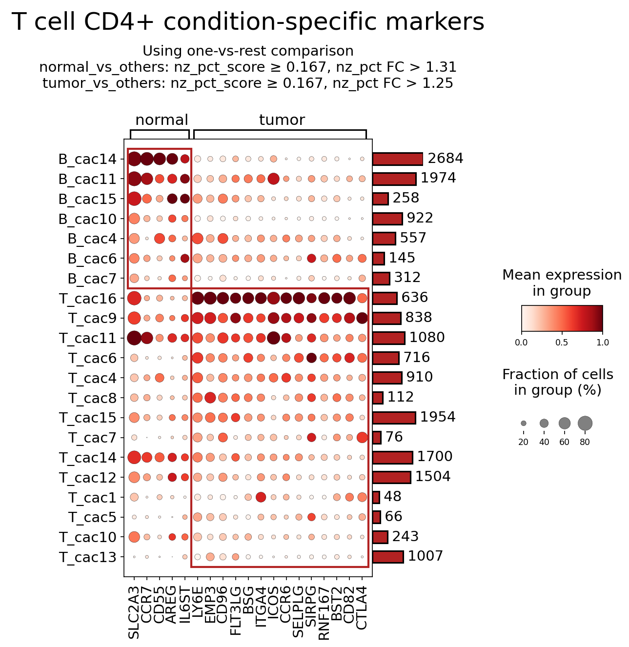
[Analysis Visualization Results]...
Analysis Overview
This analysis identifies and visualizes surfaceome markers differentially expressed in T cell CD4+ populations, comparing 'normal' and 'tumor' conditions within the colon tissue context of colorectal cancer. The dot plot illustrates the mean expression level (color intensity) and the fraction of cells expressing each marker (dot size) across various cell clusters (denoted as B_cac and T_cac clusters) under both conditions. The markers displayed were specifically identified for T cell CD4+ cells as per the query, focusing on surface proteins relevant for cell-cell interactions and therapeutic targeting.
Visual Summary
The dot plot clearly segregates markers into those predominantly expressed in normal conditions versus those enriched in tumor conditions, particularly within the T_cac (T cell-like) clusters.
- Normal-Enriched Markers: Several T_cac clusters (e.g., T_cac16, T_cac9, T_cac11, T_cac14) show high expression and prevalence of markers like SLC2A3, CCR7, and CD5 in the normal condition. These markers are largely diminished or absent in the tumor condition for these clusters. Interestingly, some B_cac clusters also express these markers in the normal state.
- Tumor-Enriched Markers: A distinct set of markers, including IL6ST, LY6E, EMP3, CD96, FLT3LG, BSG, ITGA4, ICOS, CCR6, SELPLG, SJRPG, RNF167, BST2, CD82, and CTLA4, demonstrate markedly higher expression and prevalence in almost all T_cac clusters (e.g., T_cac16, T_cac9, T_cac11, T_cac6, T_cac4, T_cac8, etc.) under tumor conditions compared to normal. This indicates a significant shift in the surface protein landscape of CD4+ T cells in the colorectal cancer microenvironment.
- Cluster-Specific Patterns: While many tumor-enriched markers are broadly expressed across tumor-associated T_cac clusters, there are subtle variations in expression intensity and prevalence among these clusters, suggesting heterogeneity within the T cell CD4+ compartment in the tumor microenvironment. For example, CTLA4 is prominently expressed across almost all T_cac clusters in the tumor.
Biological Interpretation
The differential expression of surfaceome markers in T cell CD4+ populations provides critical insights into their functional state and interaction dynamics within the colorectal cancer (CRC) tumor microenvironment (TME). T cell CD4+ cells, including helper T cells (Th1, Th2, Th17, Tfh) and regulatory T cells (Tregs), play diverse roles in anti-tumor immunity and immunosuppression.
Markers Associated with Normal T cell CD4+ State
CCR7 (C-C Motif Chemokine Receptor 7)
- Signaling Pathways: CCR7 binds chemokines CCL19 and CCL21, activating G-protein coupled receptor (GPCR) signaling pathways that regulate cell migration (e.g., Rho GTPases, PI3K/Akt, MAPK).
- Role in CRC TME: High CCR7 expression on CD4+ T cells in normal tissue typically indicates a naive or central memory phenotype, crucial for homing to secondary lymphoid organs (lymph nodes) for antigen presentation and initiation of immune responses. Its diminished expression in tumor-associated T cells suggests a loss of naive phenotype, altered trafficking, or differentiation into effector/exhausted states within the TME.
- Therapeutic Target Potential: Unlikely a direct target to enhance anti-tumor immunity in the TME, but potentially a marker for identifying functionally distinct T cell subsets.
- Relevant Therapeutic Agents: Not typically targeted in cancer, but its pathway modulation is studied in autoimmune diseases.
- Other Pertinent Information: Loss of CCR7 is often observed as T cells differentiate into effector memory phenotypes or become tissue-resident, which can be advantageous or detrimental depending on the effector function. GeneCards: CCR7
SLC2A3 (Solute Carrier Family 2 Member A3 / GLUT3)
- Signaling Pathways: SLC2A3 is a glucose transporter, regulating cellular glucose uptake. Its activity is modulated by various metabolic signals and signaling pathways involved in cell growth and metabolism (e.g., PI3K/Akt pathway).
- Role in CRC TME: High SLC2A3 expression in normal CD4+ T cells suggests active glucose metabolism, characteristic of highly proliferative or metabolically active immune cells. In the TME, metabolic competition and dysregulation are common, and shifts in glucose transporters can reflect altered T cell metabolic programming (e.g., glycolytic vs. oxidative phosphorylation) crucial for their function and survival.
- Therapeutic Target Potential: Modulating T cell metabolism is an emerging area. Targeting SLC2A3 could alter T cell metabolic fitness.
- Relevant Therapeutic Agents: No direct therapeutics targeting SLC2A3 in cancer currently, but metabolic modulators are under investigation.
- Other Pertinent Information: Glucose availability and utilization are critical for T cell activation and function. Altered SLC2A3 expression could be a biomarker of T cell metabolic state. GeneCards: SLC2A3
Markers Associated with Tumor-Associated T cell CD4+ State
CTLA4 (Cytotoxic T-Lymphocyte Associated Protein 4)
- Signaling Pathways: CTLA4 is a key immune checkpoint receptor. It binds to CD80/CD86 on antigen-presenting cells with higher affinity than CD28, leading to negative regulation of T cell activation by inhibiting IL-2 production and cell cycle progression. It also plays a critical role in the suppressive function of regulatory T cells (Tregs).
- Role in CRC TME: Upregulation of CTLA4 on T cell CD4+ populations in the tumor indicates a significant shift towards immune suppression. This is commonly observed on exhausted T cells or tumor-infiltrating Tregs, which actively dampen anti-tumor immune responses, contributing to tumor immune evasion in CRC.
- Therapeutic Target Potential: High potential. CTLA4 is a validated therapeutic target for immune checkpoint blockade.
- Relevant Therapeutic Agents: Anti-CTLA-4 monoclonal antibodies like Ipilimumab are approved for various cancers, including metastatic CRC in combination with anti-PD-1. PubMed: Ipilimumab in colorectal cancer
- Other Pertinent Information: Blocking CTLA4 can remove the inhibitory brake on T cell activation, promoting anti-tumor immunity. GeneCards: CTLA4
ICOS (Inducible T-cell COStimulator)
- Signaling Pathways: ICOS is a CD28-superfamily co-stimulatory receptor. It binds to ICOSL (ICOS ligand) on APCs and non-hematopoietic cells, activating PI3K/Akt, MAPK, and NF-κB pathways, promoting T cell proliferation, survival, and cytokine production (e.g., IL-4, IL-10, IL-17, IL-21).
- Role in CRC TME: ICOS expression on tumor-infiltrating CD4+ T cells is complex. While it can mark activated effector T cells with anti-tumor functions (e.g., Th1), it is also highly expressed on Tregs in the TME, where it sustains their suppressive function and survival, contributing to immune evasion in CRC. The specific subset of ICOS+ CD4+ T cells (effector vs. Treg) needs further characterization.
- Therapeutic Target Potential: High potential. ICOS can be targeted by agonists to boost anti-tumor immunity or by antagonists to inhibit pro-tumorigenic ICOS+ Tregs.
- Relevant Therapeutic Agents: ICOS agonists (e.g., Vopalimab) are in clinical trials for various cancers to enhance anti-tumor responses. PubMed: ICOS agonists cancer therapy
- Other Pertinent Information: ICOS is a marker of T cell activation and a key regulator of T cell-mediated immune responses, making it an attractive target for immune modulation. GeneCards: ICOS
IL6ST (Interleukin 6 Signal Transducer / gp130)
- Signaling Pathways: IL6ST is the common signal-transducing beta subunit (gp130) for receptors of the IL-6 family cytokines (e.g., IL-6, IL-11, LIF, OSM, CNTF, IL-27). It initiates downstream signaling via JAK/STAT3, MAPK, and PI3K/Akt pathways.
- Role in CRC TME: Upregulation of IL6ST on CD4+ T cells in the tumor indicates responsiveness to IL-6 family cytokines, which are often abundant in the inflammatory CRC TME. IL-6/STAT3 signaling is strongly associated with chronic inflammation, Th17 differentiation, immune suppression, and tumor cell survival/proliferation, thereby fostering tumor progression.
- Therapeutic Target Potential: Moderate potential. Targeting IL-6 signaling (receptor or cytokine) can modulate chronic inflammation and immune suppression in cancer.
- Relevant Therapeutic Agents: Antibodies against IL-6 or its receptor (e.g., Tocilizumab, Siltuximab) are used for inflammatory diseases and are explored in cancer, including CRC. PubMed: IL-6 signaling cancer therapy
- Other Pertinent Information: The IL-6/STAT3 axis is a major driver of pro-tumorigenic inflammation and immune evasion in CRC. GeneCards: IL6ST
CD96 (Cluster of Differentiation 96)
- Signaling Pathways: CD96 is an Ig superfamily receptor that binds to CD155 (PVR) and CD112 (PVRL2). Its signaling is complex and can be co-stimulatory or co-inhibitory depending on the cell type and context, often modulating PI3K and MAPK pathways.
- Role in CRC TME: In the TME, CD96 can act as an inhibitory checkpoint, contributing to T cell exhaustion and dampening anti-tumor responses. Its expression on CD4+ T cells suggests involvement in immune suppression, potentially by inhibiting effector functions.
- Therapeutic Target Potential: Emerging potential. CD96 is considered an emerging immune checkpoint.
- Relevant Therapeutic Agents: Anti-CD96 antibodies are in preclinical and early clinical development as a potential cancer immunotherapy. PubMed search: anti-CD96 cancer therapy
- Other Pertinent Information: Blocking CD96 may enhance anti-tumor immunity by releasing inhibitory signals on T cells and NK cells. GeneCards: CD96
CCR6 (C-C Motif Chemokine Receptor 6)
- Signaling Pathways: CCR6 binds to its ligand CCL20, activating GPCR signaling pathways that mediate cell migration (e.g., Rho GTPases, PI3K/Akt).
- Role in CRC TME: Upregulation of CCR6 on CD4+ T cells (especially Th17 cells and some Tregs) facilitates their recruitment to sites of inflammation and tumors, where CCL20 is often highly expressed by tumor cells or stromal cells. In CRC, Th17 cells can have pro-tumorigenic roles, promoting angiogenesis and immune suppression.
- Therapeutic Target Potential: Moderate potential. Targeting the CCR6-CCL20 axis could inhibit the recruitment of pro-tumorigenic immune cells to the CRC TME.
- Relevant Therapeutic Agents: CCR6 antagonists are under investigation in inflammatory diseases and cancer, but no approved agents currently for cancer. PubMed search: CCR6 antagonist cancer
- Other Pertinent Information: CCR6 is a marker for specific T cell subsets (e.g., Th17, some Tregs) involved in chronic inflammation and cancer. GeneCards: CCR6
BSG (Basigin / CD147)
- Signaling Pathways: BSG is a transmembrane glycoprotein involved in various cellular processes including cell adhesion, migration, proliferation, and invasion. It signals by interacting with various partners including cyclophilins, integrins, and caveolin-1, and is a key inducer of matrix metalloproteinases (MMPs).
- Role in CRC TME: High BSG expression on CD4+ T cells might indicate their altered activation state or participation in tumor-promoting interactions within the TME. BSG on tumor cells and stromal cells is known to promote tumor invasion and metastasis. Its presence on T cells could impact their function, migration, or interaction with tumor cells.
- Therapeutic Target Potential: Moderate potential. Targeting BSG could inhibit tumor progression and potentially modulate immune cell functions.
- Relevant Therapeutic Agents: Anti-CD147 antibodies and other inhibitors are under investigation for various cancers. PubMed search: anti-CD147 cancer therapy
- Other Pertinent Information: BSG is a multifaceted molecule involved in both tumor cell biology and immune cell regulation. GeneCards: BSG
Clinical or Translational Implications
The identification of these T cell CD4+ surfaceome markers has significant clinical and translational implications for colorectal cancer.
Therapeutic Targets
The marked upregulation of specific surface markers on T cell CD4+ populations in the tumor microenvironment presents several opportunities for therapeutic intervention:
- CTLA4: Its consistent high expression across tumor-associated T cell clusters strongly reinforces CTLA4 as a critical immune checkpoint in CRC. Therapeutic blockade with anti-CTLA-4 antibodies like Ipilimumab can reinvigorate anti-tumor immunity, especially in combination with anti-PD-1 therapy, offering a strategy to overcome T cell-mediated immunosuppression in CRC.
- ICOS: The dual role of ICOS (co-stimulation for effector cells vs. maintenance of Treg function) suggests a nuanced therapeutic approach. ICOS agonists aim to enhance anti-tumor effector responses, while ICOS antagonists could potentially deplete or neutralize pro-tumorigenic ICOS+ Tregs. Clinical trials are ongoing to evaluate these strategies in cancer.
- CD96: As an emerging inhibitory immune checkpoint, CD96 represents a novel target. Blocking CD96 with specific antibodies could potentially enhance anti-tumor T cell and NK cell cytotoxicity, offering a new avenue for immunotherapy in CRC.
- IL6ST: Targeting the IL-6/STAT3 signaling pathway via IL6ST (gp130) or its ligands/receptors could mitigate pro-tumorigenic inflammation and immune suppression, particularly in CRC cases characterized by high inflammatory burden. This could complement existing immunotherapies.
- CCR6: Inhibiting the CCR6-CCL20 axis could be a strategy to block the infiltration of pro-tumorigenic Th17 cells and certain Tregs into the CRC TME, thereby reducing inflammation and immunosuppression.
- BSG (CD147): Targeting BSG may offer a multi-faceted approach by not only affecting tumor cell invasion and metastasis but also potentially modulating the function of tumor-infiltrating T cells, thereby influencing the overall anti-tumor immune response.
Potential Biomarkers
The distinct expression profiles of these surfaceome markers between normal and tumor conditions suggest their utility as diagnostic, prognostic, or predictive biomarkers:
- Diagnosis/Prognosis: The presence and levels of specific tumor-enriched markers (e.g., high CTLA4, ICOS, IL6ST) on CD4+ T cells in patient biopsies could serve as indicators of disease progression, TME immunosuppression, or patient stratification for different therapeutic regimens.
- Predictive Markers for Immunotherapy: High CTLA4 expression on CD4+ T cells might predict response to anti-CTLA-4 therapies, similar to how PD-L1 expression predicts response to anti-PD-1. Further research is needed to validate this for CTLA4 and other emerging targets like ICOS and CD96 in CRC.
- Monitoring Treatment Response: Changes in the expression of these markers on circulating or tumor-infiltrating CD4+ T cells could be monitored during treatment to assess therapeutic efficacy and immune modulation.
Overall, these findings underscore the profound changes in the surface protein expression of CD4+ T cells in the colorectal cancer microenvironment, highlighting potential avenues for novel immunotherapeutic strategies and biomarker development.
18. Cell Cycle Pathway Gene Expression in Colorectal Cancer Intestinal Epithelial Cells
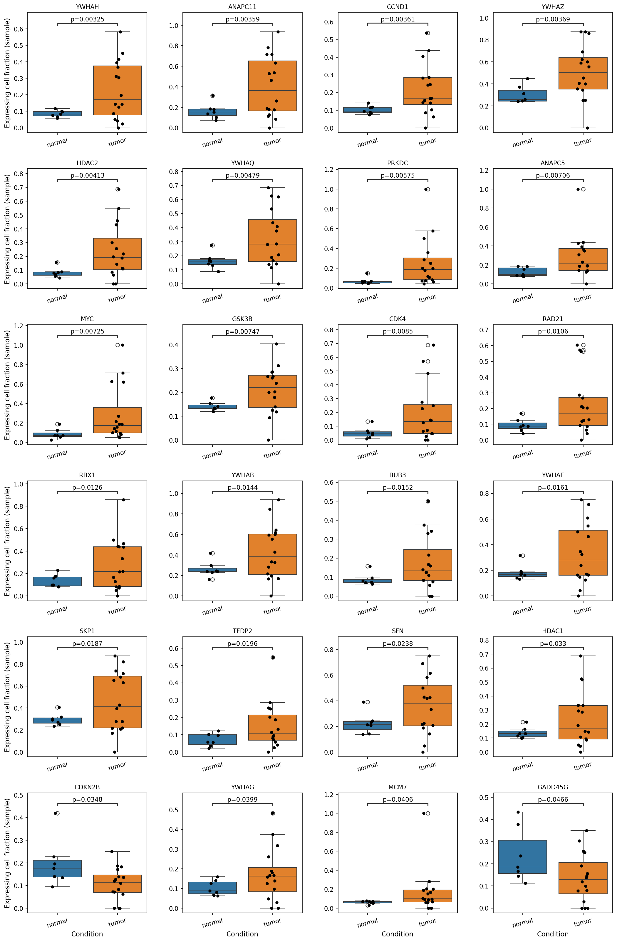
[Analysis Visualization Results]...
Analysis Overview
This analysis investigates the differential expression of a curated set of cell cycle pathway genes in Intestinal Epithelial cells, comparing cells derived from tumor tissue against those from normal colon tissue. The expression levels are represented as the fraction of cells within each sample that express a given gene. The objective is to identify cell cycle genes with statistically significant expression changes that may contribute to the progression of Colorectal Cancer (CRC).
Visual Summary
The box plots display the expressing cell fraction for 24 distinct cell cycle-related genes across normal and tumor conditions. Each black dot represents a single sample.
A predominant pattern observed is the significant upregulation of most depicted cell cycle genes in Intestinal Epithelial cells from tumor samples compared to normal samples. For instance, genes like *YWHAH*, *ANAPC11*, *CCND1*, *YWHAZ*, *HDAC2*, *MYC*, *GSK3B*, *CDK4*, *RAD21*, *MCM7*, and several other 14-3-3 family members (e.g., *YWHAQ*, *YWHAB*, *YWHA E*, *YWHA G*) show a markedly higher median expressing cell fraction and a broader distribution in tumor samples, often with p-values less than 0.01.
Conversely, a smaller subset of genes, specifically *CDKN2B* (p=0.0348) and *GADD45G* (p=0.0466), exhibit a significant downregulation in tumor Intestinal Epithelial cells. The expressing cell fraction for these genes is generally lower in tumor samples, suggesting reduced expression or fewer expressing cells compared to normal tissue.
Biological Interpretation
The observed differential expression of cell cycle genes in Intestinal Epithelial cells provides strong biological insights into the characteristics of colorectal cancer, which is inferred to originate from these cells.
- Promotion of Cell Proliferation and Cell Cycle Progression:
- Upregulation of Cyclins and CDKs: Genes like *CCND1* (Cyclin D1) and *CDK4* are key regulators of the G1-S phase transition of the cell cycle [GeneCards]. Their increased expression in tumor cells suggests an accelerated progression through the G1 phase, a hallmark of uncontrolled cell proliferation in cancer.
- Activation of Oncogenic Pathways: *MYC*, a potent proto-oncogene, is upregulated. MYC drives cell growth, proliferation, and metabolism, and its aberrant activation is a frequent event in cancer [PubMed Search]. *GSK3B* is also implicated in cell proliferation and survival, often dysregulated in cancer [GeneCards].
- Enhanced DNA Replication and Mitosis: Upregulation of genes like *MCM7* (a component of the minichromosome maintenance complex) indicates increased DNA replication activity [GeneCards]. Genes involved in the anaphase-promoting complex/cyclosome (APC/C), such as *ANAPC11* and *ANAPC5*, which are crucial for mitotic progression and exit, are also upregulated, consistent with heightened mitotic rates in tumor cells.
- DNA Repair and Genomic Instability: *PRKDC* (DNA-PKcs) and *RAD21* (a component of the cohesin complex) are involved in DNA repair and maintaining genomic integrity [GeneCards]. While often associated with repair, their upregulation can also reflect increased DNA damage inherent to rapidly dividing cancer cells, or a compensatory mechanism to handle replication stress and protect genomic stability under dysregulated cell cycle conditions.
- Role of 14-3-3 Proteins: Multiple 14-3-3 family members (*YWHAH, YWHAZ, YWHAQ, YWHAB, YWHAE, YWHAG*) are upregulated. These proteins are adaptable signaling molecules that regulate cell cycle checkpoints, apoptosis, and cellular growth [PubMed Search]. While some 14-3-3 isoforms can act as tumor suppressors (*SFN* / 14-3-3 sigma was upregulated), many are found to be overexpressed in various cancers, contributing to tumor progression, resistance to therapy, and inhibition of apoptosis, depending on the specific isoform and cellular context.
- Loss of Cell Cycle Control and Tumor Suppression:
- Downregulation of Tumor Suppressors: The significant downregulation of *CDKN2B* (p15INK4b) is particularly noteworthy. CDKN2B is a cyclin-dependent kinase inhibitor that negatively regulates the cell cycle by blocking CDK4/6 activity and inhibiting G1 progression, functioning as a potent tumor suppressor [GeneCards]. Its reduced expression removes a critical brake on cell division, contributing directly to uncontrolled proliferation.
- Evasion of Growth Arrest: *GADD45G* (Growth Arrest and DNA Damage-inducible gamma) is involved in stress responses and can induce cell cycle arrest [GeneCards]. Its downregulation suggests a compromised ability of tumor cells to respond to stress signals and induce appropriate cell cycle checkpoints or apoptosis, allowing them to evade growth control.
In summary, the gene expression profile of Intestinal Epithelial cells in tumor samples clearly indicates a shift towards enhanced proliferation, accelerated cell cycle progression, compromised DNA damage response mechanisms, and evasion of growth arrest, all characteristic features of malignant transformation and consistent with the inferred Colorectal Cancer context.
Clinical or Translational Implications
The identified cell cycle genes exhibiting significant differential expression in tumor Intestinal Epithelial cells represent potential candidates for clinical applications in Colorectal Cancer:
- Biomarkers: Genes such as *CCND1*, *MYC*, *MCM7*, and *CDKN2B* could serve as diagnostic or prognostic biomarkers. Elevated expression of pro-proliferative markers or reduced expression of tumor suppressors could indicate tumor presence, aggressiveness, or likelihood of recurrence.
- Therapeutic Targets: The upregulated genes involved in driving cell cycle progression (e.g., *CDK4*, *MYC*, *CCND1*) represent potential therapeutic targets. CDK4/6 inhibitors are already approved in other cancers (e.g., breast cancer) and could be investigated for CRC [PubMed Search]. Targeting specific components of the anaphase-promoting complex or DNA replication machinery could also hinder tumor growth.
- Understanding Treatment Resistance: The dysregulation of DNA repair genes like *PRKDC* could influence response to chemotherapy or radiation, which often work by inducing DNA damage. Understanding these dynamics might lead to strategies to overcome resistance.
- Targeting 14-3-3 Proteins: Given the broad upregulation of multiple 14-3-3 isoforms, further investigation into their specific roles in CRC could reveal novel therapeutic avenues, as these proteins are considered "chameleon" regulators and their inhibition might resensitize cancer cells to therapy or re-establish growth control [PubMed Search].
19. Gene Ontology (GSA) Analysis for Intestinal Epithelial Cells in Colon Tissue
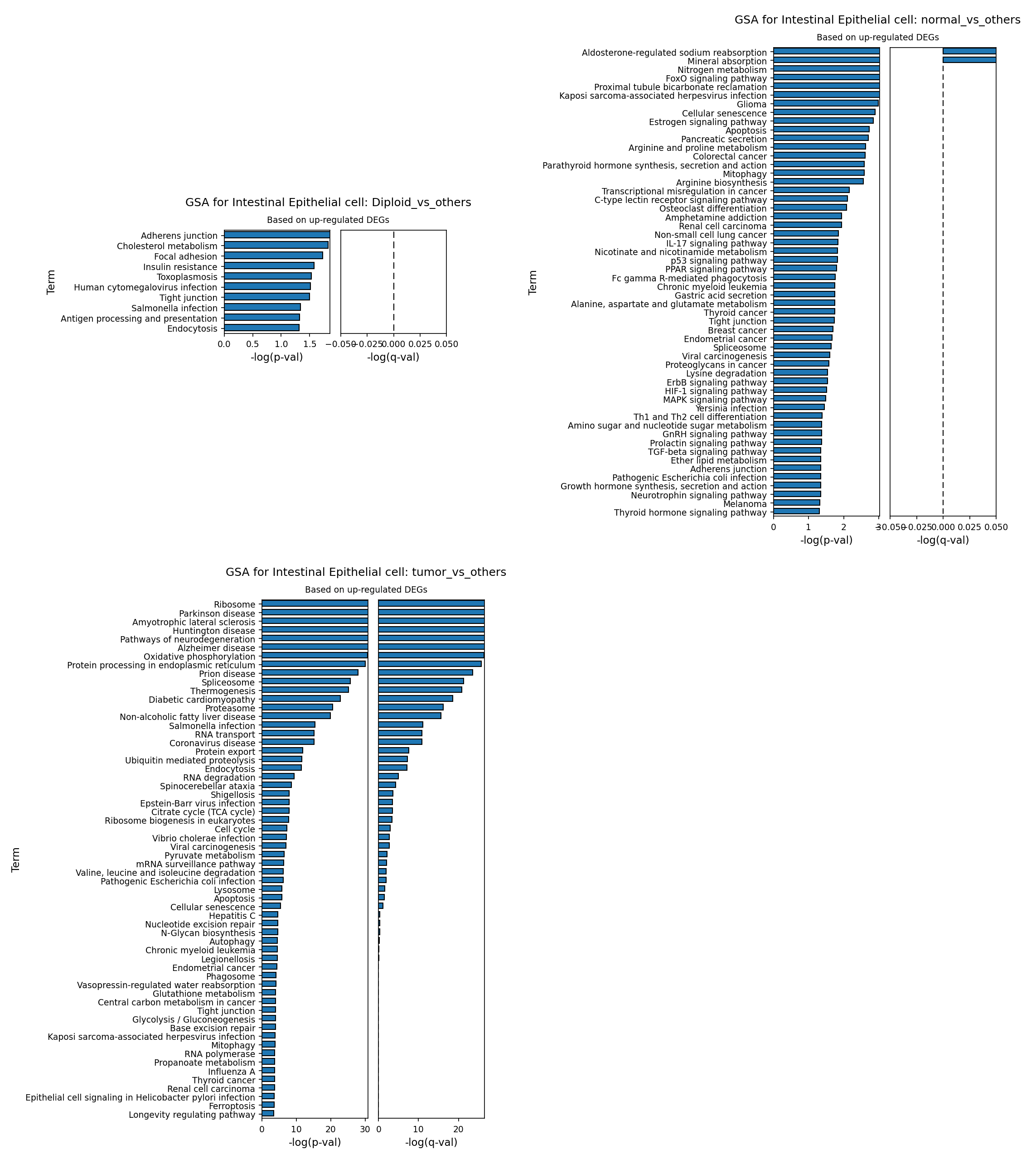
[Analysis Visualization Results]...
Analysis Overview
This analysis presents Gene Ontology (GO) enrichment results (Gene Set Analysis, GSA) for Intestinal Epithelial cells, comparing different cellular conditions based on their up-regulated differentially expressed genes (DEGs). The analysis was performed using 'go' mode, focusing on genes upregulated in the specified condition versus all other conditions in the dataset. Three comparisons are shown:
- Diploid_vs_others: Genes upregulated in diploid Intestinal Epithelial cells compared to all other Intestinal Epithelial cells (which include aneuploid and unclear ploidy states).
- normal_vs_others: Genes upregulated in Intestinal Epithelial cells from the 'normal' condition compared to all other Intestinal Epithelial cells (which include tumor cells, and potentially normal cells from other contexts if the "others" group is broad).
- tumor_vs_others: Genes upregulated in Intestinal Epithelial cells from the 'tumor' condition compared to all other Intestinal Epithelial cells (including normal cells).
The dataset context indicates human Colon tissue, with 'Intestinal Epithelial cell' identified as the 'Resolved tumor-origin cell type', strongly suggesting the study of Colorectal Cancer.
Visual Summary
The visualization consists of three bar plots, each representing GO enrichment for Intestinal Epithelial cells under a specific comparison, based on up-regulated DEGs. The x-axis shows -log(p-val) and -log(q-val) for each enriched term, indicating the statistical significance (higher values mean greater significance).
- GSA for Intestinal Epithelial cell: Diploid_vs_others: This plot shows enrichment for terms like "Adherens junction," "Cholesterol metabolism," and "Focal adhesion." However, the -log(p-val) values are relatively low (around 1.5), and crucially, the -log(q-val) values (after FDR correction) are all very close to zero, indicating that these enrichments are not statistically significant once multiple testing is accounted for.
- GSA for Intestinal Epithelial cell: normal_vs_others: This plot displays a broader range of enriched terms, including pathways related to metabolism ("Nitrogen metabolism," "Arginine and proline metabolism"), signaling ("FoxO signaling pathway," "p53 signaling pathway," "ErbB signaling pathway," "HIF-1 signaling pathway," "MAPK signaling pathway"), and organ-specific functions ("Aldosterone-regulated sodium reabsorption," "Proximal tubule bicarbonate reclamation"). While some terms show moderate -log(p-val) values, the corresponding -log(q-val) values are generally low (near zero or slightly above), suggesting that most of these enrichments, similar to the "Diploid_vs_others" comparison, do not pass stringent multiple-testing correction.
- GSA for Intestinal Epithelial cell: tumor_vs_others: In stark contrast to the other two plots, this visualization reveals a highly significant and extensive enrichment of numerous GO terms. Both -log(p-val) and -log(q-val) values are substantially higher, with many terms reaching values above 20, indicating robust statistical significance for these enrichments after FDR correction. The enriched pathways span categories such as "Ribosome," "Oxidative phosphorylation," "Protein processing in endoplasmic reticulum," "Cell cycle," "Metabolism" (e.g., "Citrate cycle (TCA cycle)," "Glycolysis / Gluconeogenesis"), and various disease-associated terms including some related to neurodegeneration, infections, and other cancers. This plot clearly indicates a profound biological shift in tumor-derived Intestinal Epithelial cells.
Biological Interpretation
The GSA results provide critical insights into the biological changes occurring in Intestinal Epithelial cells under different conditions, particularly highlighting the dramatic transformation in tumor cells.
- Basal Epithelial Cell State (Diploid and Normal Conditions): The lack of statistically significant GO term enrichment in the "Diploid_vs_others" and "normal_vs_others" comparisons (after FDR correction) suggests that while there might be subtle differences in gene expression, there aren't strong, coherent GO pathway enrichments when focusing solely on upregulated genes in these "non-tumor" contexts compared to a heterogeneous "others" group. The low-significance terms in normal cells, such as basic metabolic processes, certain signaling pathways (e.g., p53 signaling), and cell adhesion (Adherens junction, Tight junction), likely represent fundamental homeostatic functions that are typically active in healthy intestinal epithelium. The presence of 'Colorectal cancer' as a low-significance term in normal_vs_others might reflect normal physiological pathways that are dysregulated in cancer or protective mechanisms in healthy cells.
- Profound Reprogramming in Tumor Intestinal Epithelial Cells: The "tumor_vs_others" comparison reveals a highly significant and extensive set of up-regulated pathways, consistent with the known hallmarks of cancer and the identification of Intestinal Epithelial cells as the tumor-origin cell type in this colorectal cancer context.
- Enhanced Biosynthesis and Proliferation: Highly significant enrichment for "Ribosome," "Protein processing in endoplasmic reticulum," "Spliceosome," "RNA transport," "Protein export," and "Cell cycle" points to a massive upregulation of protein synthesis, processing, and cellular machinery necessary for rapid growth and proliferation characteristic of cancer cells.
- Metabolic Reprogramming: Pathways like "Oxidative phosphorylation," "Citrate cycle (TCA cycle)," "Glycolysis / Gluconeogenesis," "Central carbon metabolism in cancer," "Valine, leucine and isoleucine degradation," and "N-Glycan biosynthesis" indicate a significant shift in cellular metabolism. This often includes increased aerobic glycolysis (Warburg effect) and altered amino acid and lipid metabolism to support biomass accumulation and energy demands of proliferating cancer cells.
- Cellular Stress and Quality Control: Terms such as "Proteasome," "Ubiquitin mediated proteolysis," "Autophagy," "Apoptosis," "DNA repair" (e.g., "Nucleotide excision repair," "Base excision repair"), and "Ferroptosis" suggest active mechanisms for protein degradation, stress response, and cell death regulation, which are frequently dysregulated in cancer for survival advantage.
- Unexpected Overlap with Neurodegenerative Disease Pathways: The enrichment of terms like "Parkinson disease," "Amyotrophic lateral sclerosis," "Huntington disease," "Pathways of neurodegeneration," and "Alzheimer disease" is notable. While these are not directly related to colorectal cancer etiology, they often share underlying mechanisms involving protein misfolding, aggregation, mitochondrial dysfunction, and oxidative stress, which are also prevalent in aggressive cancers. This could indicate a general cellular stress response or dysregulation of protein quality control systems.
- Immune and Infection Response: The presence of terms related to various infections ("Salmonella infection," "Coronavirus disease," "Epstein-Barr virus infection," "Viral carcinogenesis," "Pathogenic Escherichia coli infection") suggests potential interactions with the gut microbiome or viral pathogens, or the co-option/dysregulation of host defense mechanisms by tumor cells. Chronic inflammation, often triggered by infections or immune dysregulation, is a known driver of colorectal cancer progression. PubMed search: "colorectal cancer inflammation microbiome"
- Cell Adhesion and Motility: "Tight junction" appearing with higher significance in tumor cells compared to normal could reflect altered cell-cell adhesion dynamics, which is crucial for cancer cell invasion and metastasis.
Clinical or Translational Implications
The highly significant and distinct biological pathways enriched in tumor-derived Intestinal Epithelial cells offer several potential clinical and translational implications for Colorectal Cancer:
- Therapeutic Targets: The extensive upregulation of pathways involved in protein synthesis (Ribosome), metabolism (Glycolysis, Oxidative phosphorylation, Central carbon metabolism), and cell cycle provides a rich landscape for identifying novel therapeutic targets. For instance, inhibitors of specific metabolic enzymes or components of the ribosomal machinery could selectively target rapidly proliferating cancer cells.
- Biomarker Discovery: The identified gene sets and pathways represent robust biological signatures of colorectal tumor epithelial cells. Genes within these up-regulated pathways could serve as diagnostic or prognostic biomarkers for disease detection, progression monitoring, or predicting treatment response.
- Understanding Cancer Biology: The results underscore the complex metabolic and cellular reprogramming that intestinal epithelial cells undergo during malignant transformation. The unexpected enrichment of neurodegenerative disease pathways warrants further investigation into shared molecular mechanisms of cellular stress and protein handling in cancer.
- Role of Microbiome and Inflammation: The enrichment of infection-related pathways highlights the potential role of the gut microbiome and chronic inflammation in driving colorectal cancer. This could open avenues for therapeutic interventions targeting microbiome modulation or anti-inflammatory strategies in conjunction with conventional cancer therapies. PubMed search: "colorectal cancer microbiome"
- Targeting Cell Survival and DNA Repair: Upregulation of proteasome activity and DNA repair pathways suggests that tumor cells are actively managing protein quality control and genomic instability, which are critical for their survival. Inhibiting these processes could enhance sensitivity to chemotherapy or radiation. GeneCards: Proteasome
20. Gene Set Enrichment Analysis (GSEA) of Colon Tissue: Uncovering Pathway Dynamics in Colorectal Cancer
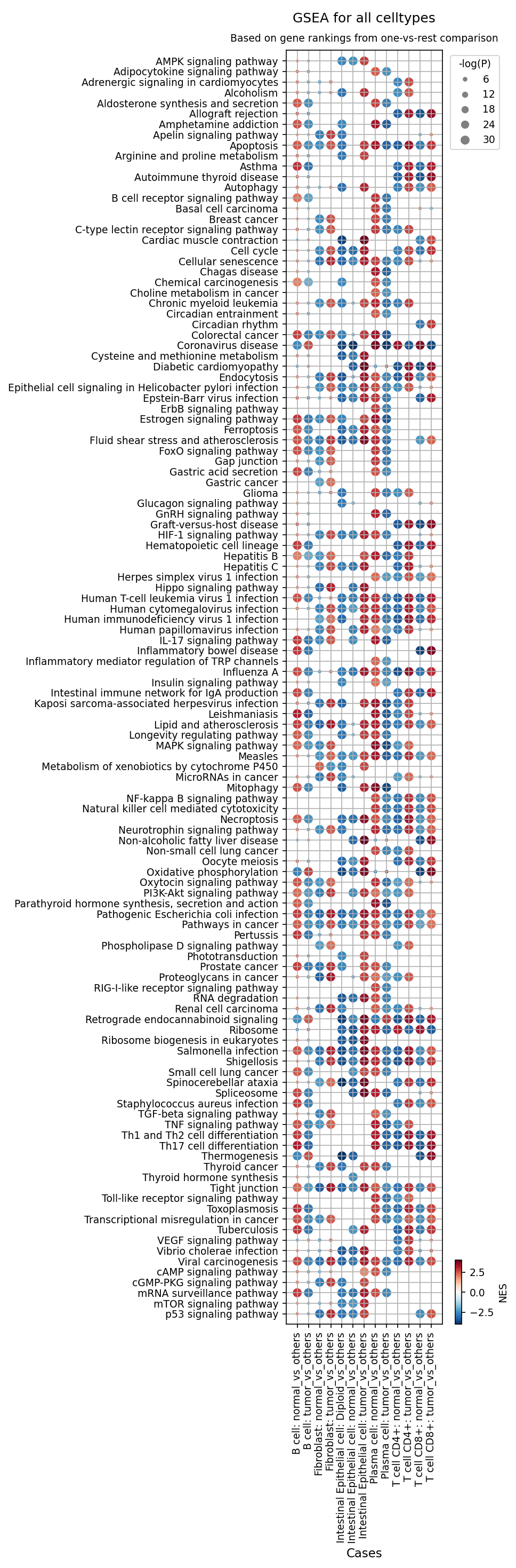
[Analysis Visualization Results]...
Analysis Overview
This analysis presents Gene Set Enrichment Analysis (GSEA) results across various cell types found in colon tissue, comparing conditions like "normal" vs. "others," "tumor" vs. "others," and "Diploid" vs. "others" for Intestinal Epithelial cells. The dot plot visualizes the Normalized Enrichment Score (NES) and statistical significance (-log(P)) for a wide array of gene sets (pathways/biological processes). The inferred biological context is Colorectal Cancer, with Intestinal Epithelial cells identified as the tumor-origin cell type. This analysis aims to highlight key cellular processes and pathways dysregulated in tumor and adjacent microenvironments.
Visual Summary
The dot plot effectively displays the GSEA results, with each row representing a gene set and each column representing a specific cell type and comparison condition.
- Color Scale: The color of each dot indicates the Normalized Enrichment Score (NES), ranging from dark blue (highly negatively enriched, i.e., downregulated) to dark red (highly positively enriched, i.e., upregulated). The RdBu_r colormap is used.
- Dot Size: The size of each dot corresponds to the negative logarithm of the p-value (-log(P)), with larger dots indicating greater statistical significance. Only statistically significant enrichments (p-val < 0.05, NES cutoff 1) are shown, as implied by the tool parameters.
Key Observations:
- Intestinal Epithelial Cells (Tumor): Show widespread positive enrichment (red, large dots) for cancer-related pathways such as "Pathways in cancer," "Colorectal cancer," "Cell cycle," and crucial signaling pathways like PI3K-Akt, HIF-1, ErbB, MAPK, mTOR, and p53 signaling pathways. Conversely, pathways related to normal epithelial function, like "Intestinal immune network for IgA production," are strongly negatively enriched (blue, large dots).
- Intestinal Epithelial Cells (Diploid): Even diploid epithelial cells within the tumor context exhibit enrichment for "Colorectal cancer," "Pathways in cancer," "Cell cycle," and key oncogenic signaling pathways (HIF-1, PI3K-Akt, p53), suggesting their active participation in or response to the tumor environment.
- Fibroblasts (Tumor): Exhibit strong positive enrichment for pathways critical in tumor microenvironment remodeling and angiogenesis, including "HIF-1 signaling pathway," "PI3K-Akt signaling pathway," "VEGF signaling pathway," and "TGF-beta signaling pathway."
- Immune Cells (B cell, Plasma cell, T cell CD4+, T cell CD8+ in Tumor): These populations display a complex pattern, with enrichments for various inflammatory responses (e.g., "IL-17 signaling pathway," "TNF signaling pathway," "Inflammatory bowel disease") and a notable number of viral infection-related pathways. This suggests an altered and potentially dysregulated immune landscape within the tumor.
- Normal Cells: Generally show an inverse pattern to their tumor counterparts, with cancer pathways being negatively enriched or not significantly enriched, and normal physiological pathways potentially being preserved or positively enriched in normal_vs_others comparisons.
Biological Interpretation
The GSEA results provide clear biological insights into the cellular and molecular landscape of Colorectal Cancer:
- Tumor Cell Intrinsic Pathways: The strong enrichment of "Colorectal cancer," "Pathways in cancer," "Cell cycle," and multiple oncogenic signaling cascades (e.g., PI3K-Akt, HIF-1, ErbB, MAPK, mTOR, p53) in Intestinal Epithelial cell: tumor_vs_others confirms the epithelial cells as the primary neoplastic component. These pathways drive uncontrolled proliferation, survival, altered metabolism, and resistance to apoptosis, which are hallmarks of cancer development and progression PubMed search: hallmarks of cancer signaling pathways. The depletion of "Intestinal immune network for IgA production" highlights a loss of normal epithelial differentiation and barrier function in tumor cells.
- Role of Diploid Epithelial Cells: The observation that Intestinal Epithelial cell: Diploid_vs_others also shows enrichment in "Colorectal cancer," "Pathways in cancer," and associated oncogenic signaling pathways is crucial. This suggests that not all tumor-associated epithelial cells are aneuploid, or that even diploid epithelial cells within the tumor microenvironment are undergoing malignant transformation or are significantly influenced by tumor-promoting signals. These could represent early transforming cells or a specific subset of tumor cells.
- Cancer-Associated Fibroblasts (CAFs): The robust enrichment of "HIF-1 signaling pathway," "PI3K-Akt signaling pathway," "VEGF signaling pathway," and "TGF-beta signaling pathway" in Fibroblast: tumor_vs_others strongly implicates these cells as activated CAFs. CAFs are known to play critical roles in remodeling the extracellular matrix, promoting angiogenesis, fostering immune evasion, and contributing to tumor growth and metastasis in CRC PubMed search: cancer associated fibroblasts colorectal cancer.
Immune Microenvironment Remodeling:
- B cells: Enrichment of "B cell receptor signaling pathway" and various viral infection pathways in tumor B cells suggests an active, potentially chronic, inflammatory or pathogen-driven B cell response, which can be pro-tumorigenic (e.g., through B regulatory cells) or anti-tumorigenic depending on the context in CRC PubMed search: B cells colorectal cancer immunology.
- Plasma cells: Depletion of "Intestinal immune network for IgA production" suggests a shift away from protective IgA immunity in the tumor, possibly contributing to immune evasion.
- T cells (CD4+ and CD8+): The enrichment of "Th1 and Th2 cell differentiation," "Th17 cell differentiation," "IL-17 signaling pathway," and "TNF signaling pathway" points to a highly inflammatory T cell response in the tumor microenvironment. While some of these pathways can be anti-tumorigenic (Th1), others (Th17, IL-17) can be pro-tumorigenic in CRC, highlighting a complex and potentially dysregulated immune response rather than an effective anti-tumor immunity PubMed search: T cell subsets colorectal cancer tumor microenvironment. The frequent enrichment of "Apoptosis" in both T cell subsets within the tumor context could indicate T cell exhaustion or activation-induced cell death, further hindering effective anti-tumor immunity.
Clinical or Translational Implications
The GSEA results provide several potential clinical and translational implications for Colorectal Cancer:
- Therapeutic Targets: The consistent upregulation of specific oncogenic signaling pathways (e.g., PI3K-Akt, MAPK, HIF-1, ErbB) in tumor-originating Intestinal Epithelial cells and supportive fibroblasts suggests these pathways as promising therapeutic targets for drug development or repurposing.
- Biomarkers of Disease Progression: The distinct pathway enrichment patterns in diploid vs. aneuploid epithelial cells could offer insights into different stages of tumor progression or subsets of tumor cells with varying malignant potential. This might lead to identifying novel biomarkers for early detection or predicting treatment response.
- Immunotherapy Strategies: The complex and often dysregulated immune responses observed in B cells, Plasma cells, and T cells within the tumor microenvironment (e.g., inflammatory pathways, viral response, T cell apoptosis/exhaustion) highlight the need for nuanced immunotherapy approaches. Understanding the specific functional states of these immune cells could guide the development of therapies that re-educate or reactivate anti-tumor immunity in CRC. For instance, addressing T cell exhaustion or modulating pro-tumorigenic inflammatory pathways could enhance therapeutic efficacy.
- Stromal Targeting: The clear role of CAFs, indicated by the enrichment of pathways like TGF-beta and VEGF signaling, reinforces the importance of targeting the tumor stroma to inhibit tumor growth, metastasis, and angiogenesis.
- Risk Stratification: Differential pathway activation patterns across various cell types could contribute to a more precise risk stratification of CRC patients, potentially identifying those who would benefit most from specific targeted therapies or intensified surveillance.
21. Discussion
The single-cell analyses reveal a profoundly altered landscape in Colorectal Cancer (CRC), extending beyond the malignant epithelial cells to encompass significant remodeling of the tumor microenvironment (TME). Intestinal Epithelial cells, confirmed as the tumor origin and largely aneuploid, exhibit a hyperactive cell cycle with pronounced upregulation of genes like CCND1, CDK4, and MYC, alongside downregulation of tumor suppressors such as CDKN2B and GADD45G. This intrinsic proliferative drive is further supported by broad enrichment of pathways related to protein synthesis, metabolism, and cell cycle, with unexpected overlaps with neurodegenerative disease pathways, potentially reflecting shared cellular stress responses. These malignant epithelial cells also display a distinct surfaceome signature, including high expression of CEACAM5, ERBB3, CD44, and CD147 (BSG), underscoring their aggressive phenotype and potential for targeted intervention.
The immune compartment of the CRC TME is heavily skewed towards immune suppression. We observe a dramatic increase in T cell (CD8 Exhausted) and T cell (Treg) populations, coupled with high expression of immune checkpoints like CTLA4 and ICOS on tumor-infiltrating CD4+ T cells, indicating widespread immune evasion. Macrophages show a statistically significant shift towards pro-tumorigenic Mac (M2B) and Mac (M2D) phenotypes, despite some samples maintaining a high proportion of Mac (M1) cells, suggesting a complex and heterogeneous macrophage polarization state that ultimately favors tumor progression. The stromal compartment is characterized by expanded and activated Cancer-Associated Fibroblasts (CAFs), which upregulate a suite of surface markers such as PDGFRB, ANTXR1, CDH11, and MMP14, indicative of their active role in extracellular matrix remodeling, angiogenesis, and supporting tumor growth.
Cell-cell interaction analyses reveal a striking disruption of normal epithelial-immune/stromal crosstalk in the tumor, replaced by specific pro-tumorigenic interactions. Notable tumor-specific communications include CEACAM5/6-mediated interactions between aneuploid epithelial cells and immune/epithelial cells, CD147 (PPIA_BSG) interactions with macrophages, and PGE2 signaling influencing T CD4+ cells. Additionally, elevated CD93_IFNGR1 (macrophage-macrophage) and CD86_CD28 (macrophage-T CD4+) interactions in the tumor suggest altered macrophage activity and T cell co-stimulation dynamics that likely contribute to an immunosuppressive environment. Together, these findings highlight a multi-faceted and highly dysregulated TME that actively promotes CRC progression and offers numerous points of therapeutic vulnerability across malignant, stromal, and immune cell types.
Hypotheses:
- Aberrant activation of PI3K-Akt, MAPK, HIF-1, ErbB, mTOR, and p53 signaling pathways in malignant Intestinal Epithelial cells drives uncontrolled proliferation, survival, and metabolic reprogramming in Colorectal Cancer.
- The expansion of exhausted CD8+ T cells, regulatory T cells, and pro-tumorigenic M2B/M2D macrophages creates an immunosuppressive tumor microenvironment that enables immune evasion in Colorectal Cancer.
- Cancer-associated fibroblasts (CAFs), identified by distinct surface markers and activated oncogenic pathways, actively remodel the extracellular matrix and promote angiogenesis, thus facilitating tumor growth and metastasis in Colorectal Cancer.
- Specific tumor-associated cell-cell interactions (e.g., CEACAM5/6, CD147, PGE2, CD93, CD86-CD28) involving malignant epithelial cells, immune cells, and stromal cells orchestrate a pro-tumorigenic and immunosuppressive crosstalk, contributing to Colorectal Cancer progression.
Potential therapeutic targets:
- CEACAM5: Highly overexpressed on malignant Intestinal Epithelial cells in CRC, promotes proliferation, survival, invasion, and immune evasion. Acts as a key oncofetal antigen. Evidence: Section 13: CEACAM5_CEACAM6 interactions are highly frequent and strong between aneuploid Intestinal Epithelial cells and various immune cells. Section 15: High surface expression on tumor Intestinal Epithelial cells; established biomarker and therapeutic target. Validation: Validate CEACAM5 expression by IHC in CRC patient tissues. Test anti-CEACAM5 antibody-drug conjugates (e.g., tusamitamab ravtansine) in patient-derived organoids or xenograft models to assess tumor growth inhibition and selectivity.
- ERBB3: Overexpressed on malignant Intestinal Epithelial cells, mediates PI3K/Akt signaling, and implicated in resistance to anti-EGFR therapies, driving cell growth and survival. Evidence: Section 4: ERBB2 amplification (a heterodimerization partner). Section 15: High surface expression on tumor Intestinal Epithelial cells. Section 20: GSEA shows PI3K-Akt and ErbB signaling pathways are strongly upregulated in tumor epithelial cells. Validation: Confirm ERBB3 expression by IHC in CRC biopsies. Test anti-ERBB3 antibodies (e.g., patritumab deruxtecan) in CRC cell lines and patient-derived organoids, especially those resistant to anti-EGFR therapy, to assess synergistic effects on tumor proliferation and survival.
- CD147 (BSG): Overexpressed on malignant Intestinal Epithelial cells and potentially CD4+ T cells. It stimulates matrix metalloproteinase (MMP) production, promoting tumor invasion, angiogenesis, and can modulate immune cell function. Evidence: Section 13: PPIA_BSG interaction is robustly observed between aneuploid Intestinal Epithelial cells and macrophages/plasma cells. Section 15: High surface expression on tumor Intestinal Epithelial cells. Section 17: Detected on CD4+ T cells in tumor conditions. Validation: Validate CD147 expression by IHC in CRC tissues and on immune subsets (e.g., T cells, macrophages) by flow cytometry. Test anti-CD147 antibodies or small molecule inhibitors in preclinical models to assess impact on invasion, metastasis, angiogenesis, and immune cell functions.
- CTLA4: A key immune checkpoint receptor, highly upregulated on CD4+ T cells (including regulatory T cells) in the tumor microenvironment, contributing significantly to immune suppression and T cell exhaustion. Evidence: Section 8: Statistically significant increase in Treg and CD8 Exhausted T cell populations in tumor. Section 17: High surface expression across almost all tumor-associated CD4+ T cell clusters. Validation: Validate CTLA4 expression on tumor-infiltrating lymphocytes by flow cytometry/IHC. Clinically, anti-CTLA4 antibodies (e.g., Ipilimumab) are approved; further investigate patient stratification and combination strategies based on CTLA4 expression and T cell subset dynamics.
- PDGFRB: A canonical marker for Cancer-Associated Fibroblasts (CAFs), mediating CAF activation, recruitment, desmoplasia, and angiogenesis, thus significantly contributing to CRC progression. Evidence: Section 16: High surface expression (PDGFRB) on tumor-associated fibroblasts, contrasting with normal fibroblasts. Section 20: GSEA shows HIF-1, PI3K-Akt, VEGF, and TGF-beta signaling pathways are strongly upregulated in tumor fibroblasts. Validation: Confirm PDGFRB expression on CAFs by IHC/immunofluorescence in CRC tissues. Test PDGFR inhibitors (e.g., imatinib, sunitinib, regorafenib) in preclinical models of CRC (e.g., co-culture with CAFs, xenografts) to assess reduction in CAF activation, stromal remodeling, and tumor growth.
- CDK4/6: CDK4, along with Cyclin D1 (CCND1), is significantly upregulated in malignant Intestinal Epithelial cells, driving uncontrolled G1-S phase progression, a hallmark of cancer proliferation. CDKN2B, an inhibitor, is downregulated. Evidence: Section 18: CDK4 and CCND1 are significantly upregulated in tumor Intestinal Epithelial cells, while CDKN2B (p15INK4b), a negative regulator of CDK4/6, is significantly downregulated. Section 20: 'Cell cycle' pathway is highly enriched in tumor epithelial cells in GSEA. Validation: Validate CDK4/CCND1 expression in CRC by IHC. Test CDK4/6 inhibitors (e.g., palbociclib, ribociclib, abemaciclib) in CRC cell lines and patient-derived organoids, potentially in combination with other agents, and assess impact on proliferation and cell cycle arrest.
Follow-up validation ideas:
- Flow Cytometry/Immunostaining: Validate T cell exhaustion (CTLA4, ICOS, PD-1 on CD8/CD4 T cells), Treg infiltration (FOXP3, CD25 on CD4 T cells), macrophage polarization markers (M2B/M2D markers), and CAF markers (PDGFRB) in human CRC tissue cohorts using immunohistochemistry, immunofluorescence, or multiparametric flow cytometry.
- Spatial Transcriptomics/Proteomics: Map the spatial distribution and interaction niches of malignant epithelial cells, CAFs, and distinct immune subsets (exhausted T cells, Tregs, M2 macrophages) in human CRC to confirm predicted cell-cell interaction patterns and their spatial relevance.
- In vitro/in vivo Perturbation Assays: Use 3D organoid cultures (patient-derived Intestinal Epithelial cells), co-culture models, or xenograft models to functionally validate key identified therapeutic targets (e.g., CEACAM5, CD147, PDGFRB, CTLA4, ICOS, MMP14, CDK4/6) by genetic knockdown/CRISPR or pharmacologic inhibition and assess their impact on tumor growth, immune evasion, and TME remodeling.
- Single-cell Multi-omics: Perform combined single-cell RNA-seq with CITE-seq (surface protein expression) or ATAC-seq (chromatin accessibility) on fresh CRC samples to further validate surface marker expression, T cell states, and regulatory landscapes.
- Clinical Cohort Validation: Validate prognostic and predictive biomarker potential of identified cell type proportions (Treg, exhausted CD8 T cells, M2 macrophages) or gene expression signatures (e.g., cell cycle genes, CAF markers) in independent, larger CRC patient cohorts with associated clinical outcomes and treatment responses.
Limitations:
This report is based on single-cell RNA-sequencing data, providing a snapshot of cellular states and interactions rather than dynamic processes. Inferences regarding CNVs, ploidy, and cell type annotations are computational and not clinical diagnoses, requiring further validation. A notable 'unassigned' cell population in tumor samples warrants future characterization. Predicted cell-cell interactions are computational and require experimental validation. Gene Set Analysis and Gene Set Enrichment Analysis highlight pathway correlations but do not establish direct causation. The observed heterogeneity across tumor samples suggests that findings may not generalize universally, emphasizing the need for personalized approaches. This analysis did not include direct functional assays, limiting definitive conclusions about cause-and-effect relationships.
22. Query List
- Show and save UMAPs for condition, sample, major cell type, minor cell type, ploidy_dec, and celltype_subset in two columns.
- Show major cell type scores on UMAP and save the result.
- Show and save a marker expression dot plot for celltype_subset. SET target_cell = None and var_group_rotation = 45. Leave all other arguments at their default values.
- Select Intestinal Epithelial cell as tumor-origin cells, show a CNV heatmap grouped by sample together with a summary of regions with significantly amplified copy numbers, and save the results.
- Show and save UMAPs of CNV patterns colored by major cell type, minor cell type, ploidy results, condition, and sample in two columns.
- Show and save a population bar plot of minor cell types.
- Show and save a population bar plot of T cell subsets.
- Show and save box plots of T cell subset populations with statistically significant differences between conditions, if any. Choose ncols based on the total number of panels.
- Show and save a population bar plot of macrophage subsets.
- Show and save box plots of macrophage subset populations with statistically significant differences between conditions, if any. Choose ncols based on the total number of panels.
- Select Intestinal Epithelial cell as tumor-origin cells, show their ploidy populations as a bar plot, and save the result.
- Show and save cell-cell interaction patterns involving Intestinal Epithelial cell, Fibroblast, Macrophage, T cell, and other relevant cell types. Select at most 80 cell-cell interactions per group.
- Find cell-cell interactions involving Intestinal Epithelial cell, major immune and stromal cells that differ significantly between conditions, show them as a dot plot, and save the result. Set max_n_items_per_group to 60. For each interaction identified under each condition, using the provided dataset context, describe the associated signaling pathways and mechanisms, its potential as a therapeutic target, and relevant therapeutic agents.
- Find cell-cell interactions containing immune checkpoint and cell cycle pathway genes and involving Intestinal Epithelial cell, major immune and stromal cells that differ significantly between conditions, show them as a dot plot, and save the result. Set max_n_items_per_group to 60. For each interaction identified under each condition, using the provided dataset context, describe the associated signaling pathways and mechanisms, its potential as a therapeutic target, and relevant therapeutic agents.
- Show marker expressions for Intestinal Epithelial cell in a dot plot, and save the result. Use only surfaceome markers, with at most 60 markers per condition. For the markers identified under each condition, distinguish between established therapeutic targets and less well-characterized markers with potential as therapeutic targets. For each marker, using the provided dataset context, describe the associated signaling pathways and mechanisms, relevant therapeutic agents, and supporting evidence.
- Show marker expressions for Fibroblast in a dot plot, and save the result. Use only surfaceome markers, with at most 40 markers per condition. For each marker identified under each condition, using the provided dataset context, describe the associated signaling pathways, the role of the corresponding cell type in the tumor microenvironment, the marker’s potential as a therapeutic target, relevant therapeutic agents, and other pertinent information.
- Show marker expressions for T cell CD4+ in a dot plot, and save the result. Use only surfaceome markers, with at most 40 markers per condition. For each marker identified under each condition, using the provided dataset context, describe the associated signaling pathways, the role of the corresponding cell type in the tumor microenvironment, the marker’s potential as a therapeutic target, relevant therapeutic agents, and other pertinent information.
- Among cell cycle pathway genes, select those with statistically significant expression differences between conditions in Intestinal Epithelial cell, show box plots, and save the result. Set max_n_items_to_plot to 24 and choose ncols based on the total number of panels for an overall width-to-height ratio of approximately 2:3.
- Show and save Gene Ontology (GSA) analysis results for Intestinal Epithelial cell as a bar plot.
- Show Gene Set Enrichment Analysis results as a dot plot, and save it. Set the color map to RdBu_r, n_pws_to_show to 120, and target_cells to None.