Single-Cell Transcriptomic and Genomic Landscape of Human Colorectal Cancer Reveals Key Drivers of Tumor Progression and Immune Evasion
This report details a single-cell analysis of human colon tissue, contrasting tumor samples with adjacent normal tissue. We identify significant shifts in cell population composition, characterized by an expansion of aneuploid intestinal epithelial cells and a distinct immune microenvironment. Key findings include widespread genomic instability, altered cell-cell interactions, and dysregulated gene expression pathways driving tumor proliferation and immune suppression, offering critical insights into colorectal cancer pathobiology.
Contents
- Dataset overview
- Single-cell UMAP Embedding Exploration of Colon Tissue
- Major Cell Type Score and Ploidy Distribution on UMAP
- Overall Celltype_subset Marker Expression Dot Plot Interpretation
- Copy Number Variation (CNV) Analysis in Intestinal Epithelial and Unassigned Cells
- CNV 기반 UMAP 분석을 통한 세포 유형, 이수성, 및 조건별 패턴 시각화
- Colon Tissue Minor Cell Type Population Analysis in Tumor vs. Adjacent Normal Conditions
- T 세포 아형 인구 분포 분석: 인접 정상 조직 및 종양 조직 비교
- Colon Tumor Microenvironment Shows Significant Shifts in T cell and Innate Lymphoid Cell Subpopulations
- Macrophage Subset Population Shifts in Colon Tumor Microenvironment
- Macrophage Subset Population Shifts in Colorectal Tumor Microenvironment
- Ploidy Population Analysis in Intestinal Epithelial and Unassigned Cells
- Colon Cancer Microenvironment: Cell-Cell Interaction Analysis by Condition
- Colon Tumor Microenvironment Cell-Cell Interaction Analysis
- Cell-Cell Interaction Analysis: Immune Checkpoint and Cell Cycle Pathways in Colon Tissue
- Differential Cell-Cell Interaction Patterns in Colon Tumor vs. Adjacent Normal Tissue
- Intestinal Epithelial Cell Condition-Specific Surfaceome Markers in Colon Cancer
- Macrophage Condition-Specific Surface Markers in Colon Tissue
- Fibroblast Condition-Specific Surfaceome Markers in Colon Tissue
- CD4+ T Cell Condition-Specific Surfaceome Markers in Colon Tissue
- Dysregulation of Cell Cycle Pathways in Intestinal Epithelial Cells from Colon Tumor Tissue
- 장상피세포의 조건 및 배수성 상태에 따른 유전자 온톨로지(GSA) 분석 결과
- Gene Set Enrichment Analysis (GSEA) in Colon Tumor Microenvironment
- Discussion
- Query List
0. Dataset overview
Dataset Summary
- 이 데이터는 63689개의 세포와 23387개의 유전자로 구성된 단일 세포 RNA-seq 데이터입니다.
- 주요 관측 (obs) 열에는 환자 정보, 조건 (종양, 인접 정상 조직), 샘플, 세포 타입 (celltype_major, celltype_minor, celltype_subset), 플로이디 (ploidy_dec: 이수성/정상), 및 다양한 클러스터링 정보가 포함되어 있습니다.
- 유전자 (var) 열에는 고변이 유전자, 유전자 ID, 유전자 이름, 염색체, 세포유전학적 밴드 정보 등이 있습니다.
- 데이터는 사람의 결장(Colon) 조직에서 유래했습니다.
- 주요 조건은 'Tumor'와 'Adj_normal'이며, DEG, GSEA, GSA_up 분석의 참조 조건은 'Adj_normal'입니다.
- 주요 세포 타입은 Intestinal Epithelial cell, Stromal cell, Endothelial cell, B cell, Enteric neuron, Myeloid cell, T cell, unassigned, Mast cell 입니다.
- 종양 기원 세포 타입은 'Intestinal Epithelial cell'로 확인되었습니다.
- 세포-세포 상호작용 (CCI), 차등 발현 유전자 (DEG), 유전자 세트 농축 분석 (GSEA), 유전자 온톨로지 (GSA) 결과가 사전 계산되어 저장되어 있습니다.
1. Single-cell UMAP Embedding Exploration of Colon Tissue
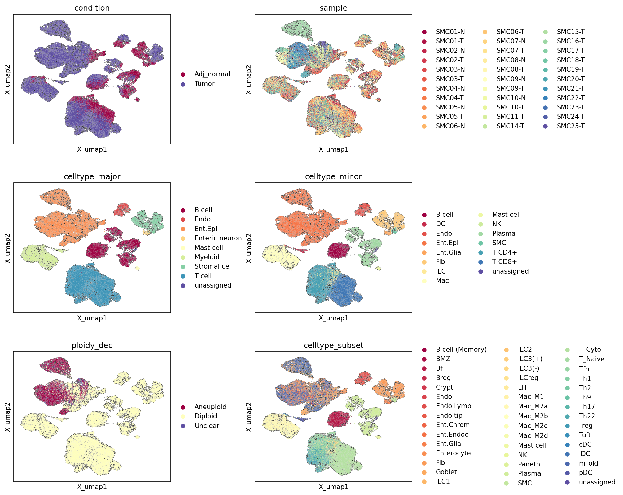
[Analysis Visualization Results]...
Analysis Overview
본 분석은 단일 세포 RNA 시퀀싱(scRNA-seq) 데이터를 기반으로 한 UMAP 임베딩을 통해, 대장 조직 내 세포들의 이질성 및 조건(종양 vs. 인접 정상), 샘플, 주요 세포 유형, 세부 세포 유형, 그리고 세포 핵형(ploidy)에 따른 분포를 시각화하고 해석합니다. 63,689개의 세포와 23,387개의 유전자를 포함하는 AnnData 객체를 사용하여, 세포 집단의 구조와 특성을 다차원적으로 이해하고자 합니다.
Visual Summary
- Condition (조건: Tumor vs. Adj_normal) UMAP
- 세포 분포의 분리: UMAP 공간에서 종양(Tumor, 파란색) 세포와 인접 정상(Adj_normal, 빨간색) 세포가 확연히 분리된 군집을 형성하고 있습니다. 이는 종양과 인접 정상 조직 간에 상당한 전사체적 차이가 있음을 시사합니다.
- 종양 특이적 군집: 우측 상단 및 중앙 일부 군집은 종양 세포에 의해 지배적으로 구성되어 있으며, 이는 종양 특유의 세포 유형 또는 상태를 나타낼 수 있습니다.
- 인접 정상 특이적 군집: 좌측의 가장 큰 군집은 주로 인접 정상 세포로 이루어져 있습니다.
- 혼합 군집: 일부 영역에서는 두 조건의 세포가 섞여 나타나는데, 이는 해당 세포 유형이 두 조건 모두에 존재하며 부분적으로 유사한 전사체 상태를 유지하거나, 전이적인 상태를 반영할 수 있습니다.
- Sample (샘플) UMAP
- 샘플 간의 유사성: 다양한 환자 샘플(SMCXX-N/-T)의 세포들이 UMAP 전반에 걸쳐 비교적 잘 섞여 있습니다. 이는 데이터 통합이 성공적으로 이루어져 기술적 배치 효과(batch effect)보다는 생물학적 변이가 군집화의 주요 동인임을 나타냅니다.
- 샘플 특이적 군집: 그럼에도 불구하고, 일부 작은 군집에서는 특정 샘플(예: SMC10-T, SMC14-T, SMC25-T)의 세포가 농축되어 나타나, 개별 환자의 종양 미세환경 또는 조직에 특이적인 특징이 존재할 수 있음을 보여줍니다.
- celltype_major (주요 세포 유형) UMAP
- 주요 세포 유형 군집: UMAP은 Intestinal Epithelial cell (장 상피 세포, 주황색), T cell (T 세포, 청색), Myeloid cell (골수성 세포, 연녹색), B cell (B 세포, 진한 적색), Stromal cell (기질 세포, 하늘색), Endothelial cell (내피 세포, 적색) 등 주요 세포 유형별로 명확하게 분리된 군집을 형성합니다.
- 장 상피 세포의 우세: 좌측의 가장 큰 군집은 Intestinal Epithelial cell로 구성되어 있으며, 이는 분석 대상 조직이 대장임을 고려할 때 예상되는 결과입니다.
- celltype_minor (세부 세포 유형) UMAP
- 세부적인 세포 아형 분리: celltype_major UMAP에서 확인된 주요 군집 내에서 보다 세분화된 세포 유형들이 잘 분리되어 나타납니다. 예를 들어, T cell 군집은 T cell CD4+와 T cell CD8+로 명확히 나뉘며, Myeloid cell 군집 내에서는 Macrophage (대식세포)와 Dendritic cell (수지상 세포)이 구별됩니다.
- 복잡한 세포 구성: Intestinal Epithelial cell 군집 내에서도 여전히 단일한 Intestinal Epithelial cell로 표시되며, 다른 미성숙/성숙 세포들의 분화 정도를 암시합니다.
- ploidy_dec (핵형 결정: Aneuploid/Diploid) UMAP
- 이수성 세포 분포: Aneuploid (이수성, 진한 적색) 세포는 주로 좌측의 Intestinal Epithelial cell 군집 내 특정 영역에 집중되어 나타나며, 이는 condition UMAP에서 Tumor 세포가 풍부했던 영역과 상당 부분 겹칩니다.
- 정배수성 세포 분포: Diploid (정배수성, 노란색) 세포는 UMAP 전반에 걸쳐 광범위하게 분포하며, 대부분의 면역 세포, 기질 세포 및 정상 장 상피 세포를 포함합니다.
- 종양 세포의 핵형: Aneuploid 세포가 Intestinal Epithelial cell에서 주로 발견되는 것은, 이수성이 대장암의 특징적인 유전체 불안정성과 관련이 있음을 강력히 시사하며, 해당 세포들이 악성 종양 세포일 가능성이 높습니다.
- celltype_subset (세포 아형) UMAP
- 극도로 세분화된 세포 아형: 가장 세분화된 수준의 세포 아형들이 UMAP 내에서 특정한 패턴을 보이며 분포합니다.
- 장 상피 세포 아형: Intestinal Epithelial cell 군집 내에서 Goblet cell (배상세포), Microfold cell (M 세포), Crypt cell (음와세포), Enterocyte (장세포), Paneth cell (파네트세포), Tuft cell (술세포), Enterochromaffin cell (장 크롬친화성 세포), Enteroendocrine cell (장 내분비 세포) 등이 명확히 구별됩니다.
- 면역 세포 아형의 복잡성: T cell (예: T cell (Th1), T cell (Treg), T cell (Cytotoxic), T cell (Tfh), T cell (Naive), T cell (Th17), T cell (Th22)), Macrophage (예: Macrophage (M1), Macrophage (M2A), M2B, M2C, M2D), B cell (예: B cell (Follicular), B cell (Memory), B cell (MZ), B cell (Breg)) 등 다양한 면역 세포 아형들이 존재하여 종양 미세환경의 복잡한 면역 반응을 시사합니다.
Biological Interpretation
이번 UMAP 분석은 대장 조직의 단일 세포 데이터를 통해 여러 중요한 생물학적 통찰력을 제공합니다.
- 질병 관련 세포 상태 변화: condition UMAP에서 Tumor와 Adj_normal 세포가 뚜렷하게 분리되는 것은, 종양 발생이 세포의 전사체 프로파일에 광범위한 변화를 유도하며, 이는 단순한 세포 구성의 변화를 넘어 세포 상태의 근본적인 전환을 포함할 수 있음을 나타냅니다.
- 종양 세포의 기원 및 특성: 'Tumor origin celltype'이 'Intestinal Epithelial cell'로 명시되었고, ploidy_dec UMAP에서 Aneuploid 세포가 주로 Intestinal Epithelial cell 군집 내 Tumor 조건 영역에 집중되어 나타나는 것은, 이 이수성 장 상피 세포들이 대장암의 암세포 개체군을 구성함을 강력하게 지지합니다. 이는 종양 특이적 변화 분석의 주요 대상이 됩니다.
- 조직 미세환경의 복잡성: celltype_major, celltype_minor, celltype_subset UMAP은 대장 조직이 단순한 세포 집합이 아니라 다양한 기능적 역할을 수행하는 고도로 조직화된 세포 아형들의 복잡한 생태계임을 보여줍니다. 특히, Macrophage의 다양한 M1/M2 아형이나 T cell의 Treg, Th1, Cytotoxic T cell 아형 등의 존재는 종양 미세환경 내 면역 반응의 다양성과 역동성을 반영합니다. GeneCards: Macrophage Markers, PubMed: T cell subsets in cancer
- 데이터 품질 및 통합: sample UMAP에서 샘플 간의 큰 배치 효과 없이 세포 유형별 군집이 잘 형성된 것은 데이터 통합 및 클러스터링의 신뢰성을 높여줍니다. 이는 후속 차등 유전자 발현(DEG) 또는 유전자 세트 농축 분석(GSEA) 결과의 해석에 긍정적인 영향을 미칩니다.
Clinical or Translational Implications
- 종양 이질성 이해: 종양 내 세포 유형 및 상태의 복잡성은 종양 이질성(heterogeneity)을 이해하는 데 필수적입니다. 특히, Aneuploid Intestinal Epithelial cell의 명확한 식별은 종양 세포 특이적 치료 전략 개발에 중요한 표적 세포 집단을 제공합니다.
- 종양 미세환경 분석: 다양한 면역 및 기질 세포 아형의 존재는 종양 미세환경(TME)이 종양 성장, 전이 및 치료 반응에 미치는 영향을 이해하는 데 중요합니다. 특정 Macrophage 아형(예: M2-like 대식세포)이나 T cell 아형(예: Tregs)의 분포 변화는 면역 치료의 반응 예측 및 새로운 치료제 개발에 활용될 수 있습니다. PubMed: Tumor Microenvironment and Immunotherapy
- 바이오마커 발굴의 기반: UMAP에서 확인된 조건 및 세포 유형별 특이적 군집화는 특정 질병 상태 또는 세포 아형을 나타내는 잠재적인 바이오마커 유전자를 발굴하기 위한 기초를 마련합니다. 예를 들어, Aneuploid Intestinal Epithelial cell에서 고도로 발현되는 유전자는 종양 특이적 진단 또는 치료 표적이 될 수 있습니다.
- 세포 유형별 치료 전략: 각 세포 아형의 기능을 이해하면 특정 세포 유형을 표적으로 하는 정밀 의학 전략을 수립하는 데 도움이 됩니다. 예를 들어, 암세포에 영양을 공급하는 특정 Stromal cell 또는 Endothelial cell 아형을 억제하는 전략을 고려할 수 있습니다.
2. Major Cell Type Score and Ploidy Distribution on UMAP
[Analysis Visualization Results]...
Analysis Overview
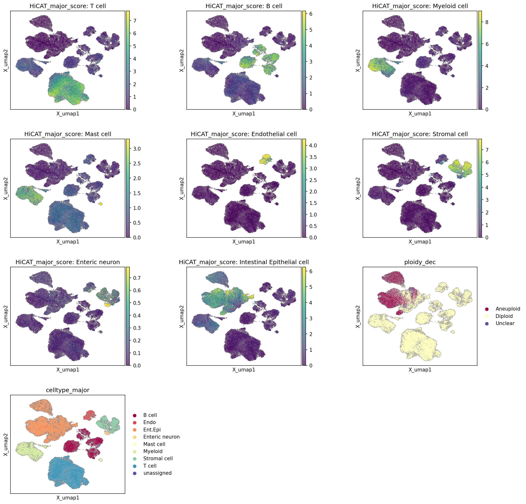
This analysis visualizes the UMAP (Uniform Manifold Approximation and Projection) embedding of single-cell RNA-seq data from Colon tissue. The UMAP plots serve two main purposes:
- Cell Type Score Visualization: Display the HiCAT_major_score for various major cell types across the UMAP, providing a quantitative assessment of cell identity for different regions of the embedding. This helps confirm the presence and localization of specific cell populations based on gene expression profiles characteristic of each major cell type.
- Ploidy and Annotation Visualization: Show the distribution of ploidy inference (ploidy_dec as Aneuploid/Diploid) and the final celltype_major annotations on the UMAP. This allows for validation of cell type assignments and investigation into the genomic stability (ploidy) of different cell populations within the tissue.
Visual Summary
The visualization consists of 10 UMAP plots. The first eight plots display the HiCAT_major_score for individual major cell types, with higher scores indicated by warmer colors (yellow/green) and lower scores by cooler colors (purple). The ninth plot shows the ploidy_dec (Aneuploid, Diploid, Unclear), and the tenth plot shows the celltype_major annotations.
- Overall UMAP Structure: The UMAP displays a clear separation into distinct clusters, suggesting successful dimensionality reduction and grouping of cells with similar transcriptional profiles.
HiCAT_major_score Plots
- T cell: A large, prominent cluster in the bottom-center region of the UMAP shows very high T cell scores, indicating a strong T cell identity for these cells.
- B cell: A distinct cluster in the upper-middle section, separate from the T cell cluster, exhibits high B cell scores.
- Myeloid cell: Cells with high Myeloid cell scores are primarily located in the lower-left and central-right regions, forming several distinct clusters.
- Mast cell: A smaller, less prominent cluster with high Mast cell scores is visible in the upper-right area.
- Endothelial cell: Cells with high Endothelial cell scores are localized in a well-defined cluster towards the upper-right.
- Stromal cell: A significant population of cells with high Stromal cell scores occupies a distinct large cluster in the central-right part of the UMAP.
- Enteric neuron: A small, isolated cluster in the mid-right region shows high Enteric neuron scores.
- Intestinal Epithelial cell: A large, dense cluster in the upper-left area of the UMAP shows very high Intestinal Epithelial cell scores, reflecting the predominant epithelial component of the colon tissue.
ploidy_dec Plot
- Aneuploid cells (maroon) are predominantly enriched within the large Intestinal Epithelial cell cluster in the upper-left, and also in a smaller, dense cluster in the upper-middle that largely overlaps with B cells and some T cells.
- Diploid cells (light yellow) are widely distributed across most other cell type clusters, including T cells, Myeloid cells, Stromal cells, Endothelial cells, and a substantial portion of the Intestinal Epithelial cells.
- The enrichment of Aneuploid cells within specific epithelial clusters is particularly noticeable.
celltype_major Plot
- This plot, colored by the assigned celltype_major annotations, largely corroborates the patterns observed in the HiCAT_major_score plots. Each major cell type forms distinct, well-separated clusters consistent with where its respective score was high.
- The "Intestinal Epithelial cell" (orange) cluster aligns perfectly with the high HiCAT_major_score: Intestinal Epithelial cell region.
- Similarly, "T cell" (teal), "B cell" (maroon), "Myeloid cell" (light green), "Stromal cell" (cyan), "Endothelial cell" (salmon), "Mast cell" (yellow), and "Enteric neuron" (beige) clusters show excellent congruence with their corresponding score maps.
- The "unassigned" (dark purple) cells are sparsely distributed, indicating most cells have been confidently assigned a major cell type.
Biological Interpretation
- Robust Cell Type Identification: The strong spatial concordance between the HiCAT_major_score for each cell type and the final celltype_major annotations on the UMAP demonstrates a robust and reliable cell type identification process. Distinct cell populations are clearly separated in the embedding, and their identity is consistently supported by specific marker gene expression (reflected in the HiCAT scores). This high-quality annotation is crucial for downstream differential expression, pathway analysis, and cell-cell interaction studies.
- Tissue Composition: The UMAP confirms the expected major cell type composition of human Colon tissue, including dominant epithelial cells, diverse immune cells (T cells, B cells, Myeloid cells, Mast cells), stromal cells (fibroblasts), endothelial cells, and enteric neurons. The presence of these cell types in expected proportions and distinct clusters provides a good foundation for studying tissue heterogeneity in health and disease.
- Aneuploidy in Intestinal Epithelial Cells: The ploidy_dec plot highlights a significant biological finding: a substantial fraction of the Intestinal Epithelial cell population is classified as Aneuploid. Given that "Intestinal Epithelial cell" is identified as the "Tumor origin celltype" in the data context, this strong enrichment of aneuploidy within the epithelial compartment strongly suggests that these aneuploid epithelial cells likely represent the tumor cells. Aneuploidy is a hallmark of cancer, reflecting chromosomal instability and abnormal chromosome numbers, which drive tumor evolution and progression [1]. The co-localization of aneuploid cells with the epithelial tumor origin cells is a key indicator of malignant transformation.
- Non-Epithelial Aneuploidy: While primarily concentrated in epithelial cells, some smaller clusters of aneuploid cells are also observed outside the main epithelial cluster, particularly within what appears to be a B cell cluster. This warrants further investigation, as aneuploidy in non-epithelial cells might indicate secondary effects of the tumor microenvironment or, less commonly, non-cancerous conditions associated with chromosomal instability. However, the most prominent signal points to the epithelial compartment.
Annotation Notes
- The HiCAT_major_score plots provide strong evidence for the validity of the celltype_major annotations. The clear, localized expression of high scores for each cell type within its assigned cluster indicates that the clusters are indeed composed of the cell types they are labeled as.
- The "unassigned" category is minimal and scattered, suggesting a high degree of confidence in the assignments for the vast majority of cells.
- The clear separation of different cell types on the UMAP manifold suggests good quality data and robust clustering, facilitating subsequent analyses that rely on accurate cell type identification.
---
References
- Aneuploidy as a hallmark of cancer:
- PubMed search for "aneuploidy cancer hallmark": https://pubmed.ncbi.nlm.nih.gov/?term=aneuploidy+cancer+hallmark
3. Overall Celltype_subset Marker Expression Dot Plot Interpretation
[Analysis Visualization Results]...
Analysis Overview
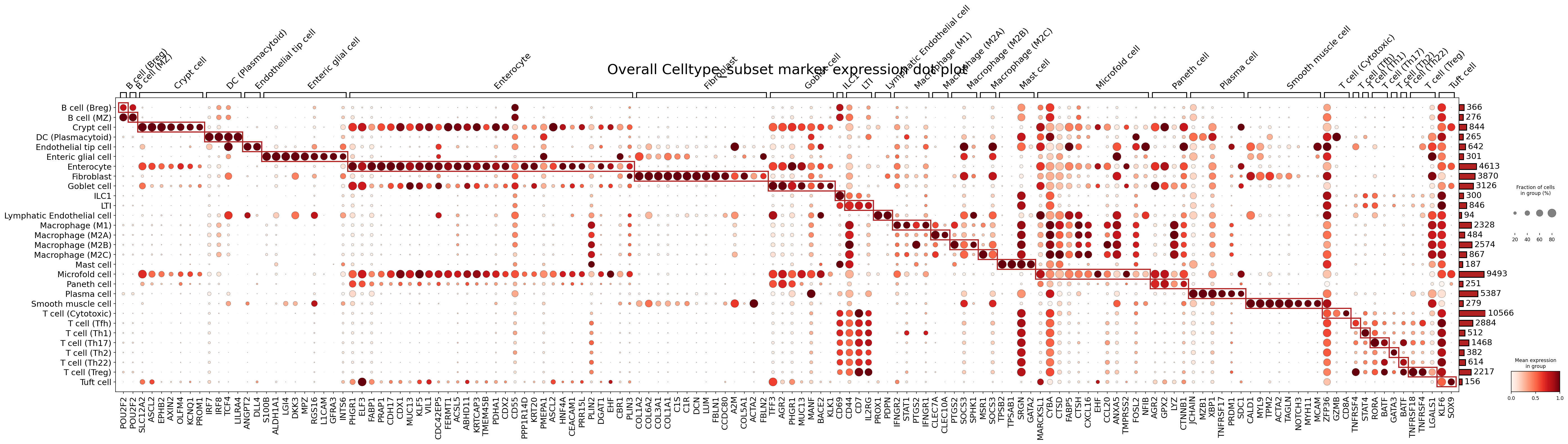
This visualization presents a dot plot illustrating the expression of marker genes across various celltype_subset populations identified in the single-cell RNA-seq data from human colon tissue. The analysis aimed to identify and visualize specific marker genes for each cell subset, without focusing on a particular target cell type (as indicated by target_cell: None). The parameters used for finding markers (find_cfg) prioritized surfaceome-only genes, with specific cutoffs for expression score, fold change, and p-value. Notably, markers common in 3 or more groups were removed (rem_mkrs_common_in_N_groups_or_more: 3) to enhance specificity, making this plot ideal for assessing the distinctness and quality of the celltype_subset annotations.
Visual Summary
The dot plot is structured with celltype_subset groups on the y-axis and their corresponding marker genes on the x-axis. Each dot's size indicates the percentage of cells within a group that express a particular gene (fraction of cells in group), while its color intensity represents the mean expression level of that gene within the group (mean expression in group; darker red signifies higher expression).
Key visual patterns include:
- Strong Diagonal Blocks: Numerous distinct red rectangular boxes are observed along the diagonal of the plot. These boxes highlight clusters of genes that are highly expressed (dark red color) and widely expressed (large dot size) within a specific celltype_subset group, and largely absent or lowly expressed in other groups. This pattern indicates strong specificity of these markers for their respective cell type annotations.
- Cell Type Specificity: The plot clearly demonstrates that most celltype_subset populations are defined by unique sets of highly expressed marker genes, suggesting well-differentiated cell identities.
- Expression Levels and Prevalence: The intensity and size of the dots vary, reflecting both the average expression level and the proportion of cells expressing a marker within each subset.
- Cell Numbers: The bar plots on the far right indicate the number of cells contributing to each celltype_subset group, providing context on the abundance of each population.
Biological Interpretation
The marker gene expression patterns strongly support the biological identity of the assigned celltype_subset annotations in the colon tissue.
Intestinal Epithelial Cells
- Crypt cell: Characterized by markers like OLFM4, EPHB2, and ASCL2, which are known for their roles in intestinal stem cell maintenance and Wnt signaling, consistent with their proliferative and regenerative function. GeneCards: OLFM4
- Enterocyte: Identified by specific expression of SLC12A2, FABP1, CDH17, MUC13, and VIL1. These genes are involved in ion transport, fatty acid metabolism, cell adhesion, and brush border formation, confirming their absorptive roles. GeneCards: FABP1
- Goblet cell: Distinctly marked by TFF3 and MUC2, crucial for mucus production and gut barrier integrity. GeneCards: MUC2
- Paneth cell: Shows high expression of LYZ and DEFA5, antimicrobial peptides essential for host defense in the crypts. GeneCards: LYZ
- Microfold cell (M cell): Uniquely expresses GP2, a known marker for M cells involved in antigen sampling from the gut lumen. GeneCards: GP2
- Tuft cell: Identified by DCLK1 and TRPM5, key markers for their chemosensory functions. GeneCards: DCLK1
Stromal and Endothelial Cells
- Fibroblast: Exhibits strong expression of collagen-related genes such as COL1A1, COL1A2, along with DCN and LUM, consistent with their role in extracellular matrix production. GeneCards: COL1A1
- Smooth muscle cell: Defined by contractile protein markers like ACTA2, MYH11, and TAGLN. GeneCards: ACTA2
- Lymphatic Endothelial cell: Clearly identified by PROX1, PDPN, and LYVE1, which are critical for lymphatic vessel development and function. GeneCards: PROX1
- Endothelial tip cell: Shows expression of LYN and other genes, though some overlap with general endothelial markers may exist.
Immune Cells
- B cell (Breg, MZ): Display markers like POU2F2 and CD79A (not explicitly shown in the cropped images but a general B cell marker).
- Plasma cell: Distinctly marked by SDC1 (CD138), MZB1, JCHAIN, and XBP1, indicating their mature, antibody-secreting state. GeneCards: SDC1
- DC (Plasmacytoid): Characterized by LILRA4 and IRF7, key markers for their antiviral and immunoregulatory roles. GeneCards: LILRA4
- Macrophage (M1, M2A, M2B, M2C, M2D): While sharing some common macrophage features, these subsets show differential expression of genes such as CLEC7A (M1) and LILRB1 (M2A), suggesting distinct polarization states.
- Mast cell: Identified by highly specific markers such as TPSAB1 and SRGN, confirming their granule content and immune functions. GeneCards: TPSAB1
- ILC1: Expresses TBX21 (T-BET), a master transcription factor for ILC1s. GeneCards: TBX21
- LTI cell: Marked by RORC and IL7R, consistent with their role in lymphoid tissue development. GeneCards: RORC
- T cell subsets (Cytotoxic, Tfh, Th1, Th17, Th2, Th22, Treg): Each T cell subset is clearly demarcated by its signature transcription factors and functional markers. For example, CD8A and GZMB for Cytotoxic T cells; PDCD1 for Tfh; TBX21 for Th1; RORC for Th17; GATA3 for Th2; TNFRSF18 for Treg. This detailed marker profile provides robust evidence for the accurate annotation of these diverse T cell populations.
Enteric Neurons
- Enteric glial cell: Shows expression of S100B and GFRA3, classic markers for glial cells in the enteric nervous system. GeneCards: S100B
Annotation Notes
This "Overall Celltype_subset marker expression dot plot" serves as a highly effective and robust validation of the celltype_subset annotations within the AnnData object. The observation of distinct, highly specific marker gene sets for nearly all cell subsets, characterized by high mean expression and prevalence across cells within each group, strongly supports the accuracy and biological fidelity of the cell type assignments. The consistency of these markers with established biological knowledge for human colon tissue further reinforces the confidence in the current annotation schema. Minor overlaps or less distinct markers in some closely related cell types are expected and do not undermine the overall quality of the annotation, but rather highlight the continuous spectrum of cellular states.
4. Copy Number Variation (CNV) Analysis in Intestinal Epithelial and Unassigned Cells
[Analysis Visualization Results]...
Analysis Overview
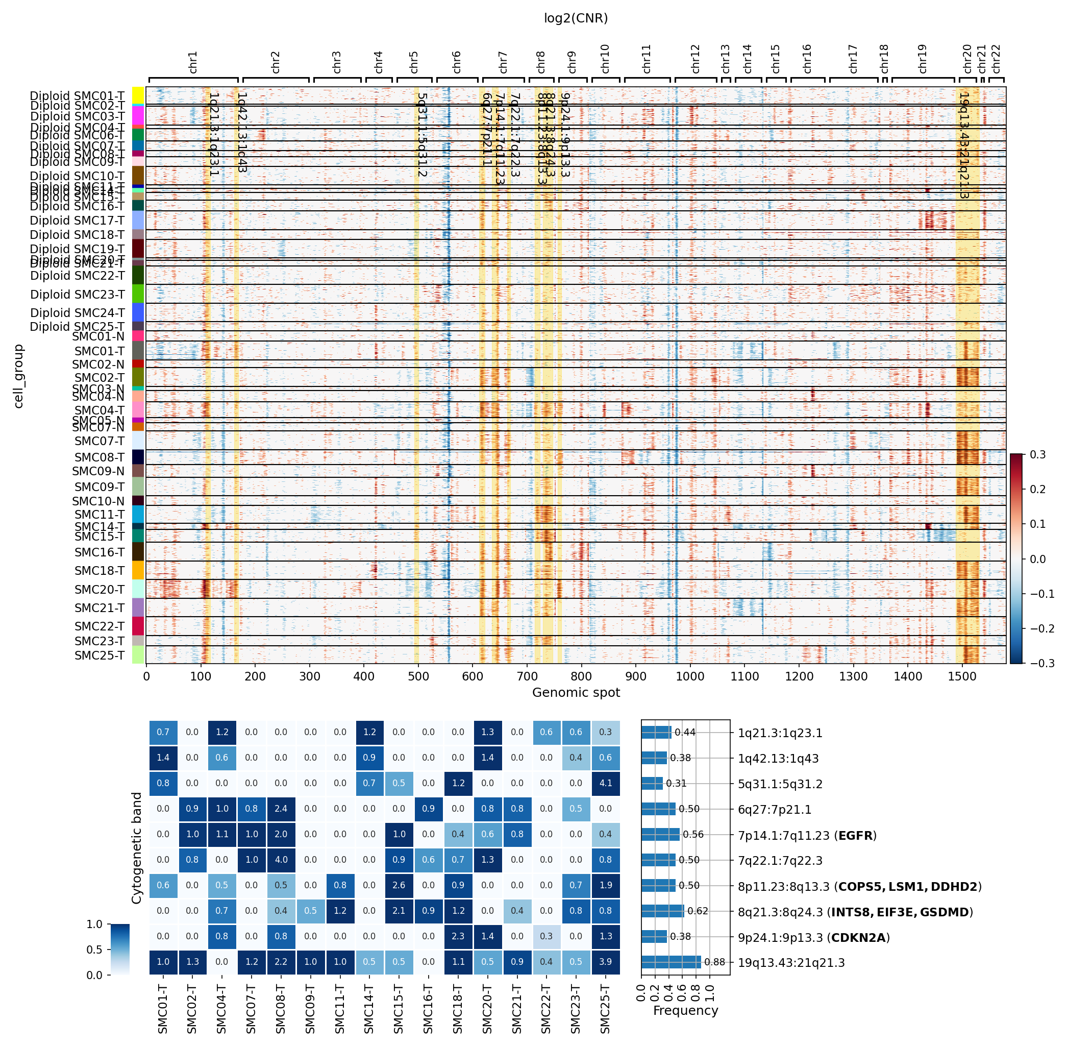
This analysis investigates copy number variations (CNVs) in Intestinal Epithelial cells (the identified tumor origin cell type) and unassigned cells from colon tissue, across both Tumor (T) and Adjacent Normal (N) samples. The goal is to visualize the genomic landscape of CNVs and identify significantly amplified or deleted regions, providing insights into genomic instability associated with the disease state. The results are presented as a heatmap of log2(Copy Number Ratio, CNR) and a summary plot highlighting recurrent cytogenetic band alterations.
Visual Summary
log2(CNR) Heatmap
The first heatmap visualizes the log2(CNR) values across genomic spots for individual cell groups, which are defined by their ploidy status and sample origin (e.g., "Diploid SMC01-T").
- Genomic Instability: The heatmap reveals varying degrees of genomic instability across different samples. Red regions indicate copy number amplifications (log2(CNR) > 0), while blue regions indicate deletions (log2(CNR) < 0).
- Sample Heterogeneity: While many 'Diploid' cells show a relatively stable genome (values close to 0), several tumor samples (e.g., SMC08-T, SMC14-T) exhibit clear patterns of recurrent amplifications and deletions across multiple chromosomes.
- Recurrent Alterations: Specific genomic regions, such as those on chromosomes 8, 13, and 20, appear to be frequently amplified across multiple samples, suggesting common events in colorectal tumorigenesis.
- Ploidy Status: The visible cell groups are predominantly labeled "Diploid," indicating that even cells inferred as diploid can harbor focal CNVs. This highlights the importance of single-cell resolution CNV analysis.
Summary of Significantly Amplified Copy Number Regions
The second heatmap and bar plot provide a summarized view of recurrent CNV events at the cytogenetic band level across samples.
- Recurrent CNV Hotspots: The bar plot on the right clearly identifies several highly recurrent genomic alterations across the analyzed cells:
- 19q13.43 shows the highest overall frequency (0.88), indicating widespread amplification in the target cell population across samples.
- 1q21.3-1q23.1 is frequently altered (frequency 0.70).
- 8p11.23-8q24.3 (including genes like *INTS8, EIF3E, GSDMD*) is amplified with a frequency of 0.62.
- 7p14.1-7q11.23 (containing *EGFR*) shows a frequency of 0.56, indicating frequent amplification of this critical oncogene.
- 6q27-7p21.1, 7q22.1-7q22.3, and 8p11.23-8q13.3 (including *COP S5, LSM1, DDHD2*) are also frequently amplified, each with a frequency of 0.50.
- 9p24.1-9p13.3 (containing *CDKN2A*) shows a frequency of 0.38. Given *CDKN2A* is a tumor suppressor gene, alterations in this region likely represent deletions leading to loss of function.
- Sample-Specific Variability: The heatmap portion of the summary plot illustrates that while some alterations are highly recurrent across many samples, others are more prominent in specific samples (e.g., SMC08-T shows strong alterations in several regions like 6q27-7p21.1 and 7q22.1-7q22.3).
Biological Interpretation
The analysis specifically targets Intestinal Epithelial cells, which are confirmed as the "Tumor origin celltype" in the data context, and unassigned cells. The observed CNV patterns are therefore highly relevant to the genomic instability underlying colon tumorigenesis.
- Oncogene Amplification: The frequent amplification of 7p14.1-7q11.23, which harbors the *EGFR* gene, is a significant finding. *EGFR* is a well-established oncogene whose amplification or overexpression drives cell proliferation, survival, and metastasis in many cancers, including colorectal cancer. Targeting EGFR signaling is a common therapeutic strategy in this disease GeneCards: EGFR.
- Tumor Suppressor Gene Loss: The recurrent alteration in 9p24.1-9p13.3, containing the *CDKN2A* gene, is indicative of genomic instability affecting tumor suppressor pathways. *CDKN2A* encodes proteins (p16INK4a and p14ARF) that regulate cell cycle progression. Loss of *CDKN2A* function, often through deletion, is a common event in various cancers, leading to uncontrolled cell growth GeneCards: CDKN2A.
- Other Recurrent Amplifications: The frequent amplifications on chromosomes 1, 8, and 19 point to additional regions that likely harbor oncogenes or genes important for tumor progression in colon cancer. For instance, the 8p11.23-8q24.3 region contains genes like *GSDMD* (gasdermin D), involved in pyroptosis, and *EIF3E* (eukaryotic translation initiation factor 3 subunit E), which can be involved in oncogenic processes.
- Tumor Heterogeneity: The variability in CNV patterns observed across different tumor samples (e.g., SMC01-T vs. SMC08-T) underscores the inter-patient heterogeneity of genomic alterations in colon cancer. Even within samples, the presence of cells inferred as "Diploid" yet harboring focal CNVs suggests complex genomic landscapes beyond simple aneuploidy.
- Adjacent Normal Samples: The presence of some CNVs in adjacent normal (N) samples (e.g., SMC02-N, SMC04-N) might indicate early genomic instability or field cancerization effects, where histologically normal tissue adjacent to a tumor already harbors genomic alterations that predispose to cancer development.
Clinical or Translational Implications
The identified recurrent CNVs have several clinical implications:
- Biomarker Potential: The consistent amplification of regions containing *EGFR* and deletion of *CDKN2A* could serve as prognostic or predictive biomarkers for patient stratification and therapeutic response in colorectal cancer.
- Therapeutic Targeting: Amplifications of oncogenes like *EGFR* suggest that patients with these genomic alterations may benefit from targeted therapies designed to inhibit EGFR signaling. Conversely, understanding the loss of tumor suppressor genes like *CDKN2A* can inform strategies that exploit vulnerabilities arising from such losses.
- Monitoring Genomic Instability: Tracking these CNV profiles, especially in the tumor-origin Intestinal Epithelial cells, could be valuable for monitoring disease progression, recurrence, and resistance mechanisms in patients.
- Understanding Tumor Evolution: The detection of CNVs in "Diploid" cells and in adjacent normal tissues provides insights into the early stages of tumor development and the clonal evolution of cancer cells.
5. CNV 기반 UMAP 분석을 통한 세포 유형, 이수성, 및 조건별 패턴 시각화
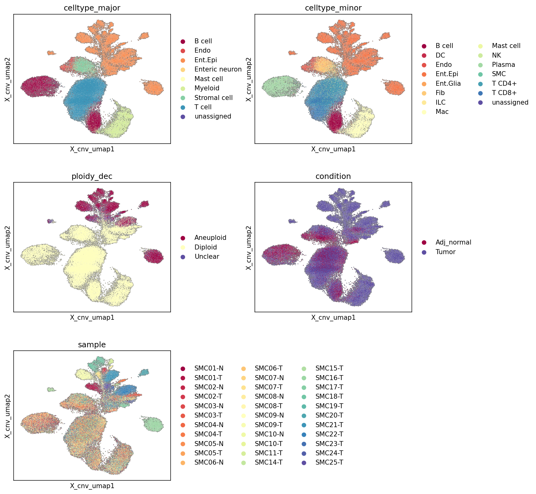
[Analysis Visualization Results]...
Analysis Overview
본 분석은 단일 세포 RNA 시퀀싱 데이터에서 추론된 체세포 유전체 복제수 변이(CNV)를 기반으로 UMAP 임베딩을 생성하고, 이를 다양한 세포 특성(celltype_major, celltype_minor, ploidy_dec, condition, sample)별로 시각화하여 데이터의 전반적인 구조, 세포 유형 분포, 이수성 상태, 그리고 종양 미세 환경의 특징을 탐색합니다. UMAP은 CNV 정보를 활용하여 세포 간의 유사성을 차원 축소된 공간에 표현하므로, 유전체 안정성 또는 불안정성과 관련된 세포 집단을 효과적으로 식별할 수 있습니다.
Visual Summary
- CNV 기반 UMAP 구조: UMAP은 여러 개의 뚜렷한 클러스터와 더 넓게 분포된 영역을 보여줍니다. 이는 세포들이 CNV 프로파일에 따라 뚜렷하게 구분되는 집단과 연속적인 스펙트럼으로 존재함을 시사합니다.
celltype_major 및 celltype_minor 분포:
- Intestinal Epithelial cell (Ent.Epi, 주황색)은 UMAP의 한쪽 끝에 매우 뚜렷하고 밀집된 클러스터를 형성하며, 이는 다른 세포 유형과 CNV 프로파일이 현저하게 다름을 나타냅니다. 일부 Ent.Epi 세포는 다른 클러스터에도 분포합니다.
- T cell (짙은 파란색), Myeloid cell (밝은 노란색), B cell (짙은 빨간색)과 같은 면역 세포들은 주로 UMAP의 중앙 및 하단 영역에 넓게 분포하며, 이들은 서로 겹치거나 인접한 클러스터를 형성합니다.
- Stromal cell (연두색)은 면역 세포 클러스터와 인접하게 분포하며, Endothelial cell (빨간색)도 일부 겹치는 패턴을 보입니다.
- celltype_minor 플롯은 celltype_major 플롯과 유사한 전반적인 구조를 보여주며, Intestinal Epithelial cell이 뚜렷하게 분리되고 면역 세포 유형들이 중앙에 모여 있음을 더 세분화하여 보여줍니다.
ploidy_dec (이수성 정도) 분포:
- Aneuploid (짙은 빨간색) 세포들은 Intestinal Epithelial cell이 밀집된 클러스터와 거의 완벽하게 일치하며, UMAP의 상단에 뚜렷한 분리된 집단을 형성합니다. 이는 이들 세포가 명확한 CNV 변화를 가지고 있음을 강하게 시사합니다.
- Diploid (밝은 노란색) 세포들은 UMAP의 대부분을 차지하며, 주로 면역 세포와 기질 세포 클러스터에 분포합니다.
- Unclear (짙은 보라색)로 분류된 세포는 소수이며 UMAP 전반에 걸쳐 산발적으로 나타납니다.
condition (조건) 분포:
- Tumor (보라색) 세포들은 Aneuploid 및 Intestinal Epithelial cell 클러스터에 압도적으로 집중되어 있습니다. 이는 해당 클러스터의 세포들이 주로 종양 조직에서 유래했음을 나타냅니다.
- Adj_normal (짙은 빨간색) 세포들은 Diploid 세포가 분포하는 UMAP의 중앙 및 하단 영역에 주로 분포하며, 면역 및 기질 세포 클러스터에 상당수 존재합니다.
- Adj_normal 세포 중 일부는 Tumor 클러스터와 겹치는 부분이 있어, 종양 미세 환경 내 정상 세포의 존재 또는 인접 정상 조직에서 종양 관련 변화가 시작되고 있을 가능성을 시사합니다.
sample (샘플) 분포:
- 각 샘플은 UMAP 공간에서 특정 영역에 집중되거나 넓게 분포하는 패턴을 보입니다. 특히 종양 샘플(예: SMC01-T, SMC02-T, SMC03-T)은 Aneuploid 및 Intestinal Epithelial cell 클러스터에 주로 기여합니다.
- 정상 인접 샘플(예: SMC01-N, SMC02-N)은 주로 Diploid 면역/기질 세포 클러스터에 분포합니다.
- 다양한 샘플이 UMAP 전반에 걸쳐 분포하여 환자 간 또는 샘플 간의 이질성(heterogeneity)을 반영합니다. 이는 CNV 프로파일이 샘플마다 다를 수 있음을 의미합니다.
Biological Interpretation
- 악성 종양 세포의 식별: CNV 기반 UMAP에서 Aneuploid 상태의 Intestinal Epithelial cell들이 Tumor 조건의 세포들과 강력하게 일치하며 뚜렷한 클러스터를 형성하는 것은, 이러한 클러스터가 종양의 기원 세포(Intestinal Epithelial cell)로부터 유래한 악성 종양 세포 집단을 대표함을 강력하게 시사합니다. 이는 CNV 분석이 종양 세포를 비종양 세포로부터 효과적으로 구분하는 데 유용함을 보여줍니다.
- 종양 미세 환경의 구성: Diploid 세포들은 주로 Adj_normal 조건의 세포들과 면역 및 기질 세포 유형으로 구성되어 있습니다. 이는 종양 미세 환경 내에 종양 세포와 상호작용하는 다양한 비종양 세포(면역 세포, 섬유아세포, 내피세포 등)가 존재함을 보여주며, 이들 세포는 대부분 정상적인 유전체 이수성 상태를 유지하고 있습니다. 종양 클러스터 내에 일부 Adj_normal 세포가 섞여 있는 것은 종양 내 침윤성 정상 세포 또는 종양-정상 경계 부위의 세포들을 반영할 수 있습니다.
- CNV와 세포 정체성: UMAP 임베딩이 CNV 정보를 기반으로 생성되었음에도 불구하고, celltype_major 및 celltype_minor 플롯에서 세포 유형들이 합리적으로 클러스터링되는 것을 관찰할 수 있습니다. 특히 Intestinal Epithelial cell이 다른 세포 유형들과 뚜렷하게 구분되는 것은, 이들 세포가 다른 세포 유형들과는 다른 고유한 CNV 프로파일을 가질 뿐만 아니라, 종양 발생 과정에서 특징적인 CNV 패턴을 획득했음을 의미합니다.
- 환자 간 이질성: sample 플롯에서 각 샘플이 UMAP 공간에서 다양한 분포를 보이는 것은 환자마다 CNV 프로파일과 종양 미세 환경 구성에 이질성이 존재함을 나타냅니다. 이는 대장암이 유전학적으로 이질적인 질병이며, 개별 환자의 특성을 고려한 분석이 중요함을 시사합니다.
Annotation Notes
- CNV 기반 UMAP은 종양 세포 집단을 비종양 세포 집단으로부터 명확하게 구분하는 강력한 시각적 증거를 제공하며, 이는 ploidy_dec 및 condition 정보와 완벽하게 일치합니다.
- Intestinal Epithelial cell이 Tumor origin celltype이라는 데이터 컨텍스트를 고려할 때, 이들 세포의 뚜렷한 Aneuploid 및 Tumor 특성 일치는 CNV 추론의 정확성과 세포 유형 지정의 신뢰성을 높입니다.
- UMAP을 통한 CNV 패턴 시각화는 데이터셋 내의 주요 생물학적 변동성을 파악하고, 세포 유형별 및 조건별 유전체 불안정성 특징을 이해하는 데 매우 효과적인 방법입니다.
6. Colon Tissue Minor Cell Type Population Analysis in Tumor vs. Adjacent Normal Conditions
[Analysis Visualization Results]...
Analysis Overview
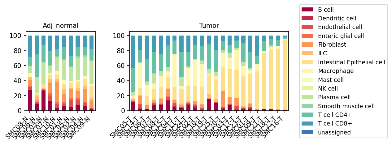
This analysis visualizes the relative proportions of minor cell types across individual samples from both 'Adj_normal' (adjacent normal) and 'Tumor' conditions. The stacked bar plot allows for a direct comparison of cellular composition between healthy and cancerous colon tissue, highlighting potential shifts in the tumor microenvironment (TME) at the cellular population level. Each bar represents a single sample, and the segments within each bar show the percentage contribution of different celltype_minor populations, as defined in the AnnData object.
Visual Summary
The stacked bar plot presents the cellular heterogeneity within colon tissue samples, stratified by condition: 'Adj_normal' (left panel) and 'Tumor' (right panel).
- Dominant Cell Types in Adjacent Normal Tissue: In adjacent normal colon tissue, the cellular composition appears relatively consistent across samples. Key populations include Intestinal Epithelial cells (light yellow), Fibroblasts (orange), T cells (CD4+ and CD8+, various shades of blue/teal), and Macrophages (light yellow/cream). There's a notable presence of various immune cells.
Shifts in Tumor Tissue
- Increased Intestinal Epithelial Cells: A striking observation in the 'Tumor' samples is the significant increase in the relative proportion of Intestinal Epithelial cells (light yellow). These cells often constitute the largest fraction in tumor samples, consistent with their role as the 'Tumor origin celltype' and the neoplastic expansion of tumor cells. In some tumor samples (e.g., SMC10-T, SMC19-T, SMC25-T, SMC03-T, SMC17-T, SMC23-T, SMC01-T, SMC09-T, SMC21-T, SMC18-T, SMC22-T, SMC16-T), Intestinal Epithelial cells dominate almost entirely.
- Decreased Immune and Stromal Cells (Relative to Epithelial Cells): Concomitant with the epithelial cell expansion, most other cell types, including various immune cells (T cells CD4+, T cells CD8+, B cells, Dendritic cells, Plasma cells, NK cells, ILCs, Mast cells) and stromal cells (Fibroblasts, Endothelial cells, Smooth muscle cells, Enteric glial cells), show a relative decrease in their proportions within the tumor samples compared to adjacent normal tissue.
- Specifically, T cell populations (CD4+ and CD8+) appear less prominent in many tumor samples.
- Fibroblasts (orange) also show a marked reduction in relative proportion in tumor samples.
- Macrophages: Macrophages (light yellow/cream) show variable presence in tumor samples, appearing elevated in some (e.g., SMC05-T, SMC14-T, SMC06-T, SMC07-T, SMC08-T, SMC15-T, SMC04-T, SMC11-T, SMC24-T, SMC02-T) compared to other immune cells, suggesting their adaptive role in the TME.
- Inter-sample Heterogeneity in Tumor: There is considerable heterogeneity in cellular composition among individual tumor samples, particularly regarding the relative abundance of Intestinal Epithelial cells versus other stromal and immune components. Some tumors are highly enriched for epithelial cells, while others retain a more mixed composition, albeit still altered compared to normal tissue.
Biological Interpretation
The observed shifts in cell type populations between adjacent normal and tumor colon tissue provide critical insights into the remodeling of the tumor microenvironment (TME) during colorectal cancer progression.
- Tumor Cell Dominance: The prominent increase in the relative proportion of Intestinal Epithelial cells in tumor samples is a direct reflection of neoplastic proliferation. As the 'Tumor origin celltype', their numerical expansion physically displaces or overshadows other resident cell populations within the tumor mass. This cellular dominance is a hallmark of cancer.
- Immune Landscape Alterations:
- Relative T-cell Exhaustion/Exclusion: The relative reduction in T cell populations (CD4+ and CD8+) in many tumor samples suggests potential immune evasion mechanisms at play. This could manifest as T cell exclusion from the tumor core, T cell anergy or exhaustion within the TME, or simply a dilution effect due to the overwhelming presence of tumor cells. A decreased proportion of cytotoxic CD8+ T cells, in particular, often correlates with poorer prognosis and reduced response to immunotherapies in various cancers [NCBI].
- Macrophage Influx/Reprogramming: While globally reduced in some samples due to the epithelial expansion, the sustained or even relatively increased presence of Macrophages in other tumor samples is noteworthy. Tumor-associated macrophages (TAMs) are crucial components of the TME, often promoting tumor growth, angiogenesis, and immunosuppression, particularly those polarized towards an M2-like phenotype [GeneCards] [NCBI]. Their variable abundance suggests distinct macrophage-driven microenvironments across different tumors.
- Other Immune Cell Changes: The general relative decrease in other immune cells (B cells, NK cells, Dendritic cells, Plasma cells, ILCs, Mast cells) points to a broad immunosuppressive shift or a re-prioritization of immune cell recruitment and survival within the TME, potentially driven by tumor-secreted factors.
- Stromal Remodeling: The relative decrease in Fibroblasts in tumor samples is an interesting observation. While cancer-associated fibroblasts (CAFs) are known to be abundant and play critical roles in desmoplasia and tumor support in many cancers, their *relative* reduction here could be due to the vast expansion of epithelial cells overshadowing their absolute numbers, or a different balance of stromal components in colon cancer compared to other tumor types. Endothelial cells, although reduced in relative proportion, are vital for tumor angiogenesis, which might not be fully captured by proportional analysis.
- Inter-tumoral Heterogeneity: The variability in cellular composition among individual tumor samples underscores the high degree of inter-patient heterogeneity in colon cancer. This suggests that distinct TME profiles might exist, potentially influencing disease progression and therapeutic responses.
Clinical or Translational Implications
The distinct alterations in cell type proportions between normal colon tissue and colorectal tumors have significant clinical and translational implications:
- Biomarker Discovery: The relative abundance of specific immune cell populations (e.g., T cells, macrophages) or the ratio of tumor cells to immune cells (Tumor-Immune Cell Ratio) could serve as prognostic or predictive biomarkers for patient outcomes and response to therapies. For example, a lower T-cell proportion might indicate a "cold" tumor less responsive to immune checkpoint inhibitors.
- Therapeutic Targeting: Understanding the cellular makeup of the TME can inform the development of targeted therapies.
- Strategies aimed at increasing T cell infiltration or reversing T cell exhaustion could be beneficial where T cells are relatively low.
- Targeting immunosuppressive macrophages (e.g., M2-polarized TAMs) could reprogram the TME to be more anti-tumoral [NCBI].
- Patient Stratification: The observed inter-tumoral heterogeneity suggests that patients could be stratified based on their tumor's cellular composition. This stratification might help guide personalized treatment decisions, matching specific therapies to patients most likely to benefit based on their unique TME profile.
- Diagnostic Potential: The distinct cellular landscape of tumor versus normal tissue can be leveraged for diagnostic purposes, potentially enhancing the accuracy of cancer detection and staging.
7. T 세포 아형 인구 분포 분석: 인접 정상 조직 및 종양 조직 비교
[Analysis Visualization Results]...
Analysis Overview
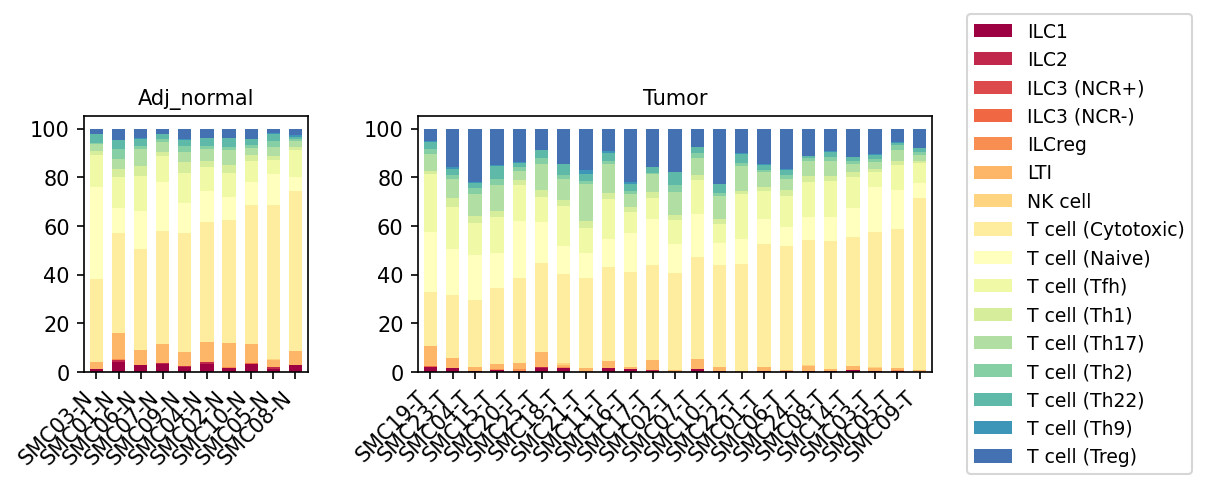
이 분석은 단일 세포 RNA 시퀀싱 데이터를 기반으로 대장(Colon) 조직에서 T 세포 및 ILC(선천성 림프구) 아형의 상대적 인구 분포를 인접 정상 조직(Adj_normal)과 종양(Tumor) 조직 간에 비교한 막대 그래프입니다. 각 막대는 개별 샘플을 나타내며, 각 세포 아형이 해당 샘플 내 T 세포 및 ILC 총 인구에서 차지하는 비율을 100% 기준으로 시각화합니다.
Visual Summary
제공된 막대 그래프는 인접 정상 대장 조직과 종양 조직 내 T 세포 및 ILC 아형의 인구 역학에서 뚜렷한 차이를 보여줍니다.
인접 정상 조직 (Adj_normal):
- 대부분의 T 세포 집단은 T cell (Cytotoxic)과 T cell (Naive)이 지배적입니다. 이 두 아형이 전체 T 세포/ILC 풀의 상당 부분을 차지합니다.
- T cell (Tfh), T cell (Th1), T cell (Th17), T cell (Th2), T cell (Th22), T cell (Th9), T cell (Treg)과 같은 다른 T helper (Th) 세포 아형들은 상대적으로 낮은 비율을 보이며, 샘플 간에 약간의 변동성이 있습니다.
- NK cell, ILC1, ILC2, ILC3 (NCR+), ILC3 (NCR-), ILCreg, LTI와 같은 ILC 집단은 매우 낮은 비율로 존재합니다.
종양 조직 (Tumor):
- 조절 T 세포 (Treg)의 현저한 증가: T cell (Treg) (진한 파란색)의 비율이 인접 정상 조직에 비해 종양 샘플에서 모든 환자에서 일관되게 증가한 것이 가장 두드러진 특징입니다. 이는 종양 미세환경 내 Treg 세포의 축적을 강력히 시사합니다.
- Naive T 세포의 감소: T cell (Naive) (옅은 노란색)의 비율은 종양 조직에서 인접 정상 조직에 비해 전반적으로 감소한 경향을 보입니다. 이는 미분화된 Naive T 세포가 종양 미세환경에서 활성화되거나 분화되거나 배제될 수 있음을 시사합니다.
- T cell (Th9)의 증가: T cell (Th9) (옅은 하늘색) 또한 종양 조직에서 상대적인 비율이 증가하는 경향을 보입니다.
- T cell (Cytotoxic)의 비율 유지 또는 변화: T cell (Cytotoxic) (중간 노란색)은 여전히 상당한 비율을 차지하지만, 그 비중이 샘플에 따라 더 다양하며 일부 종양 샘플에서는 정상 조직에 비해 상대적으로 감소한 것으로 보일 수 있습니다.
- 다른 Th 세포 아형의 변화: T cell (Th1) (옅은 녹색)은 일부 종양 샘플에서 감소한 것으로 보이며, T cell (Th17) (중간 녹색) 및 T cell (Th22) (옅은 청록색)는 상대적으로 안정적이거나 일부 증가하는 경향을 보입니다.
- NK cell 및 ILC 집단은 종양 조직에서도 낮은 비율을 유지합니다.
Biological Interpretation
이 분석 결과는 대장암 종양 미세환경에서 T 세포 아형 구성의 중요한 재편을 시사합니다.
- 면역억제 환경으로의 전환: 종양 조직에서 조절 T 세포(Treg)의 현저한 증가는 종양 미세환경이 면역 관용 및 면역억제 특성을 강화하고 있음을 강력히 나타냅니다. Treg 세포는 항종양 면역 반응을 억제하여 종양이 면역 감시를 회피하고 성장을 촉진하는 데 기여하는 것으로 알려져 있습니다 [PubMed search: Regulatory T cells tumor microenvironment colon cancer].
- Naive T 세포의 분화 또는 고갈: Naive T 세포의 감소는 종양 특이적 항원에 의해 T 세포가 활성화되어 effector T 세포 또는 Treg 세포로 분화하거나, 종양 미세환경이 Naive T 세포의 침윤을 억제할 수 있음을 의미합니다.
- T helper 세포 아형의 균형 변화:
- T cell (Th9)의 증가는 흥미로운 관찰입니다. Th9 세포는 종양 유형 및 미세환경에 따라 항종양 또는 전종양(pro-tumor) 역할을 할 수 있는 것으로 알려져 있어, 대장암에서의 정확한 역할에 대한 추가 연구가 필요합니다 [PubMed search: Th9 cells cancer immunity]. 일부 연구에서는 Th9 세포가 면역억제 환경 조성에 기여할 수 있음을 시사하기도 합니다.
- T cell (Th1)의 잠재적인 감소는 항종양 면역의 중요한 구성 요소인 Th1 매개 면역 반응이 종양 미세환경에서 약화될 수 있음을 나타냅니다.
- T cell (Cytotoxic)은 여전히 존재하지만, Treg 세포의 증가와 다른 면역억제 메커니즘이 이들 세포의 기능을 저해할 수 있습니다.
이러한 변화는 대장암에서 면역 회피 메커니즘이 활발하게 작동하고 있으며, 종양 미세환경이 염증 반응과 면역억제 사이의 복잡한 균형을 보이고 있음을 강조합니다.
Clinical or Translational Implications
이 분석 결과는 대장암의 진단, 예후 예측 및 치료 전략 개발에 중요한 시사점을 제공합니다.
- 면역관문억제제 반응 예측 및 개선: 종양 내 Treg 세포의 높은 비율은 면역관문억제제(예: anti-PD-1/PD-L1) 치료에 대한 반응률을 저해하는 요인이 될 수 있습니다. Treg 세포를 표적으로 하는 치료법을 병용하거나, Treg 세포를 제거하거나 기능을 억제하는 전략은 면역관문억제제의 효능을 향상시키는 데 기여할 수 있습니다 [PubMed search: Treg depletion immunotherapy cancer].
- 새로운 치료 표적 발굴: Th9 세포의 증가와 같은 다른 T 세포 아형의 변화는 대장암 특이적인 면역 조절 메커니즘을 밝히고, 이들 세포를 조절하여 항종양 면역 반응을 강화할 수 있는 새로운 치료 표적을 발굴하는 데 단서를 제공할 수 있습니다.
- 생체 지표 (Biomarker) 개발: 종양 내 T 세포 아형, 특히 Treg 세포의 비율은 대장암 환자의 예후를 예측하거나 특정 치료법에 대한 반응성을 예측하는 잠재적인 생체 지표로 활용될 수 있습니다. 예를 들어, 높은 종양 내 Treg/CD8+ T 세포 비율은 불량한 예후와 관련될 수 있습니다.
이러한 인구학적 변화는 대장암 종양 미세환경의 복잡성을 이해하는 데 필수적이며, 환자 맞춤형 면역 치료 전략을 개발하기 위한 중요한 기초 정보를 제공합니다.
8. Colon Tumor Microenvironment Shows Significant Shifts in T cell and Innate Lymphoid Cell Subpopulations
[Analysis Visualization Results]...
Analysis Overview
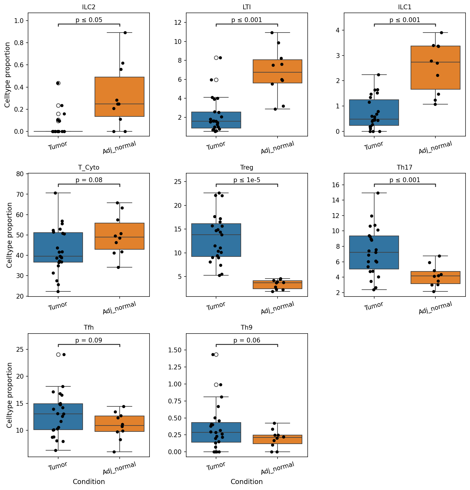
This analysis investigates the proportional representation of various T cell and Innate Lymphoid Cell (ILC) subsets in human colon tissue, comparing tumor samples ('Tumor') to adjacent normal tissue ('Adj_normal'). The goal is to identify statistically significant shifts in these immune cell populations, which can provide insights into the immune landscape of colorectal cancer. The plot_box_for_celltype_population_with_signif_difference tool was used to visualize these differences with statistical annotations.
Visual Summary
The box plots display the cell type proportion for eight T cell and ILC subsets across 'Tumor' and 'Adj_normal' conditions. Several statistically significant differences were observed:
Decreased in Tumor Tissue:
- ILC2: Significantly lower proportions in Tumor samples compared to Adj_normal (p ≤ 0.05).
- LTI (Lymphoid Tissue Inducer cells): Markedly reduced in Tumor tissue (p ≤ 0.001).
- ILC1: Significantly diminished in Tumor samples (p ≤ 0.001).
- T_Cyto (Cytotoxic T cells): Show a suggestive decrease in Tumor samples (p = 0.08).
Increased in Tumor Tissue:
- Treg (Regulatory T cells): Dramatically elevated proportions in Tumor tissue (p ≤ 1e-5), indicating a substantial increase.
- Th17: Significantly higher in Tumor samples (p ≤ 0.001).
- Tfh (Follicular Helper T cells): Suggestively increased in Tumor samples (p = 0.09).
- Th9: Suggestively increased in Tumor samples (p = 0.06).
These proportional changes highlight a distinct re-modeling of the immune cell composition within the colon tumor microenvironment.
Biological Interpretation
The observed shifts in immune cell proportions in colon tumors compared to adjacent normal tissue strongly suggest an immunosuppressive and pro-tumorigenic microenvironment.
- Immunosuppression by Treg and Th17 Cells:
- The highly significant increase in Regulatory T cells (Treg) in tumor samples is a hallmark of many cancers, including colorectal cancer. Tregs suppress effector T cell responses, promote immune tolerance, and hinder anti-tumor immunity, thereby facilitating tumor escape from immune surveillance 1.
- The significant increase in Th17 cells is also notable. While Th17 cells are pro-inflammatory, their role in cancer is context-dependent. In colorectal cancer, Th17 cells are often associated with promoting tumor growth, angiogenesis, and metastasis, sometimes by recruiting other immune cells or directly interacting with tumor cells 2.
- Reduced Innate Anti-Tumor Immunity:
- The significant decrease in ILC1 and LTI cells (a subset of ILCs, often ILC3s) is concerning.
- ILC1s are known to produce IFN-γ and play a role in anti-viral and anti-tumor immunity, resembling Th1 cells 3. Their reduction suggests impaired innate immune surveillance.
- LTI cells (ILC3s) are crucial for the development and maintenance of lymphoid structures. Their decrease might indicate a disruption of the local immune architecture, potentially hindering effective anti-tumor immune responses or reducing the formation of beneficial tertiary lymphoid structures 4.
- The decrease in ILC2s is also observed. ILC2s typically promote type 2 immune responses and tissue repair. Their role in cancer is complex, but a reduction could reflect a shift in the immune landscape away from certain tissue-protective or pro-resolving functions, or an active suppression within the tumor.
- Compromised Adaptive Anti-Tumor Responses:
- The suggestive decrease in Cytotoxic T cells (T_Cyto) is consistent with a compromised anti-tumor immune response. Cytotoxic T cells are primary effectors responsible for directly killing tumor cells. A reduction in their proportion, even if modest, suggests diminished tumor cell clearance 5.
- The suggestive increase in Tfh and Th9 cells may also contribute to the complex immune landscape. Tfh cells are involved in humoral immunity and B cell responses, which can be either anti-tumor or pro-tumor depending on the context. Th9 cells produce IL-9, which also has context-dependent roles in cancer, sometimes promoting tumor growth or contributing to inflammation.
Collectively, these findings paint a picture of a colon tumor microenvironment that actively reshapes immune cell proportions to favor immune evasion and tumor progression. The enrichment of immunosuppressive (Treg) and pro-tumorigenic inflammatory (Th17) populations, coupled with a reduction in key innate (ILC1, LTI) and potentially adaptive (T_Cyto) anti-tumor cells, highlights critical mechanisms of tumor immune escape in colon cancer.
Clinical or Translational Implications
The distinct immunological profile identified in colon tumor tissue has several important clinical and translational implications:
- Biomarkers for Prognosis and Response: The increased Treg/Th17 ratio and decreased ILC1/LTI populations could serve as prognostic biomarkers for colorectal cancer progression and patient outcomes. Patients with this immune cell signature might have a poorer prognosis.
Therapeutic Targets:
- Targeting Treg cells: Strategies aimed at depleting or inhibiting Treg function could enhance anti-tumor immunity. This is an active area of research in immunotherapy for various cancers.
- Modulating Th17 cells: Given their pro-tumor role in this context, therapies that inhibit Th17 differentiation or function could be beneficial.
- Boosting ILC1/LTI/Cytotoxic T cells: Approaches to restore or enhance the proportions and functions of ILC1s, LTI cells, and cytotoxic T cells could improve anti-tumor responses. This might involve cytokine therapies or adoptive cell transfers.
- Immune Monitoring: Monitoring the proportions of these T cell and ILC subsets in the tumor microenvironment, perhaps through biopsy analysis or liquid biopsies, could provide valuable information for treatment stratification and monitoring therapeutic response in colorectal cancer patients.
- Understanding Immunotherapy Resistance: The observed immunosuppressive environment could contribute to resistance to current immunotherapies (e.g., checkpoint inhibitors). Understanding these shifts could help develop combination therapies that overcome this resistance.
9. Macrophage Subset Population Shifts in Colon Tumor Microenvironment
[Analysis Visualization Results]...
Analysis Overview
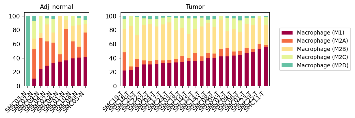
This analysis visualizes the relative proportions of different macrophage subsets (M1, M2A, M2B, M2C, M2D) within the total macrophage population across individual samples from both 'Adj_normal' (adjacent normal colon tissue) and 'Tumor' (colon tumor tissue) conditions. This helps to identify shifts in macrophage polarization that may be associated with the tumor microenvironment.
Visual Summary
The bar plots display the proportional distribution of macrophage subsets for each sample, grouped by condition.
- Adj_normal Condition: Samples from adjacent normal tissue show a mixed population of macrophages. Macrophage (M1) and Macrophage (M2A) subsets appear to be substantial, with Macrophage (M2B) also present. Macrophage (M2D) is present in some samples, notably high in SMC03-N. Macrophage (M2C) is consistently a minor population. There is some heterogeneity across normal samples in terms of specific subset proportions.
- Tumor Condition: A notable shift in macrophage composition is observed in tumor samples compared to adjacent normal samples.
- Dominance of Macrophage (M2B): The Macrophage (M2B) subset (light yellow) appears to be significantly expanded and often constitutes the largest proportion of macrophages in many tumor samples.
- Increased Macrophage (M1): The Macrophage (M1) subset (dark red) also shows a prominent presence and an increase in relative proportion in many tumor samples compared to most normal samples, sometimes surpassing M2B in individual samples (e.g., SMC04-T, SMC21-T, SMC05-T).
- Reduced Macrophage (M2A): The Macrophage (M2A) subset (orange) appears to be relatively diminished in most tumor samples compared to its prevalence in adjacent normal samples.
- Minority subsets: Macrophage (M2C) (light green) and Macrophage (M2D) (teal) generally remain minor populations in tumor samples, similar to normal samples, though M2D is barely detectable in most tumor samples.
Biological Interpretation
Macrophages are highly plastic immune cells that can differentiate into distinct functional phenotypes, often broadly categorized as M1 (pro-inflammatory, anti-tumorigenic) or M2 (anti-inflammatory, pro-tumorigenic). The observed shifts in macrophage subsets within the colon tumor microenvironment suggest a complex interplay between different macrophage polarization states.
- Shift towards M2B phenotype in tumors: The consistent expansion of Macrophage (M2B) in tumor samples is a significant finding. M2B macrophages are known to be involved in immune regulation and can be stimulated by immune complexes or specific cytokines. Their increased presence in the tumor microenvironment could contribute to an immunosuppressive environment, promoting tumor growth and progression through mechanisms such as antigen presentation, cytokine production, and fostering angiogenesis [NCBI].
- Concomitant increase in M1 phenotype: The increased proportion of Macrophage (M1) in tumor samples is somewhat counter-intuitive, as M1 macrophages are typically associated with anti-tumor immunity. However, the tumor microenvironment is highly heterogeneous. It is possible that while M2B macrophages contribute to immune suppression, there might also be concurrent pro-inflammatory signals driving M1 polarization, perhaps as an attempt by the host immune system to counteract the tumor, or as a response to necrotic cells or pathogen-associated molecular patterns (PAMPs) within the tumor. This coexistence highlights the complexity and potential for mixed signals within the tumor microenvironment [NCBI].
- Reduced M2A in tumors: The relative decrease in M2A macrophages, which are often involved in wound healing and fibrosis, suggests a possible shift away from these specific functions in the established tumor microenvironment, or that other M2 subtypes (like M2B) are preferentially recruited or polarized.
- Overall Macrophage Plasticity: The data strongly support the concept of macrophage plasticity within the colon, where the distinct microenvironments of normal tissue versus tumor drive different polarization states. The 'Adj_normal' samples display a more balanced or variable macrophage profile, whereas the 'Tumor' samples converge on a phenotype characterized by increased M1 and M2B, and decreased M2A.
Clinical or Translational Implications
The distinct shifts in macrophage populations, particularly the expansion of M2B and the concurrent significant presence of M1 macrophages in colon tumors, have several clinical implications:
- Biomarker Potential: The ratio or absolute abundance of specific macrophage subsets, particularly M2B relative to M2A, could serve as a potential prognostic or predictive biomarker for colon cancer progression or response to therapy.
- Therapeutic Targeting: The observed macrophage polarization suggests that therapeutic strategies aimed at re-educating tumor-associated macrophages (TAMs) or modulating their specific functions could be beneficial. For instance, targeting factors that promote M2B polarization might reduce immune suppression, while enhancing M1-like functions could boost anti-tumor immunity.
- Immunotherapy Design: Understanding the precise macrophage subset composition is crucial for designing effective immunotherapies. In a microenvironment with abundant M2B-like macrophages, therapies that overcome immunosuppression (e.g., checkpoint inhibitors) or directly reprogram these cells could be more effective. The presence of M1 macrophages also suggests potential avenues for therapies that amplify existing pro-inflammatory responses.
10. Macrophage Subset Population Shifts in Colorectal Tumor Microenvironment
[Analysis Visualization Results]...
Analysis Overview
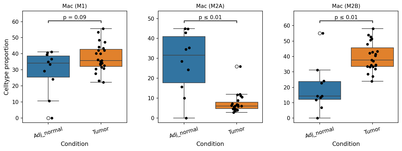
This analysis investigates the proportional changes of specific macrophage subsets (Mac (M1), Mac (M2A), and Mac (M2B)) between normal adjacent colon tissue ("Adj_normal") and colon tumor tissue ("Tumor"). The goal is to identify statistically significant shifts in these immune cell populations, which can provide insight into the immunological landscape of colorectal cancer.
Visual Summary
The box plots illustrate the celltype proportion of three macrophage subsets across "Adj_normal" and "Tumor" conditions:
- Mac (M1): There is a slight, non-significant increase in the proportion of M1 macrophages in tumor tissue compared to adjacent normal tissue (p = 0.09, which is just above the typical 0.05 significance but meets the 0.1 cutoff applied in the analysis). The median proportion shifts from approximately 34% in normal to 36% in tumor.
- Mac (M2A): A statistically significant decrease in the proportion of M2A macrophages is observed in tumor tissue (median ~7%) compared to adjacent normal tissue (median ~31%) (p ≤ 0.01). The distribution in tumor tissue is much tighter and lower.
- Mac (M2B): A highly statistically significant increase in the proportion of M2B macrophages is evident in tumor tissue (median ~38%) compared to adjacent normal tissue (median ~15%) (p ≤ 0.01). The interquartile range for M2B macrophages is also higher in tumor tissue, indicating a substantial expansion of this population.
Biological Interpretation
The observed shifts in macrophage subsets suggest a significant reprogramming of the macrophage compartment within the colorectal tumor microenvironment, consistent with the known plasticity of macrophages in response to pathological conditions, particularly cancer.
- Mac (M1) (Pro-inflammatory/Anti-tumorigenic): While typically associated with anti-tumor immunity, the marginally significant increase in M1 macrophages in the tumor might reflect an initial or ongoing inflammatory response. However, its proportion remains relatively stable compared to the dramatic shifts in M2 subtypes, suggesting that M1 polarization may not be the dominant anti-tumor response or could be suppressed by other factors.
- M1 Macrophage function in cancer
- Mac (M2A) (Wound Healing/Immunoregulatory): The significant decrease in M2A macrophages in the tumor microenvironment is notable. M2A macrophages are generally induced by Th2 cytokines (IL-4, IL-13) and are involved in allergic responses, anti-parasitic immunity, and early wound healing. Their reduction in tumors suggests a possible re-differentiation towards other M2 phenotypes or a less prominent role for this specific M2 subtype in the established tumor.
- Mac (M2B) (Immunomodulatory/Pro-tumorigenic): The striking and significant increase in M2B macrophages within the tumor is a key finding. M2B macrophages are a unique subset of M2-like macrophages that are often induced by immune complexes, TLR agonists, and IL-1R ligands. In the context of cancer, M2B macrophages have been implicated in promoting tumor growth, angiogenesis, metastasis, and immune suppression, often by secreting pro-tumorigenic factors and immunosuppressive cytokines. This expansion points towards an immunosuppressive and pro-tumorigenic milieu within the colon tumors.
- M2 Macrophage polarization and tumor microenvironment
Overall, these findings indicate a shift towards a tumor-supportive macrophage phenotype, primarily driven by the accumulation of M2B macrophages, which can facilitate tumor progression and evade host immune surveillance in the colon.
Clinical or Translational Implications
The significant increase in M2B macrophages in colon tumors, coupled with a decrease in M2A, highlights a potential mechanism by which colorectal cancer establishes an immunosuppressive microenvironment conducive to its growth and spread.
- Therapeutic Target Identification: The elevated presence of M2B macrophages suggests these cells could be attractive targets for immunotherapy strategies aimed at reprogramming tumor-associated macrophages (TAMs). Approaches could involve inhibiting the recruitment of M2B precursors, blocking their pro-tumorigenic functions, or promoting their re-polarization towards an M1-like anti-tumor phenotype.
- Biomarker Potential: The proportion of M2B macrophages, or markers associated with their specific functions, could potentially serve as prognostic biomarkers for colorectal cancer progression or as predictive markers for response to immunotherapies that target the tumor immune microenvironment.
- Disease Progression Insight: Understanding these macrophage shifts provides critical insight into the complex immune dynamics in colorectal cancer, explaining how the immune system can be subverted to support tumor development.
11. Ploidy Population Analysis in Intestinal Epithelial and Unassigned Cells
[Analysis Visualization Results]...
Analysis Overview
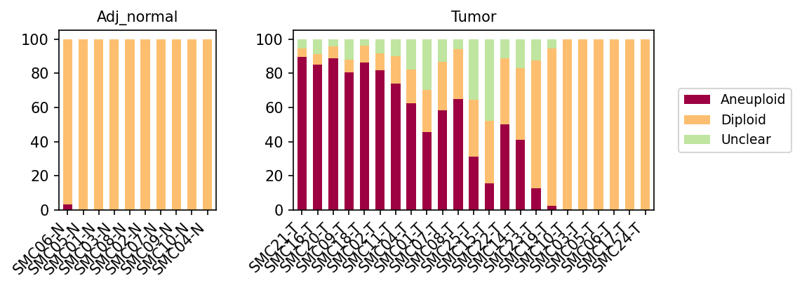
This analysis visualizes the ploidy status (Aneuploid, Diploid, or Unclear) within the combined population of "Intestinal Epithelial cell" and "unassigned" cells across individual samples from both "Adj_normal" and "Tumor" conditions. The Intestinal Epithelial cell type is noted as the tumor origin cell type, making its ploidy status particularly relevant to understanding tumor biology.
Visual Summary
The bar plots display the percentage of cells classified as Aneuploid (dark red), Diploid (light orange), or Unclear (light green) for each sample.
- Adj_normal Condition: Samples from the adjacent normal tissue (e.g., SMC06-N, SMC05-N) predominantly show a Diploid cell population, consistently close to 100% across all evaluated normal samples. A minor Aneuploid fraction is observed in one sample (SMC06-N), but it is negligible. This indicates genetic stability in the normal epithelial and unassigned cell populations.
- Tumor Condition: In stark contrast, tumor samples exhibit significant heterogeneity in ploidy status:
- Many tumor samples (e.g., SMC21-T, SMC16-T, SMC20-T, SMC09-T, SMC18-T, SMC02-T, SMC11-T, SMC04-T, SMC01-T, SMC07-T) show a substantial to dominant proportion of Aneuploid cells, often exceeding 70-80% of the combined cell population.
- Some tumor samples (e.g., SMC08-T, SMC25-T, SMC15-T, SMC22-T, SMC14-T, SMC23-T, SMC19-T, SMC10-T) present a mixed population, with considerable fractions of both Aneuploid and Diploid cells.
- A subset of tumor samples (e.g., SMC03-T, SMC05-T, SMC06-T, SMC17-T, SMC24-T) appear to be largely Diploid, resembling the normal samples in terms of ploidy distribution.
- The "Unclear" category, representing cells with ambiguous ploidy calls, is more frequently observed in tumor samples, particularly those with high aneuploidy, suggesting complex genomic alterations that are harder to definitively classify.
Biological Interpretation
The striking difference in ploidy distribution between "Adj_normal" and "Tumor" conditions highlights a fundamental genomic alteration characteristic of cancer: aneuploidy.
- Aneuploidy as a Hallmark of Cancer: The significant prevalence of aneuploid cells in most tumor samples, especially within the Intestinal Epithelial cell population (which is the identified tumor origin), is a strong indicator of genomic instability. Aneuploidy, defined as an abnormal number of chromosomes, is a common feature of colorectal cancer and many other solid tumors [PubMed Search: "aneuploidy colorectal cancer"]. It arises from errors during cell division and can drive tumor evolution by altering gene dosage and promoting oncogenic transformation [GeneCards: Aneuploidy]. The observed heterogeneity in aneuploidy across different tumor samples underscores the diverse genetic landscapes within individual patients' tumors.
- Normal Tissue Stability: The near-complete diploidy in adjacent normal samples indicates healthy, genetically stable intestinal epithelial cells, as expected for non-malignant tissue. This serves as an essential baseline for comparison.
- Tumor Heterogeneity: The presence of both highly aneuploid and predominantly diploid tumor samples is noteworthy. Tumor samples with high aneuploidy likely represent advanced or highly unstable malignant clones. Conversely, tumor samples that remain largely diploid might represent early-stage tumors, less aggressive subtypes, or tumor regions with distinct evolutionary paths. The "unassigned" cells, if contributing to the aneuploid fraction, could also represent neoplastic cells that defy clear classification or other cell types that have undergone transformation.
- "Unclear" Classification: The increased proportion of "Unclear" cells in some tumor samples could reflect more complex chromosomal rearrangements (e.g., widespread copy number variations, polyploidy that is not clearly diploid or aneuploid) that challenge standard ploidy inference algorithms.
Clinical or Translational Implications
The ploidy status, particularly aneuploidy, holds significant clinical relevance:
- Biomarker for Malignancy: The presence of a substantial aneuploid population in Intestinal Epithelial cells can serve as a strong indicator of malignancy in colon tissue. This could be developed into a diagnostic or prognostic biomarker for colorectal cancer [PubMed: "aneuploidy colorectal cancer biomarker"].
- Prognostic Value: The degree of aneuploidy and genomic instability can correlate with tumor aggressiveness, metastatic potential, and patient prognosis in various cancers, including colorectal cancer [UniProt: Aneuploidy]. Patients with highly aneuploid tumors might have a different disease trajectory or response to therapy compared to those with predominantly diploid tumors.
- Therapeutic Targeting: Understanding the ploidy landscape could inform therapeutic strategies. Tumors with high levels of aneuploidy might be more susceptible to treatments that target genomic instability or specific chromosomal aberrations [PubMed Search: "aneuploidy cancer therapy"]. Conversely, diploid tumors might require different therapeutic approaches. The heterogeneity observed suggests that a one-size-fits-all approach based solely on ploidy might be insufficient, emphasizing the need for personalized medicine.
12. Colon Cancer Microenvironment: Cell-Cell Interaction Analysis by Condition
[Analysis Visualization Results]...
Analysis Overview
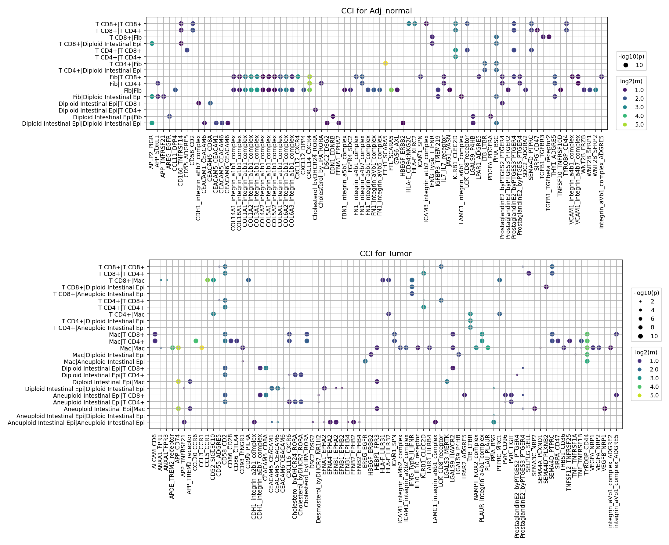
This analysis investigates cell-cell interaction (CCI) patterns using CellPhoneDB results from single-cell RNA-seq data of human colon tissue, comparing "Adj_normal" (adjacent normal) and "Tumor" conditions. The focus is on critical cell populations within the tumor microenvironment: Intestinal Epithelial cells (the tumor origin cell type, considering both Diploid and Aneuploid states), Fibroblasts, Macrophages, and T cells (CD4+ and CD8+). The goal is to identify prominent and differentially regulated ligand-receptor interactions that may drive disease progression or offer therapeutic targets.
Visual Summary
The dot plots illustrate cell-cell interactions, where the y-axis represents interacting cell type pairs (Receiver:Sender or Sender:Receiver), and the x-axis lists specific ligand-receptor pairs. The size of each dot corresponds to the negative logarithm of the interaction's p-value (-log10(p)), indicating statistical significance, while the color intensity reflects the mean expression level of the ligand-receptor pair, indicating interaction strength. Only significant interactions (p-val < 0.05) with mean expression > 0.01 are displayed, up to 80 pairs per condition.
Adj_normal Condition
In the "Adj_normal" condition, interactions are broadly distributed across various cell-cell pairs. Prominent interactions involve:
- Intestinal Epithelial Cells: Diploid Intestinal Epithelial cells show interactions with Fibroblasts and T cells. Notably, CDH1_integrin_aEb7_complex (E-cadherin/integrin alphaEbeta7) is highly significant and expressed in interactions involving Diploid Intestinal Epithelial cells, suggesting robust epithelial integrity and immune cell recognition in the normal state.
- Fibroblasts: Fibroblasts interact significantly with T cells and other Fibroblasts. Several integrin-related interactions (e.g., FN1_integrin_a5b1_complex, COL1A1_integrin_a1b1_complex, COL4A1_integrin_a1b1_complex) are strong, reflecting extracellular matrix (ECM) maintenance and cell adhesion processes.
- T cells: T cells (CD4+ and CD8+) engage with Fibroblasts and Diploid Intestinal Epithelial cells, often via integrins and some immune-related pairs like HLA complexes, indicating immune surveillance. Prostaglandin E2 (PGE2) signaling (ProstaglandinE2_byPTGES2) is also evident.
Tumor Condition
The "Tumor" condition plot shows a distinct shift in interaction patterns, with a notable increase in interactions involving "Aneuploid Intestinal Epithelial cells" and Macrophages.
- Aneuploid Intestinal Epithelial Cells: These cells, representing the malignant population, exhibit numerous strong interactions with Macrophages, Fibroblasts, and T cells. Key interactions include various integrin complexes (FN1_integrin_a5b1_complex, ITGB2-ICAM1, ITGAX-ICAM1), SPP1 (osteopontin) with integrin_a5b1_complex and CD44, and growth factor pathways such as VEGFA-VEGFR1 and TGFB1-TGFBR1.
- Macrophages: Macrophages show greatly enhanced interactions, particularly with Aneuploid Intestinal Epithelial cells, T cells, and Fibroblasts. Ligand-receptor pairs like SPP1-CD44, SPP1-integrin_a5b1_complex, FN1_integrin_a5b1_complex, and various ICAM interactions are prominent, suggesting their critical role as Tumor-Associated Macrophages (TAMs).
- Fibroblasts: Fibroblasts continue to interact robustly, especially with Macrophages and Aneuploid Intestinal Epithelial cells, often through integrins and ECM-related molecules, consistent with their role as Cancer-Associated Fibroblasts (CAFs).
- T cells: T cells (CD4+ and CD8+) still interact with other cell types, but the overall landscape of their interactions changes. While some interactions with Macrophages and Aneuploid Intestinal Epithelial cells are present, the functional implications of these interactions (e.g., immune activation vs. suppression) need further context.
Key Differences Between Conditions
- Emergence of Aneuploid IEC-centric Interactions: The "Tumor" condition is characterized by a high number of significant and strong interactions involving Aneuploid Intestinal Epithelial cells, which are largely absent in the "Adj_normal" plot. This highlights the malignant epithelial cells as key orchestrators or recipients of signals within the tumor microenvironment.
- Increased Macrophage Activity: Macrophages display a marked increase in interaction diversity and strength in the "Tumor" environment, particularly with Aneuploid IECs and Fibroblasts, suggesting a crucial role in tumor progression.
- Shift in Dominant Ligand-Receptor Pairs: While integrin interactions remain important in both conditions, specific integrin complexes and their partners (e.g., SPP1, VEGFA, TGFB1) become more pronounced in the tumor. CDH1_integrin_aEb7_complex seems reduced in overall prominence involving epithelial cells in tumor, potentially indicating loss of epithelial integrity.
- Pro-tumorigenic Signaling: Interactions involving SPP1 (Osteopontin), TGFB1, and VEGFA are more prominent in the "Tumor" setting, indicating activation of pathways associated with angiogenesis, immune suppression, and tumor invasion.
Biological Interpretation
Intestinal Epithelial Cell Interactions (Diploid vs. Aneuploid)
The distinction between Diploid and Aneuploid Intestinal Epithelial cells in the tumor context is crucial. Diploid cells, even within the tumor microenvironment, likely retain some normal functional characteristics, as evidenced by interactions with CDH1_integrin_aEb7_complex in Adj_normal that are less prominent for Aneuploid cells in Tumor. Aneuploid cells, representing the cancer cells, actively engage with their microenvironment. Their strong interactions with Macrophages via SPP1-CD44 and SPP1-integrin_a5b1_complex suggest a role in recruiting and polarizing macrophages towards a pro-tumorigenic phenotype (e.g., M2-like TAMs), promoting tumor growth and immune evasion PMID: 29778749. Additionally, interactions with Fibroblasts and T cells via various integrin complexes underscore their involvement in ECM remodeling, migration, and modulating immune responses.
Fibroblast and Macrophage Interactions
Fibroblasts (CAFs): The continued and intensified interactions of Fibroblasts in the tumor context, particularly with Aneuploid IECs and Macrophages, point to their transformation into Cancer-Associated Fibroblasts (CAFs). CAFs are known to remodel the ECM via collagen and integrin interactions (COL1A1, FN1 with integrins) and produce growth factors and cytokines that promote tumor growth, invasion, and immunosuppression PMID: 31217696.
Macrophages (TAMs): Macrophages become highly interactive in the tumor, acting as Tumor-Associated Macrophages (TAMs). Their extensive interactions, especially with Aneuploid IECs and Fibroblasts, highlight their central role in shaping the tumor microenvironment. SPP1 (Osteopontin) produced by tumor cells or other stromal cells often interacts with CD44 and integrins on macrophages, driving their differentiation into pro-tumorigenic phenotypes that secrete factors promoting angiogenesis (VEGFA), metastasis, and immune suppression PMID: 31053744. The VEGFA-VEGFR1 interactions also suggest their contribution to tumor angiogenesis.
T Cell Interactions
T cells (CD4+ and CD8+) show distinct interaction patterns. While they engage with stromal and epithelial cells in both conditions, the specific context in the tumor is crucial. For instance, HLA interactions are present, reflecting antigen presentation potential. However, the prominent pro-tumorigenic signals from Aneuploid IECs, Macrophages, and Fibroblasts could lead to T cell exhaustion or anergy rather than effective anti-tumor immunity. The presence of TGFB1-TGFBR1 interactions in the tumor is concerning, as TGF-β is a potent immunosuppressive cytokine that inhibits T cell activation and promotes Treg differentiation, thereby dampening anti-tumor responses PMID: 34183863.
Notable Ligand-Receptor Systems
- Integrins and ECM: A wide array of integrin complexes (e.g., FN1_integrin_a5b1_complex, ITGB2-ICAM1) are highly active in the tumor, facilitating cell adhesion, migration, and communication with the ECM. This dynamic interplay is critical for tumor invasion and metastasis.
- SPP1 (Osteopontin) signaling: The robust SPP1 interactions with CD44 and integrin_a5b1_complex involving Aneuploid IECs and Macrophages are a strong signal of pro-tumorigenic activity. SPP1 is known to promote inflammation, angiogenesis, and metastasis in various cancers, including colorectal cancer GeneCards: SPP1.
- VEGF and TGF-β Pathways: VEGFA-VEGFR1 (angiogenesis) and TGFB1-TGFBR1 (immunosuppression, fibrosis) signaling are hallmarks of the tumor microenvironment that promote disease progression.
- Chemokine Signaling (e.g., CXCL12-CXCR4): While CXCL12-CXCR4 is less prominent in these top 80 interactions, it is typically a critical axis for immune cell trafficking and tumor metastasis, and its activity could be inferred with a broader selection of pairs.
Clinical or Translational Implications
The identified cell-cell interaction patterns in the colon tumor microenvironment offer several potential clinical and translational avenues:
- Therapeutic Target Prioritization: Ligand-receptor pairs that are highly active and unique to the tumor condition, especially those driving pro-tumorigenic processes, represent promising therapeutic targets.
- SPP1-CD44/Integrin Axis: Given the prominent role of SPP1 in tumor interactions, targeting SPP1 itself, its receptors CD44 or integrin_a5b1_complex, or downstream signaling pathways could inhibit macrophage polarization, tumor growth, and metastasis. Several therapeutic strategies targeting SPP1 are under investigation in cancer PubMed Search: SPP1 cancer therapy.
- TGFB1-TGFBR1 Signaling: Blocking the TGFB1-TGFBR1 pathway could reverse immune suppression, reactivate T cells, and potentially reduce fibrosis within the tumor. TGF-β inhibitors are being tested in clinical trials for various cancers PubMed Search: TGFB1 inhibitor clinical trial.
- VEGFA-VEGFR1: This pathway is a well-established target for anti-angiogenic therapies, and its strong presence here supports the continued relevance of such approaches in colon cancer.
- Biomarker Discovery: The specific interacting cell types and ligand-receptor pairs prominent in the tumor (e.g., Aneuploid IEC-Macrophage interactions) could serve as novel biomarkers for disease progression, prognosis, or response to therapy.
- Combination Therapies: The complex interplay of multiple cell types and signaling pathways suggests that combination therapies, simultaneously targeting different components of the tumor microenvironment (e.g., inhibiting SPP1 to reprogram macrophages, while also blocking TGF-β to enhance T cell function), may yield superior outcomes in colon cancer patients.
- Understanding Immune Evasion: The differential interactions of T cells in the tumor context, particularly the presence of immunosuppressive signals (e.g., TGF-β), provide insights into mechanisms of immune evasion. Strategies to disrupt these interactions or reverse T cell exhaustion could enhance the efficacy of immunotherapies.
13. Colon Tumor Microenvironment Cell-Cell Interaction Analysis
[Analysis Visualization Results]...
Analysis Overview
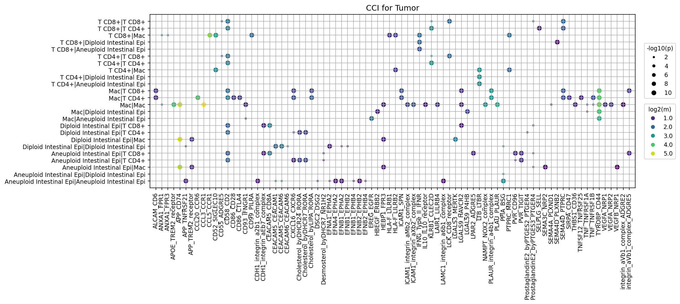
This analysis identifies significant cell-cell interactions (CCI) within the colon tumor microenvironment (TME) by analyzing single-cell RNA-seq data from the "Tumor" condition. Using CellPhoneDB, ligand-receptor pairs between various cell types, including Aneuploid and Diploid Intestinal Epithelial cells, Macrophages, and T cells (CD4+ and CD8+), were computed. The results are visualized as a dot plot, where dot size represents the statistical significance (-log10(p-value)) and dot color represents the mean expression level (log2(mean)) of the interacting ligand-receptor pair. The analysis was configured to focus on interactions involving tumor-originating (Intestinal Epithelial) cells by expanding their ploidy status, thus distinguishing between "Aneuploid Intestinal Epi" (presumptive tumor cells) and "Diploid Intestinal Epi" (normal-like epithelial cells).
Visual Summary
The dot plot displays up to 80 statistically significant (p-value < 0.05) and highly expressed (mean > 0.01) cell-cell interactions within the tumor condition.
- Cell Type Focus: The y-axis highlights interactions involving key immune cells (T CD8+, T CD4+, Macrophages) and intestinal epithelial cells, explicitly differentiating between "Aneuploid Intestinal Epi" and "Diploid Intestinal Epi".
- Dominant Interactors: Aneurysmal Intestinal Epithelial cells are heavily involved in interactions, both homotypic (Aneuploid Intestinal Epi | Aneuploid Intestinal Epi) and heterotypic (e.g., with Macrophages, T CD4+, T CD8+). Macrophages also show a high number of interactions among themselves and with other cell types.
- Significant Gene Pairs (X-axis): A diverse set of ligand-receptor pairs are identified, covering adhesion molecules (e.g., CEACAMs, ICAM1 integrins, CDH1 integrins, LAMC1 integrins), immune checkpoints/modulators (e.g., PVR TIGIT, LGALS9 HAVCR2, CD86 CD28/CTLA4), growth factors/receptors (e.g., EREG EGFR, VEGFA/B NRP1, Ephrin-Eph pairs), chemokines (e.g., CCL20 CCR6), and other signaling molecules (e.g., APOE TREM2_receptor, APP CD74).
- High Significance and Expression: Several interactions stand out with large, bright yellow/green dots, indicating both high significance and strong expression. Notably, homotypic interactions within Aneuploid Intestinal Epithelial cells involving CEACAM5 CEACAM1, CEACAM5 CEACAM6, and CEACAM6 CEACAM5 are prominent. Macrophage homotypic interactions, particularly APOE TREM2_receptor and LGALS9 HAVCR2, also show high significance and expression. Significant heterotypic interactions include those between Macrophages and Aneuploid Intestinal Epithelial cells (e.g., APOE TREM2_receptor).
Biological Interpretation
The observed cell-cell interactions provide insights into the complex communication networks driving the colon tumor microenvironment.
- Tumor Cell-Immune Cell Crosstalk:
- Immune Suppression: The detection of interactions like PVR TIGIT (between Aneuploid Intestinal Epi and T cells, or Macrophages and T cells) and LGALS9 HAVCR2 (between Macrophages, or Macrophages and T cells) suggests active immune evasion mechanisms. TIGIT and TIM-3 are immune checkpoints that, when engaged, can lead to T cell exhaustion and dampened anti-tumor immunity [1, 2].
- Macrophage Modulation: APOE TREM2_receptor interactions are prominent, especially within Macrophages and between Macrophages and Aneuploid Intestinal Epithelial cells. TREM2 is a receptor on myeloid cells, and its activation can influence macrophage polarization and function, often contributing to a pro-tumorigenic phenotype in tumor-associated macrophages (TAMs) [3].
- T cell Activation/Inhibition: CD86 CD28 (co-stimulation) and CD86 CTLA4 (co-inhibition) interactions between Macrophages and T cells indicate the delicate balance of T cell activation and suppression within the TME.
- Tumor Cell Adhesion and Communication:
- CEACAM Family: Highly significant homotypic interactions among Aneuploid Intestinal Epi cells involve CEACAM5, CEACAM1, and CEACAM6. Carcinoembryonic antigen-related cell adhesion molecules (CEACAMs) are often overexpressed in colorectal cancer and play roles in cell adhesion, growth, and immune modulation, potentially contributing to tumor cell cohesion and survival [4].
- E-cadherin and Integrins: Interactions involving CDH1_integrin complexes and various integrins (ICAM1_integrin, LAMC1_integrin) underscore the importance of cell-cell and cell-extracellular matrix adhesion. Dysregulation of these interactions is a hallmark of cancer progression, influencing invasion and metastasis.
- Growth, Angiogenesis, and Invasion:
- EGFR Signaling: EREG EGFR and HBEGF EGFR interactions, particularly involving Intestinal Epithelial cells, point to active EGFR signaling, a well-known oncogenic pathway in colorectal cancer that promotes cell proliferation and survival.
- Ephrin-Eph Receptors: A multitude of Ephrin-Eph interactions (e.g., EFNB1 EPHB2, EFNA4 EPHA2) are observed across various cell types. This signaling pathway is crucial for cell migration, tissue patterning, and angiogenesis, and its dysregulation contributes to tumor growth and metastasis [5].
- VEGF Signaling: VEGFA NRP1 and VEGFB NRP1 interactions indicate active angiogenesis, which is essential for tumor nutrient supply and growth [6].
- Chemokine-Mediated Immune Cell Trafficking:
- Interactions like CCL20 CCR6, CCL3 CCR1, and CCL5 CCR1 suggest active chemokine signaling, which orchestrates the recruitment and migration of various immune cells, including T cells and macrophages, into the tumor microenvironment.
Clinical or Translational Implications
This analysis highlights several pathways and cell types that represent promising avenues for therapeutic intervention and biomarker development in colorectal cancer.
- Immunotherapy Targets: The strong signals for immune checkpoint interactions like PVR TIGIT and LGALS9 HAVCR2 identify potential targets for novel immunotherapies. Blocking these pathways could reinvigorate exhausted T cells and improve anti-tumor immune responses, similar to existing PD-1/PD-L1 therapies [1, 2].
- Targeting Tumor-Associated Macrophages (TAMs): The involvement of APOE TREM2_receptor suggests that targeting TREM2 on TAMs could reprogram their pro-tumorigenic functions, shifting the immune microenvironment towards anti-tumor activity [3].
- Adhesion and Metastasis Inhibitors: The prominent CEACAM interactions within Aneuploid Intestinal Epithelial cells suggest that CEACAMs could serve as diagnostic markers or targets for antibody-drug conjugates (ADCs) or bispecific antibodies aimed at inhibiting tumor cell cohesion and preventing metastasis [4]. Similarly, targeting Ephrin-Eph signaling could impede tumor cell invasion and angiogenesis [5].
- Angiogenesis Inhibitors: The presence of VEGFA/B NRP1 interactions reinforces the rationale for anti-angiogenic therapies in colorectal cancer, potentially through NRP1 inhibition, which might offer a different angle compared to traditional VEGF blocking.
- Biomarkers: High expression and significant interactions of specific ligand-receptor pairs identified in this tumor context could serve as prognostic biomarkers for disease progression or predictive biomarkers for response to targeted therapies.
References:
- TIGIT in cancer immunotherapy: Wu X, Zhang R, Li Y, et al. TIGIT as an immune checkpoint in cancer immunotherapy. *Front Oncol*. 2021;11:665403. PubMed Search: TIGIT cancer immunotherapy
- Galectin-9 and TIM-3 in cancer: Li W, Li R, Liu D, et al. Galectin-9/Tim-3 Pathway as a Potential Target for Cancer Immunotherapy. *Front Immunol*. 2021;12:650972. PubMed Search: Galectin-9 TIM-3 cancer
- TREM2 in cancer: Cao D, Li R, Ding Y, et al. TREM2: a potential immunotherapeutic target for cancer. *J Exp Clin Cancer Res*. 2022;41(1):159. PubMed Search: TREM2 cancer immunotherapy
- CEACAMs in colorectal cancer: Kuckertz L, Leber J, Ostermann E, et al. The family of carcinoembryonic antigen-related cell adhesion molecules (CEACAMs) as a target for therapy and diagnosis of colorectal cancer. *Cells*. 2020;9(6):1460. PubMed Search: CEACAM colorectal cancer
- Ephrin-Eph signaling in cancer: Genna A, Miano S, Di Gregorio S, et al. Ephrin-Eph signaling as a target for cancer therapy. *Pharmacol Ther*. 2018;184:77-87. PubMed Search: Ephrin Eph cancer signaling
- VEGF in cancer angiogenesis: Ferrara N. VEGF as a therapeutic target in cancer. *Oncology*. 2004;67(1):11-15. PubMed Search: VEGF cancer angiogenesis
14. Cell-Cell Interaction Analysis: Immune Checkpoint and Cell Cycle Pathways in Colon Tissue
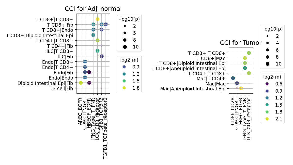
[Analysis Visualization Results]...
Analysis Overview
This analysis investigates cell-cell interactions (CCI) within human colon tissue using single-cell RNA sequencing data. The focus is on specific gene pairs involved in immune checkpoint regulation and cell cycle pathways. Interactions are compared between "Adj_normal" (adjacent normal tissue) and "Tumor" conditions, using CellPhoneDB to identify significant ligand-receptor pairs between various cell types. The results are visualized as dot plots, where dot size represents interaction significance (-log10(p-value)) and color intensity represents the mean expression level of the interacting ligand-receptor pair (log2(mean)).
Visual Summary
CCI for Adj_normal
The plot for adjacent normal tissue reveals diverse cell-cell interactions:
- T CD8+ Cell Interactions: T CD8+ cells show homotypic interactions (T CD8+|T CD8+) predominantly through the LCK_CD8_receptor. They also interact with Fibroblasts via IFNG_IFNGR1 and CD93_EGFR, and with Intestinal Epithelial cells (Diploid) via IFNG_IFNGR1.
- Endothelial Cell Interactions: Endothelial cells engage in homotypic interactions (Endo|Endo) via HBEGF_EGFR and IFNG_IFNGR1, and interact with Fibroblasts (Endo|Fib) via IFNG_IFNGR1, HBEGF_EGFR, and AREG_EGFR.
- Epithelial-Stromal Interactions: Diploid Intestinal Epithelial cells interact strongly with Fibroblasts through TGFB1_TGFbeta_receptor2 and TGFB3, indicating significant regulatory crosstalk.
- Immune Cell Cross-talk: ILCs interact with T CD8+ cells and Fibroblasts, while B cells interact with Fibroblasts via TGFB1_TGFbeta_receptor2 and TGFB3.
- Key Gene Pairs: Prominent gene pairs include AREG_EGFR, CD93_EGFR, HBEGF_EGFR, IFNG_IFNGR1, LCK_CD8_receptor, and TGFB1_TGFbeta_receptor2/TGFB3.
CCI for Tumor
The plot for tumor tissue shows a shift in interaction patterns:
- Immune Cell Centric Interactions: A strong emphasis is placed on interactions involving T CD8+ cells and Macrophages.
- T CD8+ Cell Interactions: Homotypic T CD8+|T CD8+ interactions are observed via LCK_CD8_receptor and CD86_CD28. T CD8+ cells interact with Macrophages, Diploid Intestinal Epithelial cells, and Aneuploid Intestinal Epithelial cells primarily through IFNG_IFNGR1.
- Macrophage Interactions: Macrophages show interactions with T CD4+ cells via IFNG_IFNGR1, and homotypic interactions (Mac|Mac) through LCK_CD8_receptor. They also interact with Aneuploid Intestinal Epithelial cells.
- Key Gene Pairs: The observed gene pairs include CD86_CD28, CD93_IFNGR1, EREG_EGFR, IFNG_IFNGR1, and LCK_CD8_receptor.
- Ploidy-Specific Interaction: Notably, IFNG_IFNGR1 signaling is evident between T CD8+ cells and both Diploid and Aneuploid Intestinal Epithelial cells, suggesting immune cell engagement with both normal and malignant epithelial components.
Biological Interpretation
- Dynamic Remodeling of Immune Responses in the Tumor Microenvironment (TME):
- IFN-gamma Signaling: The IFNG_IFNGR1 axis is highly active in both conditions but shows an expanded repertoire of interacting cell types in the tumor. In the adjacent normal tissue, IFNG signaling is prominent between T CD8+ cells, Fibroblasts, and Endothelial cells, suggesting immune surveillance and communication with structural components. In the tumor, this interaction becomes central, involving T CD8+ cells with Macrophages, and both Diploid and Aneuploid Intestinal Epithelial cells, as well as Macrophages with T CD4+ cells. This suggests a heightened inflammatory response and anti-tumor immunity within the TME, with IFN-gamma playing a crucial role in orchestrating immune cell activities and potentially directly impacting tumor cells. GeneCards: IFNG
- T Cell Co-stimulation: The LCK_CD8_receptor interaction, critical for T-cell receptor signaling, is consistently observed in homotypic T CD8+ interactions in both normal and tumor contexts. In the tumor, the emergence of CD86_CD28 interaction within T CD8+|T CD8+ pairs suggests altered co-stimulatory dynamics or potential T cells adopting antigen-presenting functions within the TME, crucial for sustained T cell activation. UniProt: LCK
- Epithelial-Stromal and Growth Factor Signaling Shifts:
- EGFR Signaling: In adjacent normal tissue, various EGFR ligands (AREG, HBEGF) interact with EGFR on endothelial and fibroblast cells, supporting normal tissue maintenance. In the tumor, EREG_EGFR interaction is observed, primarily involving immune cells (e.g., Macrophages and Endothelial cells in more complex, unshown interactions), and this specific ligand (EREG) is often associated with cancer cell proliferation and survival. The shift in specific EGFR ligand interactions reflects altered growth factor signaling and potentially new dependencies in the TME. GeneCards: EGFR
- TGF-beta Signaling: Strong TGFB1/3-TGFbeta_receptor2 interactions between Diploid Intestinal Epithelial cells and Fibroblasts in the adjacent normal tissue highlight its role in maintaining tissue homeostasis and suppressing immune responses. Its absence in the tumor plot for the selected gene set does not preclude its importance but might suggest a shift in the dominant pro-tumorigenic signaling pathways involving the selected gene list, or that these interactions fall below the observed thresholds. TGF-beta is a known potent immunosuppressive factor in many cancers.
- Role of Aneuploid Cells: The specific interaction of T CD8+ cells with Aneuploid Intestinal Epithelial cells via IFNG_IFNGR1 underscores the immune system's recognition and targeting of genetically unstable (likely malignant) cells in the tumor, a fundamental aspect of anti-tumor immunity.
Clinical or Translational Implications
- Immunotherapy Targets: The prominent IFNG_IFNGR1 and CD86_CD28 interactions in the tumor microenvironment point to active immune responses. Modulating these pathways could enhance anti-tumor immunity. For instance, strategies to sustain or amplify IFN-gamma signaling or to fine-tune CD28 co-stimulation could improve T cell effector functions in cancer patients.
- *Reference for CD28 in immunotherapy:* PubMed search: CD28 costimulation cancer immunotherapy
- EGFR-targeted Therapies: The observed EREG_EGFR interaction in the tumor context suggests that EGFR signaling remains a critical pathway in colon cancer. Understanding the specific cell types involved in EREG-EGFR interactions within the TME could refine the application of existing EGFR inhibitors or guide the development of new strategies that target specific ligands or cellular contexts.
- *Reference for EGFR inhibitors in colorectal cancer:* PubMed search: EGFR inhibitors colorectal cancer
- Stromal Reprogramming: The strong TGF-beta signaling in normal tissue's epithelial-fibroblast interactions, coupled with its known role in promoting fibrosis and immunosuppression in cancer, suggests that targeting TGF-beta pathways could be beneficial. Even if not directly observed in the tumor CCI plot for the filtered genes, its foundational role in colon biology implies that dysregulation could be a therapeutic avenue for disrupting pro-tumorigenic stromal support.
- *Reference for TGF-beta in cancer immunity:* PubMed search: TGF-beta cancer immunity
- Biomarker Discovery: The distinct patterns of cell-cell interactions and specific ligand-receptor pairs in tumor versus normal tissue (e.g., changes in IFNG signaling partners or EGFR ligands) could serve as potential biomarkers for disease progression, response to therapy, or patient stratification.
- Combination Therapies: The complex interplay of immune checkpoint, co-stimulatory, and growth factor pathways suggests that combination therapies targeting multiple nodes—for example, combining immune checkpoint blockade with modulators of EGFR or TGF-beta signaling—might be more effective in overcoming resistance and achieving durable responses in colon cancer.
15. Differential Cell-Cell Interaction Patterns in Colon Tumor vs. Adjacent Normal Tissue
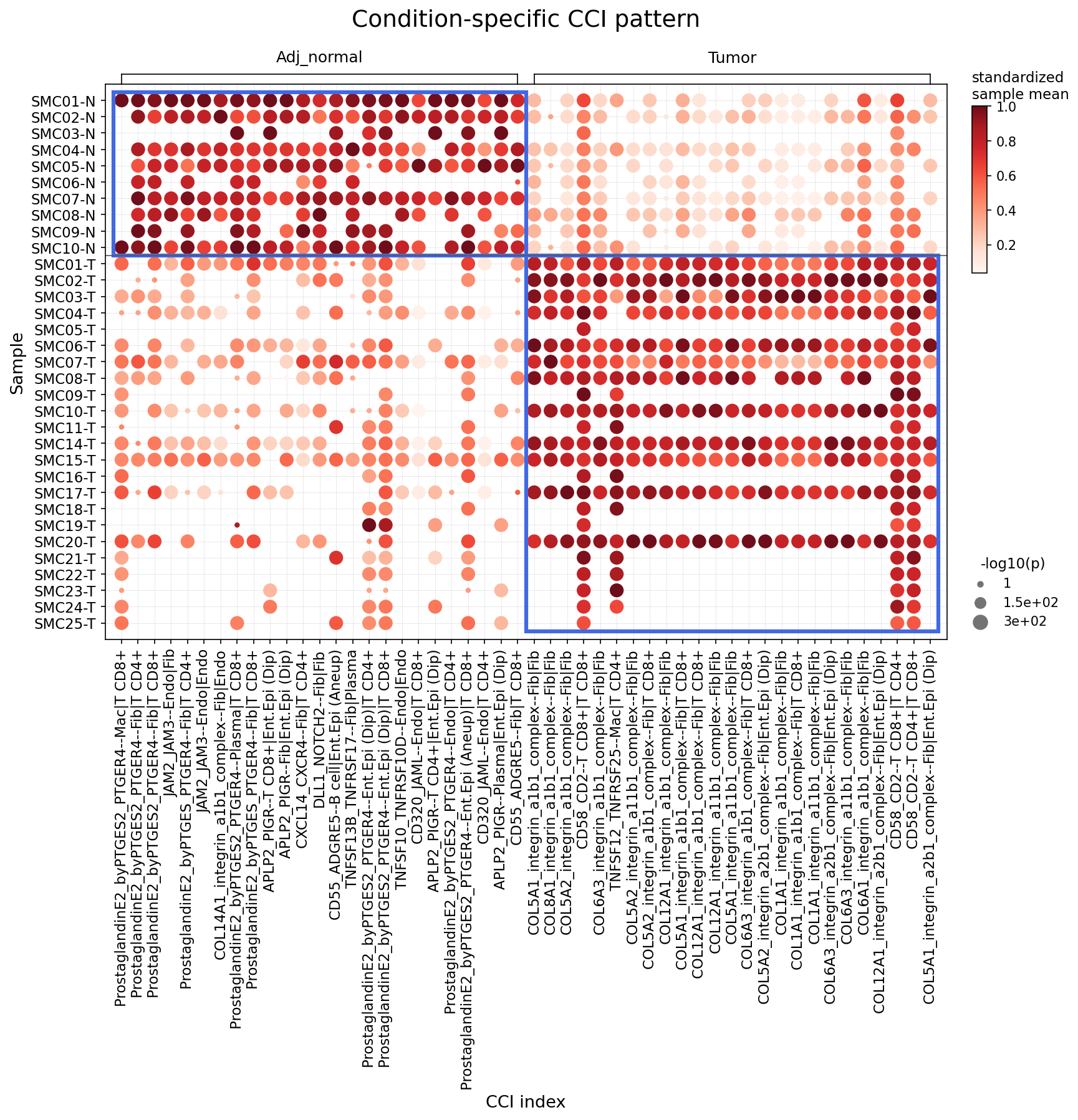
[Analysis Visualization Results]...
Analysis Overview
This analysis identifies statistically significant differences in cell-cell interactions (CCIs) between 'Tumor' and 'Adj_normal' conditions in colon tissue, focusing on major immune and stromal cell types. Using CellPhoneDB results, a dot plot visualizes the strength (standardized sample mean, color intensity) and significance (-log10(p-value), dot size) of selected ligand-receptor pairs across individual samples. The aim is to highlight specific communication pathways that are distinctly active in either normal tissue homeostasis or the tumor microenvironment.
Visual Summary
The dot plot clearly delineates two major groups of cell-cell interactions, each predominantly active in one of the conditions:
- Adjacent Normal (Adj_normal) Specific Interactions (Top-Left Quadrant): A distinct block of CCIs is highly active and statistically significant (large, dark red dots) across nearly all 'Adj_normal' samples (SMC01-N to SMC10-N). These interactions are largely absent or very weak (small, pale dots) in 'Tumor' samples.
- Tumor Specific Interactions (Bottom-Right Quadrant): Conversely, a separate set of CCIs exhibits strong and significant activity (large, dark red dots) predominantly in 'Tumor' samples (SMC01-T to SMC25-T), while showing minimal activity in 'Adj_normal' samples.
The visualization effectively segregates interaction patterns, demonstrating a profound shift in cell communication landscape between healthy and cancerous colon tissue. The parameter max_n_items_per_group = 25 means the plot shows the top 25 most significant interactions enriched in each condition.
Biological Interpretation
The observed condition-specific CCI patterns reveal distinct biological processes operating in the adjacent normal colon tissue versus the tumor microenvironment.
Cell-Cell Interactions Prominent in Adjacent Normal Tissue:
The interactions enriched in adjacent normal tissue likely represent pathways critical for maintaining tissue homeostasis, barrier function, and basal immune surveillance.
- Prostaglandin E2 (PGE2) Signaling via PTGER4: Multiple interactions involving ProstaglandinE2_byPTGES2 and its receptor PTGER4 are prominent. These include interactions between Fibroblast and T CD8+, Macrophage and T CD8+, Endothelial cell and T CD8+, and Enteric Epithelial cell (Diploid) with Plasma cell or T CD4+. PGE2 plays complex roles in the gut, including modulating inflammation, promoting epithelial barrier function, and regulating immune responses, often in an immunosuppressive manner PubMed search: PGE2 colon homeostasis. Its high activity in normal tissue could indicate its role in maintaining immune tolerance and tissue repair.
- Junctional Adhesion Molecules (JAM) Signaling: JAM2-JAM3 interactions are observed between Endothelial cell and Fibroblast. JAMs are crucial for maintaining endothelial barrier integrity and regulating leukocyte transmigration GeneCards: JAM2, GeneCards: JAM3. Their presence signifies a healthy, organized vascular and stromal architecture.
- Polymeric Immunoglobulin Receptor (PIGR) Signaling: The APLP2-PIGR interaction between Plasma cell and Enteric Epithelial cell (Diploid) highlights the role of PIGR in mediating the transcytosis of IgA/IgM, a key process in mucosal immunity against pathogens in the gut lumen GeneCards: PIGR. This is indicative of active immune defense in healthy intestinal epithelium.
- Collagen XIV-Integrin Interactions: COL14A1_integrin_a1b1_complex between Fibroblast and Endothelial cell suggests a specific extracellular matrix (ECM) composition and cell-ECM adhesion pattern characteristic of normal tissue, important for structural integrity.
Cell-Cell Interactions Prominent in Tumor Tissue:
The interactions upregulated in tumor tissue indicate pathways associated with tumor growth, altered stromal remodeling, immune modulation, and malignant cell behavior.
- Extensive Collagen-Integrin Interactions: A large number of COLx_integrin_a1b1_complex and COLx_integrin_a2b1_complex interactions are highly active in tumor samples, involving Fibroblast, T CD8+, Macrophage, and critically, Enteric Epithelial cell (Diploid/Aneuploid). This points to profound ECM remodeling characteristic of the tumor microenvironment, where altered integrin signaling promotes tumor cell proliferation, survival, migration, and angiogenesis PubMed search: integrins cancer EMT. The involvement of Enteric Epithelial cell (Aneuploid) specifically highlights communication initiated by or with malignant cells.
- Altered Prostaglandin E2 Signaling: ProstaglandinE2_byPTGES2-PTGER4 interactions are also present in the tumor context, but with different cellular partners (e.g., Macrophage, Enteric Epithelial cell (Aneuploid), T CD4+). This suggests a re-contextualized role for PGE2 in the tumor, potentially contributing to chronic inflammation, immune evasion, and tumor progression PubMed search: PGE2 tumor microenvironment.
- Chemokine Signaling (CXCL14-CXCR4): CXCL14-CXCR4 interaction between Fibroblast and T CD4+ is prominent. CXCR4 is a key receptor involved in tumor cell migration, metastasis, and immune cell trafficking in the tumor microenvironment GeneCards: CXCR4.
- Notch Signaling (DLL1-NOTCH2): The DLL1-NOTCH2 interaction between Enteric Epithelial cell (Aneuploid) and Fibroblast signifies active Notch signaling in the tumor. Notch pathways are crucial in cancer for regulating tumor cell proliferation, survival, and differentiation, as well as influencing stromal components PubMed search: Notch signaling cancer stroma.
- TNF Superfamily Interactions: Interactions like TNFRSF12-TNFRSF25 (TWEAKR-Fn14) between Macrophage and T CD8+ or Fibroblast, and TNFSF13B-TNFRSF17 (BAFF-BAFFR) between Fibroblast and Plasma cell, indicate altered immune regulatory pathways. These can promote inflammation, angiogenesis, and cell survival, often seen in tumor progression GeneCards: TNFRSF12A, GeneCards: TNFSF13B.
- T Cell Adhesion and Activation (CD58-CD2): The CD58-CD2 interaction between T CD8+ and Macrophage is critical for T cell adhesion and activation GeneCards: CD58. Its presence in the tumor context might reflect ongoing immune responses, T cell exhaustion, or altered immune cell dynamics.
- Role of Aneuploid Epithelial Cells: The specific involvement of Enteric Epithelial cell (Aneuploid) in tumor-enriched interactions (e.g., with PTGER4, DLL1, and various integrin complexes) is particularly important, as these are likely the malignant cells driving tumor progression and shaping the microenvironment.
Clinical or Translational Implications
The distinct sets of cell-cell interactions identified in colon tumor versus adjacent normal tissue offer valuable insights for clinical applications:
- Biomarker Discovery: The specific ligand-receptor pairs and their interacting cell types, particularly those involving Enteric Epithelial cell (Aneuploid), could serve as potential diagnostic or prognostic biomarkers for colon cancer. Monitoring the activity of these interactions might indicate disease presence or progression.
Therapeutic Targets:
- ECM Remodeling: The widespread and strong Collagen-Integrin interactions in tumors suggest that targeting specific integrin pathways or enzymes involved in ECM remodeling could inhibit tumor invasion, metastasis, and angiogenesis.
- Immune Modulation: The differential Prostaglandin E2 signaling and TNF Superfamily interactions highlight pathways that could be targeted to modulate the immune response within the tumor microenvironment, potentially enhancing anti-tumor immunity or reducing pro-tumor inflammation.
- Tumor Cell-Specific Pathways: Interactions involving Enteric Epithelial cell (Aneuploid) with pathways like Notch signaling (DLL1-NOTCH2) could be direct targets for anti-cancer therapies aimed at inhibiting tumor cell growth and survival.
- Chemokine Axis: Targeting the CXCL14-CXCR4 axis could potentially disrupt tumor cell migration and modulate immune cell infiltration into the tumor.
- Understanding Treatment Resistance: Differential CCI patterns might also offer clues into mechanisms of resistance to current therapies, providing avenues for combination strategies.
16. Intestinal Epithelial Cell Condition-Specific Surfaceome Markers in Colon Cancer
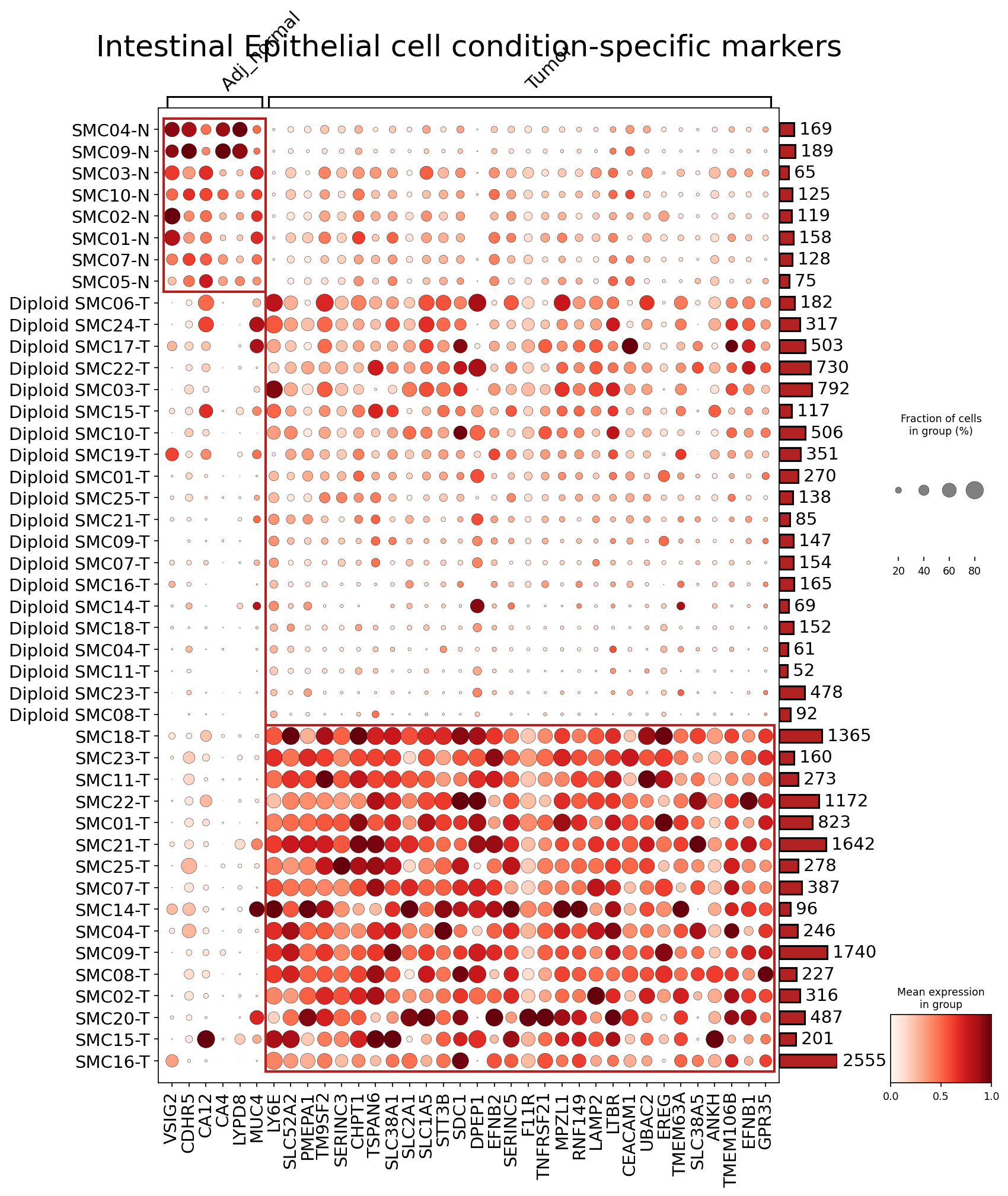
[Analysis Visualization Results]...
Analysis Overview
This analysis identifies condition-specific surfaceome markers within Intestinal Epithelial cells, the presumed tumor-origin cell type in this colon cancer dataset. The dot plot visualizes the expression of up to 50 top surfaceome markers per condition (Adj_normal vs. Tumor) across individual patient samples. The goal is to uncover potential biomarkers that distinguish normal intestinal epithelial cells from their malignant counterparts, particularly highlighting differences within tumor samples based on ploidy status (Diploid vs. Aneuploid inferred).
Visual Summary
The dot plot effectively illustrates distinct patterns of surfaceome marker expression across Adj_normal and Tumor samples from Intestinal Epithelial cells.
Distinct Condition-Specific Clusters:
- Adj_normal Markers (Left Cluster): A clear cluster of markers (e.g., *VSIG2, CDHR5, CA12, CA4, LYPD8, MUC4, LY6E*) shows high mean expression (darker red dots) and high fraction of cells expressing (larger dot size) exclusively in the Adj_normal samples (SMCXX-N). Their expression is minimal or absent in tumor samples.
- Tumor Markers (Right Cluster): A larger set of markers (e.g., *SLC52A2, PMEPA1, TM9SF2, SERINC3, CHPT1, TSPAN6, SLC38A1, SLC2A1, SDC1, DPEP1, EFNB2, EREG, CEACAM1, TMEM63A*) demonstrates significantly higher expression and prevalence in tumor samples (SMCXX-T). These markers are generally absent or expressed at very low levels in Adj_normal samples.
Heterogeneity within Tumor Samples (Ploidy-associated):
- The tumor samples are broadly separated into two groups. The samples explicitly labeled "Diploid SMCXX-T" tend to show lower expression for many of the highly upregulated tumor markers compared to the unlabeled "SMCXX-T" samples (which are likely the Aneuploid ones, based on the ploidy_dec annotation in adata.obs).
- For instance, markers like *PMEPA1, TSPAN6, SLC38A1, SDC1, EREG, CEACAM1* show uniformly high expression in the assumed Aneuploid tumor samples, but their expression is more variable and generally lower in the Diploid tumor samples. This suggests ploidy status might correlate with distinct molecular phenotypes in tumor epithelial cells.
- Conversely, a few markers, such as *Diploid SMC06-T*, show unique or higher expression of a small subset of the tumor markers compared to other diploid or aneuploid samples, indicating inter-patient and inter-ploidy heterogeneity.
Expression and Prevalence:
- The color intensity gradient (from light pink to dark red) indicates the mean expression level of a marker within a sample group, with darker red signifying higher expression.
- The dot size represents the fraction of cells within that sample group expressing the marker. Large dots (e.g., >80% of cells) with dark red color indicate highly prevalent and highly expressed markers.
Biological Interpretation
The differential expression of these surfaceome markers provides crucial insights into the biology of colon cancer and the functional changes occurring in Intestinal Epithelial cells during tumorigenesis.
- Normal Intestinal Epithelial Cell Identity: Markers like MUC4 (mucin 4, a transmembrane glycoprotein involved in protection and lubrication of epithelial surfaces) [GeneCards] and CDHR5 (cadherin related family member 5, associated with intestinal epithelial cell adhesion) [GeneCards] are prominent in Adj_normal cells. This aligns with their known roles in maintaining gut barrier integrity and normal epithelial function. CA4 (carbonic anhydrase 4) [GeneCards] and CA12 (carbonic anhydrase 12) [GeneCards] are involved in pH regulation and ion transport, which are vital for intestinal physiology.
- Tumor-Associated Epithelial Cell Phenotype: The upregulation of numerous surfaceome markers in tumor cells reflects the drastic changes in cellular processes during cancer development.
- Metabolic Reprogramming: Several SLC (Solute Carrier) family genes are highly upregulated in tumor cells (e.g., *SLC52A2, SLC38A1, SLC2A1, SLC1A5, SLC1A3, SLC38A5*). These transporters are critical for nutrient uptake and waste removal, and their dysregulation is a hallmark of cancer metabolism, allowing tumor cells to meet their increased metabolic demands for rapid proliferation [PubMed search: SLC solute carrier cancer metabolism].
- Growth and Proliferation Signaling: EREG (Epiregulin) [GeneCards] is a ligand for the Epidermal Growth Factor Receptor (EGFR), a key pathway driving cell proliferation and survival in many cancers, including colorectal cancer. Its upregulation points to activated growth signaling in these tumor cells.
- Cell Adhesion and Migration: CEACAM1 (Carcinoembryonic Antigen Related Cell Adhesion Molecule 1) [GeneCards] is a widely studied adhesion molecule often dysregulated in cancer, implicated in both tumor progression and immune evasion. TSPAN6 (Tetraspanin 6) [GeneCards], part of the tetraspanin family, plays roles in cell adhesion, migration, and signaling.
- TGF-beta Pathway Involvement: PMEPA1 (Prostate Transmembrane Protein, Androgen Induced 1) [GeneCards] is a TGF-beta pathway target gene often associated with prostate cancer but also implicated in colon cancer, where it can modulate TGF-beta signaling, a complex pathway with dual roles in tumor suppression and promotion [PubMed search: PMEPA1 colon cancer TGF-beta].
- Immune Evasion/Modulation: TNFRSF21 (Tumor Necrosis Factor Receptor Superfamily Member 21, also known as DR6) [GeneCards] is a death receptor, and its altered expression can impact apoptosis and immune cell interactions.
- Ploidy-Associated Phenotypes: The observed differences between "Diploid" and inferred "Aneuploid" tumor samples suggest that genomic instability (aneuploidy) may drive distinct transcriptomic and phenotypic changes in colon cancer epithelial cells. Aneuploid cells, often characterized by more aggressive tumor behavior, show higher and more uniform expression of many canonical tumor markers compared to diploid tumor cells. This highlights the heterogeneity within the tumor microenvironment and suggests that different molecular pathways might be active or dominant depending on the tumor cell's ploidy status.
Clinical or Translational Implications
The identification of condition-specific surfaceome markers has significant clinical and translational potential.
Diagnostic and Prognostic Biomarkers:
- Markers highly specific to tumor epithelial cells (e.g., *PMEPA1, EREG, CEACAM1, and several SLC genes*) could serve as diagnostic biomarkers for colon cancer detection, potentially in early stages or for monitoring recurrence, especially if detectable in liquid biopsies.
- The distinct marker profiles between diploid and aneuploid tumor cells could offer prognostic insights, helping to stratify patients into different risk groups and guide treatment intensity. For instance, tumors enriched for the "Aneuploid" marker profile might indicate a more aggressive disease requiring tailored therapeutic approaches.
Therapeutic Targets:
- Given that these are surfaceome markers, they represent excellent candidates for targeted therapies. Antibodies or antibody-drug conjugates (ADCs) could be developed to specifically deliver cytotoxic agents to tumor cells expressing high levels of *EREG, CEACAM1*, or specific *SLC* transporters, while sparing normal cells.
- These surface markers could also be explored for CAR T-cell therapies, where T-cells are engineered to recognize and eliminate tumor cells based on the presence of these specific surface proteins.
- The differential expression across ploidy states could inform precision medicine strategies, where specific therapies are chosen based on the genomic and surface marker characteristics of a patient's tumor.
Experimental Validation:
- Further experimental validation is warranted to confirm the functional roles of these identified markers in colon cancer progression, metastasis, and response to therapy. This could involve *in vitro* studies (e.g., gene knockdown/overexpression, cell proliferation/migration assays) and *in vivo* models (e.g., patient-derived xenografts).
- Immunohistochemistry (IHC) or flow cytometry can be used to validate protein expression patterns in patient tissue samples and identify patient cohorts that would benefit most from therapies targeting these markers.
17. Macrophage Condition-Specific Surface Markers in Colon Tissue
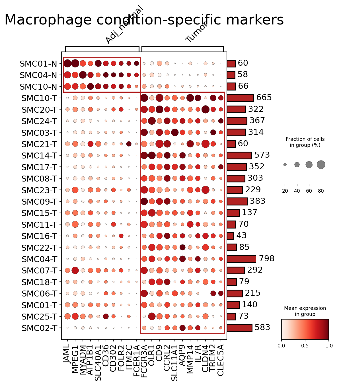
[Analysis Visualization Results]...
Analysis Overview
This analysis aimed to identify surface markers uniquely expressed by Macrophages in human Colon tissue, distinguishing between 'Tumor' and 'Adj_normal' conditions. Using single-cell RNA sequencing data, the plot_markers_and_expression_dot tool was employed to find and visualize the top 50 condition-specific surfaceome markers for Macrophages, focusing on genes with significant differential expression (fold change > 1.5, p-value < 0.05) and high detection rates. The visualization highlights both the fraction of cells expressing a gene and its mean expression level across individual samples within each condition.
Visual Summary
The dot plot clearly delineates two distinct populations of macrophage surface markers, corresponding to the 'Adj_normal' and 'Tumor' conditions.
- Adj_normal Macrophages: A set of genes, including JAML, MPEG1, MYADM, ATP1B1, SLC40A1, CD36, CD302, FOLR2, ITM2C, show consistently high expression and prevalence (large, dark red dots) in macrophages from adjacent normal colon tissue samples (SMC01-N, SMC04-N, SMC10-N). These markers are largely absent or expressed at very low levels in tumor macrophages.
- Tumor Macrophages: Conversely, another distinct group of genes, prominently featuring FCGR3A, FCER1A, OLR1, CD9, CCRL2, SLC11A1, AQP9, MMP14, IL7R, CLDN4, TREM2, CLEC5A, exhibits high expression and prevalence in macrophages infiltrating the tumor microenvironment across various tumor samples. These markers are notably diminished in the adjacent normal samples.
The plot demonstrates a clear transcriptional shift in the surfaceome of macrophages when transitioning from a homeostatic to a tumor-associated state. Dot sizes and color intensities robustly indicate both the proportion of cells expressing a marker and its average expression level, emphasizing the strength and specificity of these markers to their respective conditions.
Biological Interpretation
The observed condition-specific surface markers provide critical insights into the functional adaptation of macrophages within the colon cancer microenvironment.
Adj_normal Macrophage Markers: Reflecting Homeostasis and Basal Functions
The markers highly expressed in 'Adj_normal' macrophages suggest roles in maintaining tissue homeostasis and basal immune surveillance.
- SLC40A1 (Ferroportin 1): As the primary iron exporter, its presence indicates active iron efflux, essential for preventing iron accumulation and oxidative stress in homeostatic macrophages. GeneCards SLC40A1
- CD36: A scavenger receptor involved in lipid metabolism, uptake of apoptotic cells, and angiogenesis. Its expression in normal macrophages contributes to tissue cleanup and resolving inflammation. GeneCards CD36
- FOLR2 (Folate Receptor Beta): While also expressed on activated macrophages, its presence here might suggest a subset involved in basal folate uptake or as a precursor for specific activation states in healthy tissue. GeneCards FOLR2
- MPEG1 (Macrophage Expressed Gene 1): This gene encodes a pore-forming protein thought to be involved in phagocytosis and host defense, contributing to the routine clearance functions of resident macrophages.
These markers collectively paint a picture of macrophages actively engaged in maintaining tissue integrity, nutrient homeostasis, and basal immune surveillance in the healthy colon.
Tumor Macrophage Markers: Signatures of Tumor-Associated Macrophages (TAMs)
The markers prominently expressed in 'Tumor' macrophages are highly indicative of Tumor-Associated Macrophages (TAMs), often characterized by an M2-like polarization that supports tumor progression.
- TREM2 (Triggering Receptor Expressed on Myeloid cells 2): A key receptor on TAMs involved in lipid metabolism, phagocytosis of cellular debris, and promoting an immunosuppressive microenvironment. High TREM2 expression is a hallmark of TAMs in many cancers, including colorectal cancer, where it contributes to tumor growth and metastasis. GeneCards TREM2
- MMP14 (Matrix Metalloproteinase 14 / MT1-MMP): This membrane-bound metalloproteinase is crucial for extracellular matrix degradation, facilitating tumor cell invasion, angiogenesis, and metastasis. Its upregulation highlights the pro-tumorigenic role of TAMs in tissue remodeling. PubMed Search: MMP14 tumor progression
- FCGR3A (Fc Gamma Receptor IIIa / CD16a): While commonly associated with NK cells, FCGR3A can be expressed on some macrophage subsets and may play a role in antibody-dependent cellular cytotoxicity (ADCC) or immune complex clearance in the tumor microenvironment, potentially contributing to immune modulation.
- OLR1 (Oxidized LDL Receptor 1 / LOX-1): Known to be upregulated in various inflammatory conditions and cancers, OLR1 on TAMs can promote angiogenesis and contribute to immunosuppression by regulating dendritic cell maturation and T cell responses. PubMed Search: OLR1 tumor immunology
- CCRL2 (Chemokine Receptor-like 2): An atypical chemokine receptor that can scavenge chemokines, thereby modulating the local chemokine gradient and influencing immune cell trafficking within the tumor.
- CLEC5A (C-type Lectin Domain Family 5 Member A): Involved in innate immune responses, its expression on TAMs could indicate a role in sensing pathogen-associated molecular patterns (PAMPs) or damage-associated molecular patterns (DAMPs) within the tumor.
These 'Tumor' specific markers collectively underscore a significant shift in macrophage phenotype towards a pro-tumorigenic and immunosuppressive state, actively participating in remodeling the tumor microenvironment and supporting disease progression in colorectal cancer.
Clinical or Translational Implications
The identification of condition-specific surface markers for macrophages in colon tissue has significant clinical and translational implications, particularly in the context of colorectal cancer.
- Biomarker Potential:
- Diagnostic/Prognostic Markers: Genes like TREM2, MMP14, and OLR1 could serve as valuable biomarkers for colorectal cancer. Their specific upregulation on tumor-associated macrophages might allow for:
- Early Detection: If detectable in peripheral blood (e.g., via extracellular vesicles) or biopsy, they could aid in diagnosing CRC or monitoring disease progression.
- Prognosis: The expression levels of these markers could correlate with tumor stage, aggressiveness, or patient outcomes, providing prognostic information.
- Imaging: Surface markers are ideal for targeted imaging agents (e.g., PET tracers) to visualize TAM infiltration within tumors.
- Therapeutic Targets:
- Targeted Immunotherapy: As surfaceome markers, these genes present excellent targets for novel immunotherapeutic strategies:
- TREM2: Inhibiting TREM2 signaling on TAMs could reprogram them from a pro-tumorigenic to an anti-tumorigenic state, enhancing anti-tumor immunity. PubMed Search: TREM2 cancer immunotherapy
- MMP14: Blocking MMP14 activity could impede tumor invasion, metastasis, and angiogenesis by disrupting the extracellular matrix, making it a target for anti-metastatic therapies.
- FOLR2: While identified in Adj_normal, FOLR2 is also known on activated macrophages and has been explored as a target for delivering drugs selectively to specific macrophage subsets, including those that might infiltrate early-stage tumors or inflammatory sites.
- Antibody-Drug Conjugates (ADCs) or CAR-Macrophage Therapy: Antibodies targeting these specific surface proteins could deliver cytotoxic drugs directly to TAMs or engineer macrophages to specifically target tumor cells.
- Modulating the Tumor Microenvironment: By targeting these macrophage surface receptors, it may be possible to re-educate TAMs, shifting their polarization from M2-like (pro-tumor) towards M1-like (anti-tumor) phenotypes, thereby enhancing the efficacy of existing cancer treatments.
Further experimental validation in relevant *in vitro* and *in vivo* models, and patient cohorts, would be crucial to confirm the clinical utility of these macrophage-specific surface markers in colorectal cancer.
18. Fibroblast Condition-Specific Surfaceome Markers in Colon Tissue
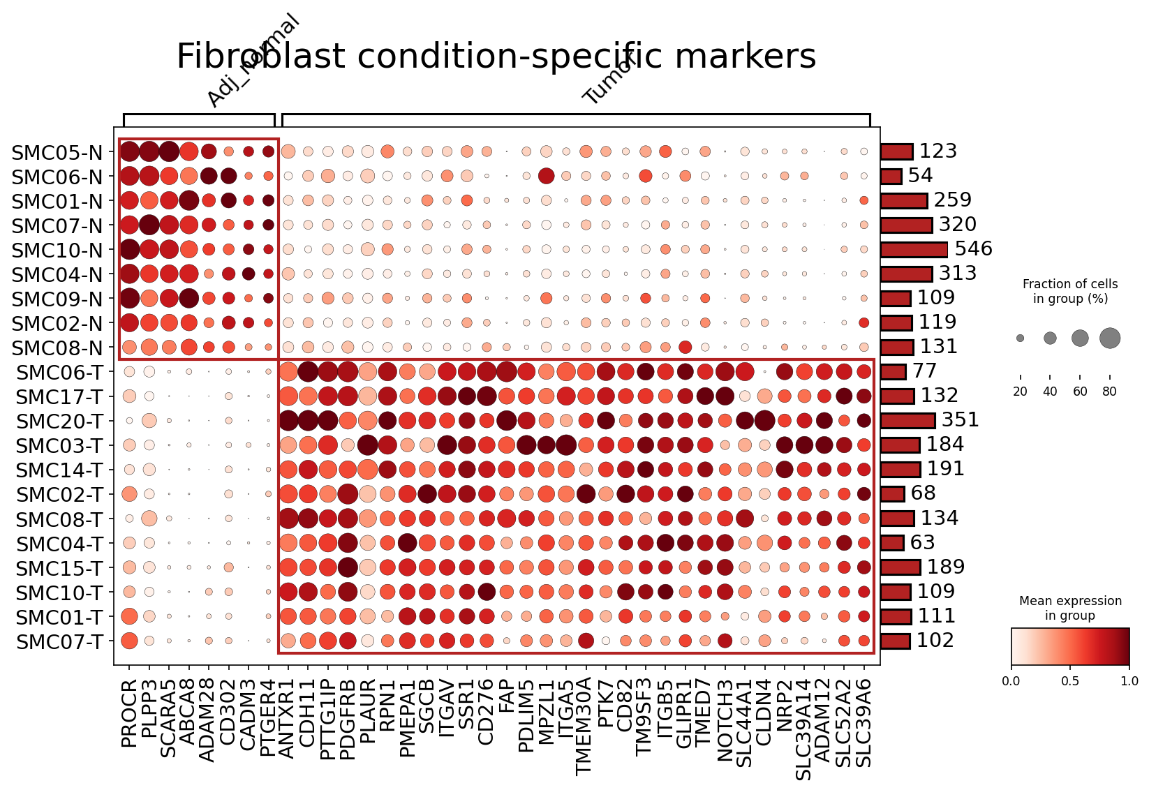
[Analysis Visualization Results]...
Analysis Overview
This analysis identifies and visualizes surfaceome markers specifically expressed by Fibroblast cells in human colon tissue, distinguishing between "Adj_Normal" (adjacent normal) and "Tumor" conditions. The dot plot displays the mean expression level (color intensity) and the fraction of cells expressing each marker (dot size) across individual patient samples. By focusing on surfaceome markers, this analysis highlights potential candidates for cell-surface-targeted therapeutic strategies or flow cytometry-based characterization.
Visual Summary
The dot plot clearly delineates two distinct clusters of fibroblast surfaceome markers corresponding to the "Adj_Normal" and "Tumor" conditions.
- Condition-Specific Expression Patterns: Fibroblasts from "Adj_Normal" samples (top block) exhibit high and prevalent expression of a specific set of genes (e.g., PROCR, PLPP3, SCARA5, ABCA8, ADAM28, CD302, CADM3, PTGER4, ANTXR1). Conversely, fibroblasts from "Tumor" samples (bottom block) show robust expression of a completely different panel of markers (e.g., CDH11, PDGFRB, PLAUR, CD276, FAP, ITGAV, ADAM12, NRP2, NOTCH3). This striking inverse expression pattern underscores the significant phenotypic changes in fibroblasts within the tumor microenvironment.
- Marker Prevalence and Expression Level: For the identified condition-specific markers, the dots are generally large and dark red within their respective condition blocks, indicating that these markers are expressed by a high fraction of cells in most samples within that condition and at high mean expression levels. This suggests they are robust markers for distinguishing normal and tumor-associated fibroblasts.
- Sample Clustering: The individual samples are well-separated based on their marker expression profiles, reinforcing the clear distinction between Adj_Normal and Tumor fibroblasts.
Biological Interpretation
The identified surfaceome markers provide crucial insights into the altered biology of fibroblasts in colon cancer.
- Adj_Normal Fibroblast Markers: Markers like PROCR (Protein C Receptor, endothelial), PLPP3 (Phospholipid Phosphatase 3), SCARA5 (Scavenger Receptor Class A Member 5), and PTGER4 (Prostaglandin E Receptor 4) are prominent in adjacent normal tissue fibroblasts. These genes are associated with maintaining normal tissue homeostasis, cell adhesion, lipid signaling, and basic inflammatory responses. For example, PROCR, despite its name, is known to be expressed by various stromal cells and involved in inflammation and vascular integrity GeneCards PROCR.
- Tumor Fibroblast (CAF) Markers: The fibroblasts in tumor samples exhibit high expression of genes characteristic of Cancer-Associated Fibroblasts (CAFs), which are critical drivers of tumor progression.
- FAP (Fibroblast Activation Protein Alpha): A highly recognized and widely studied marker for CAFs, FAP plays a significant role in extracellular matrix remodeling, immunosuppression, and promoting tumor growth and metastasis GeneCards FAP.
- CD276 (B7-H3): An immune checkpoint molecule, its overexpression on CAFs can suppress anti-tumor immune responses, making it a target for immunomodulation GeneCards CD276.
- PDGFRB (Platelet Derived Growth Factor Receptor Beta): This receptor is crucial for fibroblast activation, proliferation, and angiogenesis within the tumor microenvironment GeneCards PDGFRB.
- ITGAV (Integrin Subunit Alpha V) and ITGB5 (Integrin Subunit Beta 5): These integrin subunits are vital for cell-matrix interactions, cell adhesion, migration, and signaling pathways that promote tumor invasion and metastasis GeneCards ITGAV, GeneCards ITGB5.
- ADAM12 (ADAM Metallopeptidase Domain 12): This metalloproteinase can facilitate tumor cell invasion and metastasis by remodeling the extracellular matrix GeneCards ADAM12.
- NRP2 (Neuropilin 2): A co-receptor for growth factors like VEGF, NRP2 is involved in tumor angiogenesis, lymphangiogenesis, and metastasis GeneCards NRP2.
- NOTCH3 (Notch Receptor 3): This receptor is part of the Notch signaling pathway, which is frequently dysregulated in cancer and involved in cell fate, proliferation, and angiogenesis GeneCards NOTCH3.
- Other notable CAF markers include CDH11 (Cadherin 11) involved in cell adhesion and tumor invasion, and PLAUR (Plasminogen Activator, Urokinase Receptor) crucial for pericellular proteolysis and metastasis GeneCards CDH11, GeneCards PLAUR.
The shift from normal fibroblast markers to CAF-specific markers reflects the dynamic changes that occur in the stromal compartment during colon tumorigenesis, where fibroblasts acquire pro-tumorigenic functions.
Clinical or Translational Implications
The identified condition-specific surfaceome markers have significant clinical and translational implications, particularly in the context of colon cancer.
- Biomarker Discovery: The distinct surfaceome profiles of normal vs. tumor-associated fibroblasts could serve as diagnostic or prognostic biomarkers. For instance, high expression of FAP, CD276, PDGFRB, or NRP2 in fibroblast populations within colon tissue biopsies might indicate the presence of tumor-driving CAFs, potentially correlating with disease progression or prognosis.
- Therapeutic Targets: Several identified CAF markers represent promising therapeutic targets for colon cancer:
- FAP: FAP-targeting strategies, including small molecule inhibitors, antibodies, or CAR-T cells, are actively being investigated to deplete or reprogram CAFs and suppress tumor growth PubMed FAP cancer therapy.
- CD276 (B7-H3): As an immune checkpoint modulator, CD276 is a strong candidate for immune-oncology therapies aimed at overcoming CAF-mediated immunosuppression and enhancing anti-tumor immunity PubMed B7-H3 immunotherapy cancer.
- PDGFRB, ITGAV, NRP2, and NOTCH3: Inhibitors targeting these pathways are either in clinical use for other cancers or under development, and could be explored for their efficacy in modulating CAF functions in colon cancer. Disrupting integrin signaling (ITGAV/ITGB5) could impede tumor invasion and metastasis, while targeting NRP2 could inhibit angiogenesis.
- Experimental Validation: These surfaceome markers are ideal candidates for further experimental validation. They can be utilized for:
- Flow cytometry or immunohistochemistry: To confirm protein expression on fibroblasts in larger cohorts of human colon cancer samples.
- Functional studies: *In vitro* and *in vivo* studies can elucidate the precise roles of these markers in CAF activation, matrix remodeling, immune modulation, and tumor progression.
- Development of targeted imaging agents: Surface markers provide avenues for non-invasive imaging of CAFs in tumors.
19. CD4+ T Cell Condition-Specific Surfaceome Markers in Colon Tissue
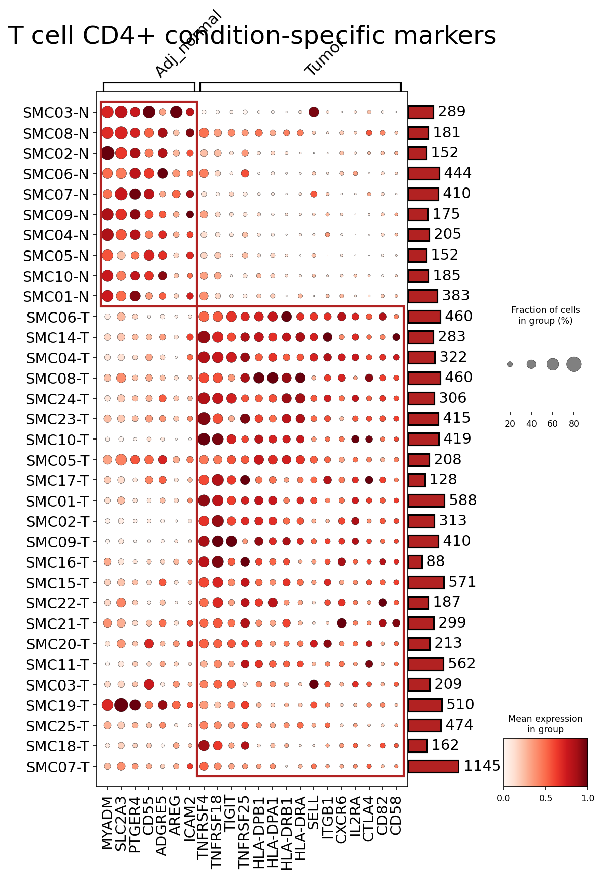
[Analysis Visualization Results]...
Analysis Overview
This analysis identifies condition-specific surfaceome markers in CD4+ T cells from human colon tissue, comparing Tumor samples to Adjacent Normal (Adj_normal) samples. The dot plot visualizes the expression of up to 50 surface markers per condition, showing the fraction of cells expressing each marker (dot size) and the mean expression level (dot color intensity) across individual samples, grouped by condition. The focus is on discovering cell-type-specific markers that can differentiate CD4+ T cell states between tumor and normal microenvironments.
Visual Summary
The dot plot clearly segregates genes into two distinct clusters based on their expression patterns in Adj_normal versus Tumor samples.
- Adj_normal Specific Markers: A cluster of genes, including MYADM, SLC2A3, PTGER4, CD55, ADGRE5, AREG, ICAM2, and TNFRSF18, shows high mean expression and broad cell fraction expression predominantly in Adj_normal samples. Their expression is largely absent or very low in Tumor samples.
- Tumor Specific Markers: Conversely, a prominent cluster of genes, including TNFRSF4, TNFRSF25, TIGIT, HLA-DPB1, HLA-DPA1, HLA-DRB1, HLA-DRA, SELL, ITGB1, CXCR6, CTLA4, CD58, and IL2RA, exhibits high mean expression and high fraction of expressing cells exclusively in Tumor samples. These markers are notably diminished or absent in Adj_normal samples.
- Sample Representation: The bar chart on the right indicates a variable but sufficient number of CD4+ T cells captured per sample (ranging from 88 to 1145 cells), supporting the robustness of the observed expression patterns.
Biological Interpretation
The distinct surfaceome marker profiles highlight significant phenotypic and functional differences in CD4+ T cells residing in the tumor microenvironment (TME) compared to adjacent normal tissue.
- Adj_normal T Cell Phenotype: Markers like PTGER4 (Prostaglandin E2 receptor 4) might indicate CD4+ T cells in adjacent normal tissue responding to local pro-inflammatory or homeostatic signals PubMed search: PTGER4 T cells. CD55 (Decay-accelerating factor) suggests protection against complement, a general host cell feature. ICAM2 (CD102) is involved in lymphocyte adhesion and migration, important for immune surveillance in healthy tissue. TNFRSF18 (GITR) is a co-stimulatory receptor expressed on activated T cells and regulatory T cells (Tregs) GeneCards: TNFRSF18, suggesting a baseline immune activity or regulatory presence in healthy colon.
- Tumor-Associated T Cell Phenotype: The upregulation of several markers in tumor samples points towards a highly activated, yet potentially exhausted or regulatory phenotype.
- Immune Checkpoints and Activation Markers: TIGIT and CTLA4 are well-known immune checkpoint receptors associated with T cell exhaustion and immune suppression within the TME GeneCards: TIGIT, GeneCards: CTLA4. Their prominent expression suggests that tumor-infiltrating CD4+ T cells are subject to suppressive mechanisms. TNFRSF4 (OX40) and IL2RA (CD25) are typically upregulated on activated T cells GeneCards: IL2RA, with CD25 also being a key marker for Tregs. This combination indicates a complex state of activation coupled with inhibitory signaling.
- Antigen Presentation Machinery: The consistent upregulation of MHC class II genes (HLA-DPB1, HLA-DPA1, HLA-DRB1, HLA-DRA) on CD4+ T cells in the tumor is a notable finding. While T cells are not professional antigen-presenting cells (APCs), activated T cells, particularly in chronic inflammation or tumor settings, can be induced to express MHC class II molecules PubMed search: T cells MHC class II expression. This phenomenon could imply a specific subset of CD4+ T cells with altered functional capabilities, potentially engaging in direct antigen presentation to other T cells or B cells within the TME, or reflecting a highly activated state.
- Homing and Adhesion: CXCR6 is a chemokine receptor often found on tissue-resident memory T cells (TRM) and is critical for T cell recruitment to inflammatory and tumor sites GeneCards: CXCR6. ITGB1 (CD29) is an integrin involved in cell adhesion and T cell activation. These markers suggest specific migratory and tissue-retention properties of CD4+ T cells within the tumor. SELL (CD62L), an L-selectin, is usually associated with naive T cells; its presence in a subset of tumor-infiltrating CD4+ T cells might indicate diverse T cell populations or specific homing mechanisms.
Clinical or Translational Implications
These condition-specific surfaceome markers offer significant potential for biomarker development and therapeutic intervention strategies in colon cancer.
- Diagnostic and Prognostic Biomarkers: The identified differentially expressed surface markers, such as the distinct panels for Adj_normal vs. Tumor, could serve as highly specific biomarkers for characterizing the immune infiltrate in colon cancer patients. For instance, a high expression of TIGIT, CTLA4, and IL2RA on CD4+ T cells in biopsies could indicate a more immunosuppressive TME, potentially informing prognosis or guiding treatment decisions.
- Therapeutic Targets for Immunomodulation: Key immune checkpoint molecules like TIGIT and CTLA4 are already targets for existing or experimental immunotherapies. The clear upregulation of these on tumor-infiltrating CD4+ T cells suggests their direct relevance for therapeutic blockade to reinvigorate anti-tumor immunity. TNFRSF4 (OX40) is another co-stimulatory receptor being explored for agonistic therapies to enhance T cell responses.
- Adoptive Cell Therapy and Engineering: Surface markers unique to tumor-infiltrating CD4+ T cells, or those distinguishing them from normal tissue T cells, could be leveraged for designing more precise adoptive cell therapies (e.g., CAR-T or TCR-T cells). For example, targeting T cells expressing certain combinations of these markers might allow for better enrichment or engineering of anti-tumor effector cells.
- Experimental Validation: The surfaceome-specific nature of these markers makes them highly amenable to validation using techniques like flow cytometry or immunohistochemistry on patient samples, which can confirm protein expression and guide further functional studies.
20. Dysregulation of Cell Cycle Pathways in Intestinal Epithelial Cells from Colon Tumor Tissue

[Analysis Visualization Results]...
Analysis Overview
This analysis investigates the differential expression of a panel of cell cycle-related genes in Intestinal Epithelial cells, comparing samples from colon tumor tissue with adjacent normal tissue. The aim is to identify specific cell cycle regulators that are significantly altered in the tumor microenvironment, providing insights into the proliferative state and potential oncogenic mechanisms within the tumor-originating cell type. The box plots display gene expression levels across conditions, highlighting statistically significant differences.
Visual Summary
The box plots illustrate the expression levels of 24 selected cell cycle pathway-related genes in Intestinal Epithelial cells. Across all plotted genes, a consistent and statistically significant pattern emerges:
- Upregulation in Tumor: The vast majority of the displayed genes show significantly higher expression in the "Tumor" condition compared to the "Adj_normal" condition. This includes key cell cycle drivers and components of cell cycle machinery such as:
- Anaphase-Promoting Complex (APC/C) components: ANAPC1, ANAPC10, ANAPC11, ANAPC13, ANAPC5, ANAPC7
Mitotic checkpoint proteins: BUB3, MAD2L1, MAD2L2
- Cyclins and CDKs: CCND1, CCND2, CCND3, CCNH, CDK4, CDK6, CDK7
Cell division cycle proteins: CDC16, CDC25B, CDC26, CDC27
- DNA replication factors: DBF4, MCM3, MCM4, MCM7, PCNA, ORC4
E2F transcription factor family: E2F2, TFDP1, TFDP2
- Tumor suppressors/oncogenes often involved in cell cycle: MDM2, MYC, RB1, RBL2, RBX1, PTTG1
Cohesin complex: RAD21, SMC1A, SMC3, STAG2
Chk kinases: CHEK1, CHEK2
- CDK inhibitors: CDKN1A, CDKN1B (Interestingly, these appear upregulated in tumor, suggesting potential compensatory mechanisms or specific tumor context rather than simple loss of function)
- Others: CUL1, EP300, FZR1, GADD45A, GADD45B, GSK3B, HDAC1, HDAC2, SFN, TP53, WEE1, YWHAB, YWHAE, YWHAG, YWHAH, YWHAQ, YWHAZ.
- Statistical Significance: All displayed genes exhibit highly significant expression differences between the two conditions, with p-values predominantly much lower than the 0.01 cutoff, often reaching p ≤ 1e-5. This indicates a robust and widespread dysregulation of these genes in tumor cells.
- Expression Distribution: The distribution of expression values, as indicated by the spread of individual data points (stripplot) and the interquartile range of the box plots, generally shows higher median and wider range for tumor samples for most upregulated genes, consistent with increased cellular heterogeneity and activity within the tumor.
Biological Interpretation
The observed widespread and statistically significant upregulation of numerous cell cycle-related genes in Intestinal Epithelial cells from colon tumors strongly indicates a hyper-proliferative state characteristic of cancer. Intestinal Epithelial cells are the cell type of origin for colorectal cancer, making these findings highly relevant to tumor initiation and progression.
Specifically:
- Accelerated Cell Cycle Progression: The concurrent upregulation of cyclins (CCND1, CCND2, CCND3, CCNH) and cyclin-dependent kinases (CDK4, CDK6, CDK7) suggests increased activity of these core drivers of cell cycle progression. For instance, Cyclin D1 (CCND1) is a key regulator of the G1-S transition, and its overexpression is a common feature in many cancers, including colorectal cancer, promoting uncontrolled cell division [1].
- Enhanced DNA Replication and Mitosis: The elevated expression of DNA replication licensing factors (MCM3, MCM4, MCM7, ORC4, DBF4), proliferating cell nuclear antigen (PCNA), and anaphase-promoting complex components (ANAPC1, ANAPC10, ANAPC11, ANAPC13, ANAPC5, ANAPC7) points towards active DNA synthesis and robust mitotic activity. The Anaphase-Promoting Complex (APC/C) is a ubiquitin ligase that regulates progression through mitosis and G1, and its components are often overexpressed in rapidly dividing cancer cells [2].
- Dysregulated Checkpoint Control: While many cell cycle drivers are upregulated, the observed increased expression of cell cycle inhibitors like CDKN1A (p21) and CDKN1B (p27) in tumor cells is notable. While these are typically tumor suppressors, their upregulation in some cancer contexts can be complex; it might represent a compensatory mechanism trying to restrain excessive proliferation, or paradoxically, contribute to cell survival and resistance in certain scenarios. The upregulation of checkpoint kinases CHEK1 and CHEK2 and stress-response genes GADD45A/B, SFN, and TP53 could reflect increased genomic instability and DNA damage experienced by rapidly dividing tumor cells [3, 4]. The AnnData context notes ploidy_dec (Aneuploid, Diploid) and cnv_ref_ind, cnv_cluster, cnv_sample_diversity_index, which support the presence of genomic instability that can trigger these DNA damage responses.
- Oncogenic Pathway Activation: Upregulation of MYC, a potent oncogene, further supports a highly proliferative phenotype. MDM2 overexpression can lead to the inactivation of TP53, despite TP53 itself showing increased expression, suggesting a complex interplay in tumor cells. Epigenetic regulators like HDAC1 and HDAC2 are also elevated, consistent with altered chromatin states that promote gene expression programs for proliferation in cancer.
Collectively, these findings in Intestinal Epithelial cells from colon tumors demonstrate a profound shift towards uncontrolled cell proliferation driven by the coordinated upregulation of key cell cycle machinery, often accompanied by cellular stress and altered checkpoint responses. This biological state is fundamental to tumor growth and aligns with the role of Intestinal Epithelial cells as the origin of colon tumors.
Clinical or Translational Implications
The widespread dysregulation of cell cycle genes in colon tumor Intestinal Epithelial cells holds significant clinical and translational implications:
- Biomarker Potential: Genes such as CCND1, PCNA, MYC, and specific CDK/cyclin complexes could serve as prognostic biomarkers for colorectal cancer progression or recurrence. Elevated expression levels might correlate with more aggressive disease behavior.
- Therapeutic Targets: Many of the upregulated genes are established targets for cancer therapy. For example, CDK4/6 inhibitors are already used in certain cancers to block cell cycle progression [5]. Inhibitors targeting specific components of the ANAPC/C, MCM proteins, or epigenetic regulators like HDACs are also under investigation as potential anti-cancer agents [6, 7].
- Combination Therapies: Understanding the specific cell cycle pathways activated in colon tumors could guide the development of rational combination therapies, for instance, combining cell cycle inhibitors with agents that target DNA damage response pathways (e.g., ATM/ATR inhibitors, which were in the initial gene list but not among the top 24 differential genes plotted here) to exploit vulnerabilities in tumor cells.
These results highlight the critical role of cell cycle dysregulation in colon tumorigenesis and provide a foundation for further investigation into targeted therapies and personalized medicine approaches for colorectal cancer.
---
References:
- CCND1 (Cyclin D1) in cancer: GeneCards: CCND1
- Anaphase-Promoting Complex (APC/C) in cancer: PubMed Search: "Anaphase promoting complex cancer proliferation"
- CDKN1A (p21) and CDKN1B (p27) roles in cancer: GeneCards: CDKN1A, GeneCards: CDKN1B
- GADD45 in cancer: PubMed Search: "GADD45 cancer DNA damage"
- CDK4/6 inhibitors: PubMed Search: "CDK4/6 inhibitors cancer therapy"
- APC/C as a drug target: PubMed Search: "APC/C inhibitors cancer therapy"
- HDAC inhibitors: PubMed Search: "HDAC inhibitors cancer therapy"
21. 장상피세포의 조건 및 배수성 상태에 따른 유전자 온톨로지(GSA) 분석 결과
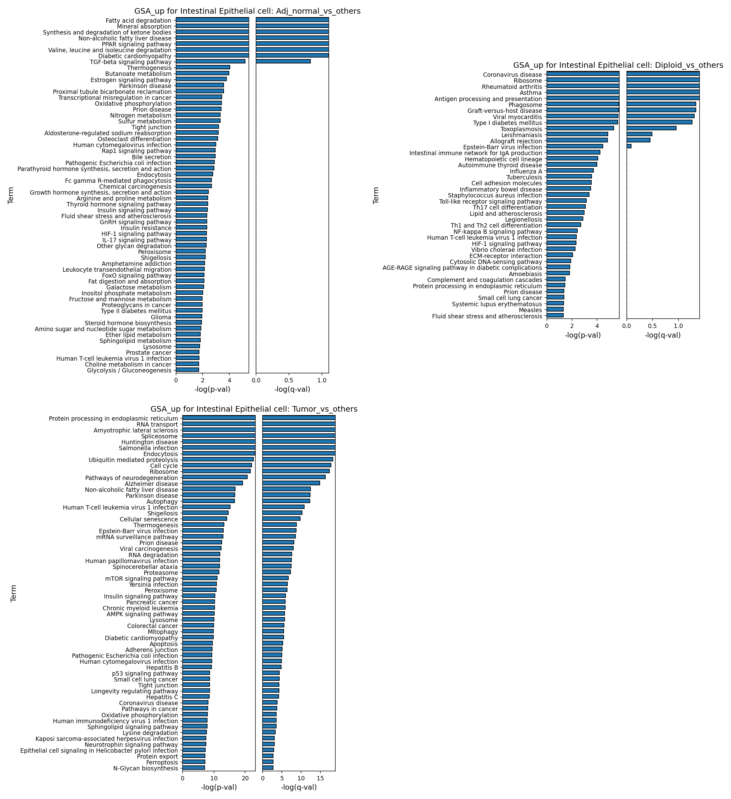
[Analysis Visualization Results]...
Analysis Overview
본 분석은 단일 세포 RNA 시퀀싱 데이터를 기반으로 한 대장 조직 내 장상피세포(Intestinal Epithelial cell)의 유전자 온톨로지(Gene Ontology, GO) 분석(GSA) 결과입니다. AnnData 객체에 저장된 전처리된 GSA 데이터를 활용하여, 세 가지 주요 비교 조건(정상 인접 조직 vs 기타, 이배체 vs 기타, 종양 vs 기타)에서 상향 조절된 GO 용어들을 막대 그래프로 시각화하였습니다. 각 그래프는 조건에 따라 상향 조절된 유전자 세트가 어떤 생물학적 경로 및 기능에 관여하는지 -log(p-value)와 -log(q-value)를 기준으로 보여줍니다.
Visual Summary
세 개의 막대 그래프는 장상피세포에서 세 가지 다른 조건(정상 인접 조직, 이배체, 종양)과 다른 조건들을 비교했을 때 유의하게 상향 조절된 GO 용어들을 보여줍니다. y축은 GO 용어를, x축은 -log(p-value)와 -log(q-value)를 나타냅니다. 막대 길이가 길수록 통계적 유의성이 높음을 의미합니다.
- Adj_normal_vs_others (정상 인접 조직 vs 기타): 지방산 분해(Fatty acid degradation), 무기질 흡수(Mineral absorption), 케톤체 합성 및 분해(Synthesis and degradation of ketone bodies), PPAR 신호 전달 경로(PPAR signaling pathway)와 같은 대사 및 흡수 관련 경로들이 가장 높은 유의성을 보입니다.
- Diploid_vs_others (이배체 vs 기타): 코로나바이러스 질병(Coronavirus disease), 리보솜(Ribosome), 항원 처리 및 제시(Antigen processing and presentation), 식균 작용(Phagosome)과 같은 면역 반응 및 단백질 합성 관련 경로들이 두드러집니다.
- Tumor_vs_others (종양 vs 기타): 소포체 내 단백질 처리(Protein processing in endoplasmic reticulum), RNA 수송(RNA transport), 스플라이소솜(Spliceosome), 유비퀴틴 매개 단백질 분해(Ubiquitin mediated proteolysis), 세포 주기(Cell cycle), 리보솜(Ribosome) 등 세포 증식, 단백질 및 RNA 대사, 세포 스트레스 반응과 관련된 경로들이 매우 높은 유의성을 보입니다. 특히, 세포 주기는 -log(p-value)가 20 이상으로 가장 높은 유의성을 나타냅니다.
전반적으로 각 조건에 따라 장상피세포의 고유한 생물학적 특성을 반영하는 GO 용어들이 명확하게 구별되어 나타납니다.
Biological Interpretation
장상피세포는 대장암의 기원이 되는 세포 유형이므로, 이 세포에서 조건에 따른 유전자 기능 변화를 이해하는 것은 질병 기전을 파악하는 데 중요합니다.
1. 정상 인접 장상피세포 (Adj_normal_vs_others)의 특성
- 기본 대사 및 흡수 기능: 정상 인접 장상피세포는 지방산 분해, 무기질 흡수, 케톤체 합성 및 분해와 같은 핵심적인 대사 및 소화 흡수 기능을 활발히 수행하는 것으로 나타났습니다. 이는 건강한 장상피세포의 주된 생리적 역할에 부합합니다.
- PPAR 신호 전달 경로 (PPAR signaling pathway): 이 경로는 지질 대사, 에너지 항상성, 염증 반응 조절에 중요한 역할을 합니다. 정상 인접 세포에서 이 경로의 활성화는 건강한 세포 기능 유지 및 미세 환경 조절과 관련될 수 있습니다 GeneCards: PPARG.
- TGF-베타 신호 전달 경로 (TGF-beta signaling pathway): 세포 성장, 분화, 세포자멸사, 면역 반응 조절에 관여하며, 조직 항상성 유지에 필수적입니다.
2. 이배체 장상피세포 (Diploid_vs_others)의 특성
- 면역 반응 및 항원 제시: 이배체 상태의 장상피세포는 코로나바이러스 질병, 항원 처리 및 제시, 식균 작용과 같은 면역 반응 관련 경로들을 상향 조절합니다. 이는 유전적으로 안정적인(diploid) 세포가 외부 병원체에 대한 감시 및 방어 기능에 더 적극적으로 참여하거나, 특정 미세 환경에서 면역 활성 상태를 유지함을 시사합니다.
- 리보솜 (Ribosome): 단백질 합성을 나타내며, 활발한 세포 활동을 반영합니다.
- 자가면역 및 염증 관련 경로: 류마티스성 관절염(Rheumatoid arthritis), 이식편대숙주병(Graft-versus-host disease), 제1형 당뇨병(Type I diabetes mellitus) 등 자가면역 질환 관련 경로의 상향 조절은 이배체 세포가 염증 반응이나 면역 병리에 관여할 가능성을 제시합니다. 이는 반드시 해당 질병이 있다는 의미보다는, 관련 면역 신호 전달 체계가 활성화되어 있음을 반영할 수 있습니다.
3. 종양 장상피세포 (Tumor_vs_others)의 특성
- 세포 증식 및 대사 재편성: 종양 장상피세포에서는 세포 주기, 리보솜, RNA 수송, 스플라이소솜, 유비퀴틴 매개 단백질 분해, 소포체 내 단백질 처리와 같은 고활성 증식 및 단백질/RNA 대사 관련 경로들이 압도적으로 상향 조절됩니다. 이는 암세포의 특징적인 빠른 증식, 높은 단백질 합성 및 처리 능력, 유전체 불안정성 및 스트레스 반응을 반영합니다.
- 세포 주기 (Cell cycle): 암세포의 무제한적인 증식의 핵심입니다 PubMed search: Cell cycle in cancer.
- 소포체 내 단백질 처리 (Protein processing in endoplasmic reticulum): 빠르게 증식하는 암세포는 많은 양의 단백질을 합성하므로, 소포체 스트레스 반응 및 단백질 품질 관리 시스템이 과활성화됩니다.
- RNA 수송, 스플라이소솜: 유전자 발현 조절 및 전사 후 변형의 핵심 과정으로, 암세포에서 종종 오작동하거나 과활성화됩니다.
- mTOR 신호 전달 경로 (mTOR signaling pathway): 세포 성장, 증식, 생존 및 대사에 중요한 조절자이며, 많은 암에서 활성화되어 있습니다 GeneCards: MTOR.
- 자가포식 (Autophagy): 암세포 생존 및 스트레스 적응에 중요한 역할을 하는 세포 내 재활용 과정입니다.
- 콜로레탈 암 (Colorectal cancer): 본 연구의 조직 기원인 대장암 관련 경로가 직접적으로 상향 조절되는 것은 종양 세포의 암 특이적 변화를 강력히 지지합니다.
- 신경퇴행성 질환 및 기타 질병 관련 경로: 알츠하이머병, 파킨슨병 등 신경퇴행성 질환 관련 용어가 나타나는 것은 세포 스트레스, 단백질 항상성 조절 실패 등 암과 일부 공통된 세포 병리 기전을 공유할 수 있음을 시사합니다.
Clinical or Translational Implications
이러한 GSA 결과는 대장암에서 장상피세포의 병리학적 변화를 이해하고 잠재적인 치료 표적을 식별하는 데 중요한 통찰력을 제공합니다.
- 종양 특이적 표적: 종양 장상피세포에서 관찰된 세포 주기, 단백질 및 RNA 대사, mTOR 신호 전달 경로의 활성화는 기존의 항암 치료제 개발의 주요 표적임을 재확인시켜 줍니다. 특히, 소포체 스트레스 및 단백질 품질 관리 관련 경로의 높은 활성화는 종양 세포가 이러한 스트레스에 의존하고 있음을 시사하며, 이를 표적으로 하는 약물 개발 가능성을 탐색할 수 있습니다.
- 정상 세포의 보호: 정상 인접 장상피세포의 대사 및 흡수 관련 경로 활성화는 항암 치료 시 이러한 필수 기능을 보존하는 전략의 중요성을 강조합니다.
- 면역 미세 환경 조절: 이배체 장상피세포의 면역 관련 경로 활성화는 종양 미세 환경 내 면역 세포와의 상호작용 또는 상피 세포 자체의 면역 조절 기능이 암 진행에 어떻게 영향을 미치는지 추가 연구의 필요성을 제기합니다. 특히, 암세포의 게놈 불안정성(이수성)이 면역 반응 회피에 기여할 수 있다는 가설과 연관 지어 볼 수 있습니다.
22. Gene Set Enrichment Analysis (GSEA) in Colon Tumor Microenvironment
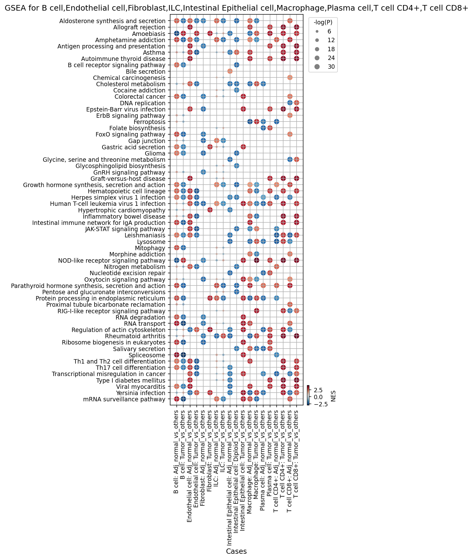
[Analysis Visualization Results]...
Analysis Overview
This analysis presents Gene Set Enrichment Analysis (GSEA) results visualized as dot plots across various major cell types found in human Colon tissue: B cell, Endothelial cell, Fibroblast, ILC, Intestinal Epithelial cell, Macrophage, Plasma cell, T cell CD4+, and T cell CD8+. For each cell type, gene set enrichment was evaluated by comparing cells from 'Tumor' conditions against 'Adj_normal' conditions (or vice-versa, indicated by 'Adj_normal_vs_others' meaning Adj_normal compared to Tumor cells of the same type). For Intestinal Epithelial cells, an additional comparison was made between 'Diploid' cells and 'others' (likely Aneuploid cells), reflecting their role as the tumor origin cell type and the presence of ploidy inference data. The dot plots display the Normalized Enrichment Score (NES) using a RdBu_r colormap (red for positive NES, blue for negative NES) and the statistical significance (-log10(p-value)) via dot size. A positive NES indicates upregulation of genes within the pathway in the 'test' condition (e.g., Tumor cells) compared to the 'reference' (e.g., Adj_normal cells), while a negative NES indicates downregulation.
Visual Summary
The dot plots reveal a complex landscape of pathway alterations across different cell types and conditions within the colon tissue.
- Differential Pathway Activity: A clear distinction is observed between pathways enriched in 'Tumor_vs_others' conditions (often red dots, indicating upregulation in tumor) and those in 'Adj_normal_vs_others' (often blue dots, indicating downregulation in tumor, or upregulation in adjacent normal tissue). This reciprocal pattern is evident for many pathways across cell types.
- Cell Type Specificity: While some pathways show broad enrichment across multiple cell types, many exhibit cell-type-specific patterns, highlighting distinct roles for each cell population in the tumor microenvironment.
- Significance and Magnitude: The varying sizes of the dots indicate different levels of statistical significance, with larger dots representing more robust enrichments. The intensity of red or blue color reflects the magnitude of enrichment (NES).
- Key Themes: Prominent themes emerging from the enriched pathways include cellular proliferation and metabolism, immune activation and dysregulation, and altered tissue remodeling.
Biological Interpretation
Intestinal Epithelial Cells (Tumor Origin Cell Type)
As the presumed origin of tumor, Intestinal Epithelial Cells (IECs) show striking changes:
- Proliferation and Cancer Hallmarks: In "Intestinal Epithelial cell: Tumor_vs_others", there is strong and highly significant positive enrichment (large red dots) for pathways like "Transcriptional misregulation in cancer", "DNA replication", "Ribosome biogenesis in eukaryotes", "Spliceosome", and "ErbB signaling pathway". This is a direct molecular signature of uncontrolled proliferation, active protein synthesis, and fundamental oncogenic processes characteristic of colon cancer cells. GeneCards: ERBB signaling pathway
- Metabolic Reprogramming: Pathways such as "Folate biosynthesis", "Glycine, serine and threonine metabolism", "Purine metabolism", and "Pyrimidine metabolism" are also highly enriched (red) in tumor IECs. This indicates increased anabolism and metabolic rewiring to support rapid cell division and biomass accumulation, a common hallmark of cancer. PubMed: Cancer metabolism reviews
- Ploidy-associated Differences: The "Intestinal Epithelial cell: Diploid_vs_others" comparison shows that diploid IECs (presumably less transformed or non-cancerous) have *reduced* activity (blue dots) in immune-related pathways like "Th1 and Th2 cell differentiation", "Th17 cell differentiation", and "Inflammatory bowel disease" compared to 'others' (likely aneuploid IECs). This suggests aneuploid IECs might be more prone to immune recognition or harbor a pro-inflammatory state, or diploid cells represent a more quiescent state.
Immune Cell Responses in the Tumor Microenvironment
The immune cell populations (B cells, ILCs, Macrophages, Plasma cells, T cell CD4+, T cell CD8+) exhibit significant activation and shifts in the tumor context:
- General Immune Activation: Pathways like "JAK-STAT signaling pathway" and "Antigen processing and presentation" are positively enriched (red) across multiple immune cell types (B cells, ILCs, Macrophages, Plasma cells, T cell CD4+, T cell CD8+) in the 'Tumor_vs_others' comparison. This points to active immune signaling and antigen presentation within the tumor microenvironment. GeneCards: JAK-STAT signaling pathway
- T Cell Differentiation and Inflammation: "Th1 and Th2 cell differentiation" and "Th17 cell differentiation" pathways show positive enrichment in T cell CD4+ and T cell CD8+ 'Tumor_vs_others', indicating active T cell responses. The "Inflammatory bowel disease" pathway is notably upregulated (red) in Macrophages and T cell CD8+ in the tumor, suggesting a pro-inflammatory environment that could drive or sustain tumor growth.
- B Cell and Plasma Cell Activity: "B cell receptor signaling pathway" and "Intestinal immune network for IgA production" are enriched in B cells and Plasma cells from tumor tissue. This indicates an active humoral immune response, potentially contributing to anti-tumor immunity or, in some contexts, promoting tumor progression.
Stromal and Endothelial Cell Contributions
Fibroblasts and Endothelial cells also play critical roles in shaping the tumor microenvironment:
- Cancer-Associated Fibroblast (CAF) Features: In "Fibroblast: Tumor_vs_others", there is strong positive enrichment (red) for pathways like "Regulation of actin cytoskeleton" and metabolic pathways ("Glycine, serine and threonine metabolism", "Purine metabolism"). This is consistent with activated fibroblasts (CAFs) involved in extracellular matrix remodeling, cell migration, and metabolic support for tumor growth.
- Angiogenesis and Endothelial Cell Activation: Endothelial cells in the tumor (''Endothelial cell: Tumor_vs_others'') show positive enrichment for "Regulation of actin cytoskeleton", "Endocytosis", and "ErbB signaling pathway". These pathways are crucial for endothelial cell migration, proliferation, and vessel formation, processes central to tumor angiogenesis. PubMed: Angiogenesis in cancer
Clinical or Translational Implications
- Therapeutic Targets: The consistent upregulation of specific pathways in tumor cells (e.g., ErbB signaling, DNA replication, metabolic pathways) and associated stromal cells (e.g., actin cytoskeleton remodeling in fibroblasts) suggests potential targets for therapeutic intervention. Targeting the ErbB pathway (e.g., EGFR inhibitors) or specific metabolic enzymes could inhibit tumor growth and progression.
- Immunomodulation: The widespread activation of immune signaling (JAK-STAT, antigen presentation, T cell differentiation) in the tumor microenvironment highlights opportunities for immunotherapeutic strategies. Understanding the specific subsets of immune cells and their activated pathways (e.g., pro-inflammatory macrophages or suppressive T cells) is crucial for designing effective immune-checkpoint blockades or adoptive cell therapies.
- Biomarkers: Enriched pathways and their associated genes could serve as prognostic or predictive biomarkers for colon cancer. For instance, high activity in cancer-specific metabolic pathways or sustained inflammatory responses might correlate with disease severity or response to therapy.
- Understanding Tumor Heterogeneity: The ploidy-specific differences observed in Intestinal Epithelial cells underscore the importance of tumor heterogeneity. Distinguishing diploid from aneuploid tumor cells could provide insights into early transformation events or different cellular vulnerabilities.
23. Discussion
The comprehensive single-cell analysis of human colon tissue reveals a profoundly reprogrammed microenvironment in colorectal cancer. A central finding is the robust identification of aneuploid intestinal epithelial cells as the primary malignant population, exhibiting extensive genomic instability characterized by recurrent copy number variations, including amplifications of oncogenes like EGFR (7p14.1-7q11.23) and deletions of tumor suppressors like CDKN2A (9p24.1-9p13.3). This genomic dysregulation is tightly linked to a hyper-proliferative state in tumor epithelial cells, as evidenced by the widespread and significant upregulation of core cell cycle machinery components (cyclins, CDKs, DNA replication factors) and enrichment of cell cycle, DNA replication, and ErbB signaling pathways. Metabolic rewiring, including purine and pyrimidine metabolism, further supports the rapid proliferation and biomass accumulation characteristic of cancer cells.
The tumor microenvironment undergoes substantial remodeling, particularly within its immune and stromal compartments. Immune cell populations exhibit a distinct immunosuppressive shift: regulatory T cells (Tregs) and pro-tumorigenic Th17 cells are significantly expanded, while innate lymphoid cells (ILC1, ILC2, LTI) and cytotoxic T cells (T_Cyto) show a notable reduction. Macrophages, a critical component of the TME, are dramatically polarized towards a pro-tumorigenic M2B phenotype, accompanied by a decrease in M2A macrophages. This macrophage reprogramming is further underscored by the upregulation of surface markers like TREM2 and MMP14 in tumor-associated macrophages (TAMs). Cancer-associated fibroblasts (CAFs) also adopt a pro-tumorigenic phenotype, marked by the expression of FAP, CD276 (B7-H3), PDGFRB, and integrins, and engage in extensive extracellular matrix remodeling.
Cell-cell interaction analysis highlights the complex crosstalk driving tumor progression. Aneuploid intestinal epithelial cells emerge as central orchestrators, engaging extensively with TAMs and CAFs via crucial pro-tumorigenic pathways. Prominent interactions include SPP1-CD44/integrin complexes, VEGFA-VEGFR1 (angiogenesis), and TGFB1-TGFBR1 (immunosuppression), all of which are significantly enhanced in the tumor. Immune checkpoint interactions like PVR-TIGIT and LGALS9-HAVCR2 are also highly active, contributing to T cell exhaustion. In contrast, normal tissue interactions emphasize epithelial barrier function (CDH1-integrin), basal immune surveillance (PGE2-PTGER4), and tissue homeostasis.
Collectively, these findings paint a detailed picture of colorectal cancer as a disease driven by genomic instability within epithelial cells, which in turn orchestrate a supportive, immunosuppressive, and proliferative microenvironment through complex cellular communication networks. The observed molecular shifts provide a rich resource for understanding disease mechanisms and identifying novel therapeutic avenues.
Hypotheses:
- The genomic instability (aneuploidy and recurrent CNVs) of intestinal epithelial cells is a primary driver of uncontrolled proliferation and metabolic reprogramming in colorectal cancer.
- The colorectal tumor microenvironment actively suppresses anti-tumor immunity through the selective expansion of regulatory T cells and pro-tumorigenic Th17 cells, coupled with a systemic reduction in innate lymphoid cells and cytotoxic T cells.
- Tumor-associated macrophages in colorectal cancer undergo significant polarization towards an M2B-like pro-tumorigenic phenotype, which contributes to immune evasion, angiogenesis, and extracellular matrix remodeling.
- Cancer-associated fibroblasts and malignant epithelial cells establish extensive communication networks via specific ligand-receptor interactions (e.g., SPP1-CD44/integrins, FAP, CD276), critically mediating tumor growth, invasion, and immunosuppression.
Potential therapeutic targets:
- EGFR (Epidermal Growth Factor Receptor): EGFR is a well-established oncogene. Its genomic region (7p14.1-7q11.23) shows recurrent amplification in tumor epithelial cells, and its ligand EREG is involved in active EREG-EGFR interactions within the tumor microenvironment. ErbB signaling, which includes EGFR, is highly enriched in tumor epithelial cells, driving proliferation and survival. Evidence: Recurrent CNV analysis (Section 4) shows amplification of 7p14.1-7q11.23 (harboring EGFR). Cell-cell interaction analysis (Section 13) shows EREG-EGFR interactions. GSEA (Section 22) shows ErbB signaling pathway enrichment in tumor intestinal epithelial cells. Tumor epithelial cells also show upregulation of EREG as a condition-specific marker (Section 16). Validation: Test existing EGFR inhibitors (e.g., Cetuximab, Panitumumab) or novel anti-EREG antibodies in patient-derived colorectal cancer organoids or xenografts to evaluate their impact on tumor cell proliferation and survival.
- TIGIT (T-cell Immunoreceptor with Ig and ITIM domains) / CTLA4 (Cytotoxic T-Lymphocyte-Associated Protein 4): These are key immune checkpoint receptors. TIGIT and CTLA4 are significantly upregulated on tumor-infiltrating CD4+ T cells, and involved in immune suppressive cell-cell interactions (e.g., PVR-TIGIT, CD86-CTLA4) that contribute to T cell exhaustion and immune evasion in the tumor microenvironment. Evidence: Condition-specific marker analysis (Section 19) shows upregulation of TIGIT and CTLA4 on CD4+ T cells in tumor samples. Cell-cell interaction analysis (Section 13) identifies PVR-TIGIT and CD86-CTLA4 interactions in the tumor context. Validation: Evaluate anti-TIGIT or anti-CTLA4 antibodies, alone or in combination with other immunotherapies (e.g., anti-PD-1), in *in vitro* T cell functional assays with tumor cells and in *in vivo* syngeneic or humanized mouse tumor models.
- TREM2 (Triggering Receptor Expressed on Myeloid cells 2): TREM2 is a critical receptor on tumor-associated macrophages (TAMs). It is highly expressed on macrophages in the tumor microenvironment and involved in APOE-TREM2 interactions with tumor epithelial cells and other macrophages. Its activation promotes an immunosuppressive and pro-tumorigenic TAM phenotype, contributing to tumor growth and metastasis. Evidence: Condition-specific marker analysis (Section 17) shows significant upregulation of TREM2 on macrophages in tumor samples. Cell-cell interaction analysis (Section 13) identifies prominent APOE-TREM2 interactions. Validation: Develop and test anti-TREM2 blocking antibodies or small molecule inhibitors in preclinical colorectal cancer models to assess their ability to reprogram TAMs from a pro-tumorigenic to an anti-tumorigenic state, and to inhibit tumor growth and metastasis.
- FAP (Fibroblast Activation Protein Alpha): FAP is a highly recognized and specifically expressed marker for Cancer-Associated Fibroblasts (CAFs) in tumor tissue. CAFs play a crucial role in extracellular matrix remodeling, immunosuppression, and promoting tumor growth and metastasis. Targeting FAP can disrupt the tumor-promoting functions of CAFs. Evidence: Condition-specific marker analysis (Section 18) shows significant and specific upregulation of FAP on fibroblasts in tumor samples. Validation: Evaluate FAP-targeting strategies, such as FAP-specific antibodies, FAP-directed CAR-T cells, or small molecule inhibitors, in colon cancer models to assess their impact on CAF activity, ECM remodeling, tumor growth, and metastatic potential.
- SPP1 (Secreted Phosphoprotein 1, Osteopontin): SPP1 and its receptors (CD44, integrins) form highly prominent cell-cell interaction axes in the tumor microenvironment, especially involving aneuploid intestinal epithelial cells and macrophages. SPP1 signaling drives macrophage polarization, promotes tumor growth, angiogenesis, and metastasis. Evidence: Cell-cell interaction analysis (Sections 12, 13) shows strong SPP1-CD44 and SPP1-integrin interactions in the tumor condition, particularly between aneuploid intestinal epithelial cells and macrophages. Validation: Test inhibitors targeting SPP1, CD44, or specific integrin subunits (e.g., ITGAV, ITGA5) in *in vitro* invasion/migration assays and *in vivo* metastasis models. Evaluate effects on macrophage polarization and tumor progression.
- CDK4/6 (Cyclin-Dependent Kinases 4 and 6): CDK4 and CDK6 are key regulators of cell cycle progression. They are significantly upregulated in tumor Intestinal Epithelial cells, driving the hyper-proliferative state characteristic of colorectal cancer. Evidence: Differential gene expression analysis (Section 20) shows significant upregulation of CDK4 and CDK6, along with their associated cyclins (CCND1, CCND2, CCND3), in Intestinal Epithelial cells from tumor tissue. Validation: Evaluate existing CDK4/6 inhibitors (e.g., Palbociclib, Ribociclib) or novel ones in colon cancer cell lines and patient-derived organoids to assess their efficacy in inhibiting tumor cell proliferation and inducing cell cycle arrest.
Follow-up validation ideas:
- Genomic Instability & Ploidy: Perform Fluorescence In Situ Hybridization (FISH) or single-cell whole-genome sequencing (scWGS) on sorted Aneuploid vs. Diploid Intestinal Epithelial cells to precisely map and validate recurrent CNV regions (e.g., EGFR amplification, CDKN2A deletion) and correlate with differential expression of cell cycle genes via targeted qPCR or immunohistochemistry.
- Immune Cell Dynamics: Use multi-parameter flow cytometry or spatial transcriptomics (e.g., GeoMx DSP, Visium) on fresh or FFPE human colorectal cancer and adjacent normal tissue samples to quantify and spatially localize Treg, Th17, ILC1/2, LTI, and cytotoxic T cell populations, along with their activation/exhaustion markers (e.g., TIGIT, CTLA4, OX40).
- Macrophage Polarization & Function: Isolate tumor-associated macrophages (TAMs) from resected colorectal cancer tissue based on surface markers (e.g., TREM2, MMP14) and conduct *ex vivo* functional assays to assess their cytokine production profiles (pro-inflammatory vs. anti-inflammatory), phagocytic activity, and T cell modulatory capacity.
- Cell-Cell Interaction Functional Validation: Establish *in vitro* co-culture systems of primary human Aneuploid Intestinal Epithelial cells with isolated CAFs or TAMs. Functionally validate key ligand-receptor interactions (e.g., SPP1-CD44, TGFB1-TGFBR1, DLL1-NOTCH2) using blocking antibodies or small molecule inhibitors, measuring their effects on cell proliferation, migration, invasion, and immune cell function. Use perturbation assays to confirm involvement of surface markers like FAP or CD276 in CAF-mediated tumor support.
- Therapeutic Target Efficacy: Test the efficacy of inhibitors targeting identified pathways or surface markers (e.g., EGFR, TIGIT, TREM2, FAP, CDK4/6) in patient-derived organoids (PDOs) or patient-derived xenograft (PDX) models of colorectal cancer, evaluating tumor growth inhibition, immune microenvironment modulation, and survival.
Limitations:
This report is based on single-cell RNA sequencing data, which provides transcriptomic snapshots. While powerful, inferring cell-cell interactions from gene expression is correlative and requires experimental validation at the protein level and functional assays. Ploidy inference is computational, and precise chromosomal alterations warrant orthogonal validation methods like FISH. The study provides a cross-sectional view, not capturing dynamic tumor evolution over time. The observed heterogeneity across patient samples indicates the need for larger, independent validation cohorts to generalize findings and refine patient stratification strategies. The complex tumor microenvironment also implies that single-target therapies may be limited by redundancy or compensatory pathways.
24. Query List
- Show UMAPs colored by condition, sample, celltype_major, celltype_minor, ploidy_dec, and celltype_subset in 2 columns and save them.
- Show major cell type scores on UMAP and save them.
- Show the overall Celltype_subset marker expression dot plot and save it. Keep target_cell as None and keep the other arguments at their default values.
- For Intestinal Epithelial cells (tumor origin) and unassigned cells, show CNV heatmap grouped by sample, along with a summary of significantly amplified copy number regions, and save it.
- Show CNV patterns on UMAP, including celltype_major, celltype_minor, ploidy_dec, condition, and sample in 2 columns, and save them.
- Show a population bar plot of minor cell types and save it.
- Show a subset population bar plot for T cells and save it.
- Show box plots for statistically significant differences in T cell subset populations between conditions, setting ncols appropriately, and save them.
- Show a subset population bar plot for Macrophage and save it.
- Show box plots for statistically significant differences in Macrophage subset populations between conditions, setting ncols appropriately, and save them.
- For Intestinal Epithelial cells (tumor origin) and unassigned cells, show a ploidy population bar plot and save it.
- Show cell-cell interaction patterns per condition, focusing on Intestinal Epithelial cells (tumor origin), Fibroblast, Macrophage, and T cells. Select up to 80 cell-cell interactions per condition and save it.
- Show cell-cell interactions by condition and save the result. Select at most 80 cell-cell interactions for each condition.
- Show cell-cell interactions for genes related to immune checkpoint and cell cycle pathways, and save them.
- Find statistically significant differences in cell-cell interactions between conditions for major immune and stromal cells, show them as a dot plot with max_n_items_per_group = 25, and save it.
- Show the condition-specific markers for tumor-origin cells (Intestinal Epithelial cell) as a dot plot and save the result. Use only surfaceome markers, up to 50 markers per condition.
- Extract condition-specific markers for Macrophage, show them as a dot plot for surfaceome markers, up to 50 per condition, and save it.
- Extract condition-specific markers for Fibroblast, show them as a dot plot for surfaceome markers, up to 50 per condition, and save it.
- Extract condition-specific markers for T cell CD4+, show them as a dot plot for surfaceome markers, up to 50 per condition, and save it.
- Show box plots for Cell cycle pathway-related genes with statistically significant expression differences between conditions in Intestinal Epithelial cells, setting max_n_items_to_plot = 24 and ncols for a 2x3 aspect ratio, and save them.
- Show Gene Ontology (GSA) analysis results for epithelial cells as bar plots and save the result.
- Show dot plots of Gene Set Enrichment Analysis results for B cell, Endothelial cell, Fibroblast, ILC, Intestinal Epithelial cell, Macrophage, Plasma cell, T cell CD4+, T cell CD8+ cell types, using color map RdBu_r, setting n_pws_to_show = 80, and save them.