Single-Cell Analysis of the Breast Cancer Tumor Microenvironment: Unveiling Genomic Instability, Cellular Reprogramming, and Therapeutic Vulnerabilities
This report presents a comprehensive single-cell RNA-sequencing analysis of breast tissue, contrasting normal and primary tumor conditions. Key findings reveal widespread genomic instability and aneuploidy predominantly within tumor-origin epithelial cells, alongside profound remodeling of the tumor microenvironment (TME) involving fibroblasts, macrophages, and T cells. We identify dysregulated cell-cell interaction networks, altered metabolic profiles, and shifts in immune cell populations, collectively highlighting critical pathways driving breast cancer progression and offering potential therapeutic targets.
Contents
- Dataset overview
- UMAP Visualization of Cell Annotations and Conditions in Breast Tissue
- UMAP Visualization of Major Cell Type Scores and Ploidy Status
- Marker Gene Expression Dot Plot for Cell Type Subsets
- Copy Number Variation Analysis in Breast Epithelial Cells
- UMAP Analysis of CNV Patterns Across Cell Types, Ploidy, and Conditions in Breast Tissue
- Minor Cell Type Population Analysis in Breast Tissue
- T Cell Subtype Population Analysis in Breast Tissue: Normal vs. Primary Tumor
- Differential T Cell Subset Proportions in Breast Cancer
- Macrophage Subset Population Analysis in Breast Tissue
- Ploidy Analysis of Epithelial Cells in Breast Tissue Samples
- Condition-Specific Cell-Cell Interaction Patterns in Breast Tissue Reveal Tumor Microenvironment Dynamics
- Condition-Specific Cell-Cell Interaction Patterns in Breast Cancer
- Condition-Specific Cell-Cell Interaction Patterns in Breast Tumor Microenvironment
- Epithelial Cell Condition-Specific Surfaceome Markers in Breast Cancer
- Macrophage Condition-Specific Surfaceome Markers in Breast Cancer
- Fibroblast Condition-Specific Surfaceome Markers in Breast Tissue
- T cell CD4+ Condition-Specific Surfaceome Marker Analysis in Breast Cancer
- Dysregulation of Cell Cycle Pathway Genes in Breast Cancer Epithelial Cells
- Gene Ontology (GSA) Analysis for Epithelial Cells in Breast Tissue
- Gene Set Enrichment Analysis (GSEA) of Cell Type-Specific Responses in Breast Tissue
- Discussion
- Query List
0. Dataset overview
Dataset Summary
- The dataset contains 88,707 cells and 25,535 genes from single-cell RNA sequencing data.
- The species is human, and the tissue is Breast.
- It includes two conditions: 'primary_tumor' and 'normal'.
- Key cell type annotations are available at major, minor, and subset levels, such as 'Stromal cell', 'Epithelial cell', 'Fibroblast', 'Macrophage', and 'T cell CD8+ '.
- Ploidy analysis shows approximately 80.0% Diploid cells and 20.0% Aneuploid cells.
- The 'Epithelial cell' has been resolved as the tumor-origin cell type.
- Copy Number Variation (CNV) estimates are available.
- 'normal' is set as the reference condition for comparative analyses like DEG, GSEA, and GSA.
Data Structure and Precomputed Results
- AnnData.obs columns: Includes metadata such as sample, condition, tissue, celltype_major, celltype_minor, celltype_subset, ploidy_dec, cluster, and cnv_cluster.
- AnnData.var columns: Contains gene_symbol, feature_type, chr, and cytogenetic_band.
- Precomputed Cell-Cell Interaction (CCI) results: Stored in uns['CCI'] (per condition) and uns['CCI_sample'] (per sample), with columns like gene_pair, cell_pair, pval, and mean.
- Precomputed Differential Expression Gene (DEG) results: Available in uns['DEG'] for each celltype_minor, with columns including log2_FC, pval_adj, gene, and Rp.
- Precomputed Gene Set Enrichment Analysis (GSEA) results: Stored in uns['GSEA'] for each celltype_minor, with columns like Name, NES, FDR q-val, and Lead_genes.
- Precomputed Gene Ontology (GO) / Gene Set Analysis (GSA) results: Accessible in uns['GSA_up'] for each celltype_minor, featuring pval_adj, genes, overlap, and Term.
Inferred Biological Context
- The dataset is strongly inferred to be related to Breast cancer. This is based on the presence of 'primary_tumor' and 'normal' conditions in 'Breast' tissue, a significant aneuploid cell population, and the identification of 'Epithelial cell' as the tumor-origin cell type.
1. UMAP Visualization of Cell Annotations and Conditions in Breast Tissue
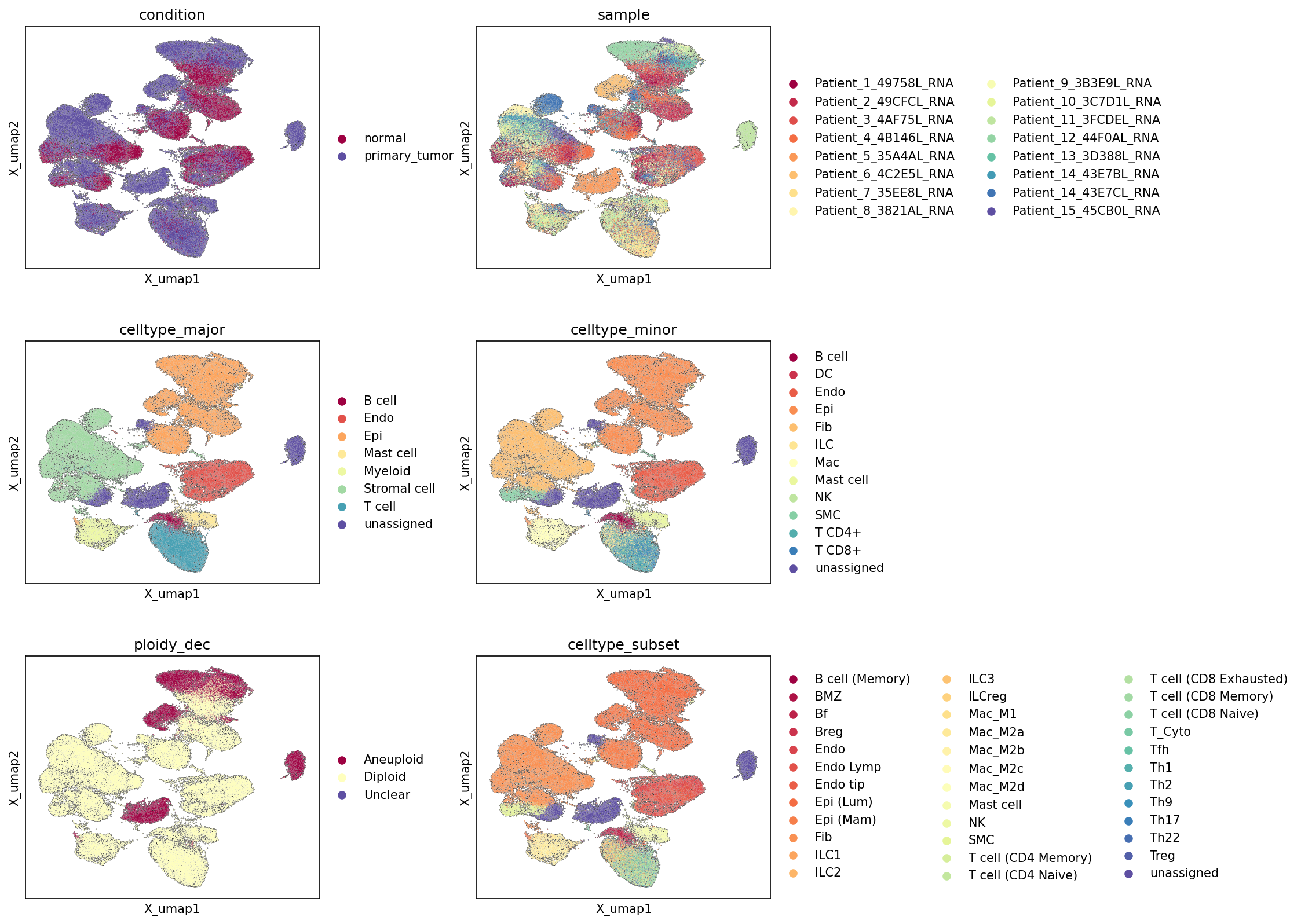
[Analysis Visualization Results]...
Analysis Overview
This analysis presents Uniform Manifold Approximation and Projection (UMAP) plots, which are dimensionality reduction visualizations of single-cell RNA-sequencing data. These plots show the global transcriptional relationships between 88,707 cells, colored by various metadata attributes: experimental condition (condition), individual patient sample (sample), hierarchical cell type annotations (celltype_major, celltype_minor, celltype_subset), and chromosomal ploidy status (ploidy_dec). The purpose is to visually assess the cellular landscape, evaluate the coherence of annotations, identify distinct cell populations, and understand the distribution of conditions and ploidy within the overall cell atlas.
Visual Summary
Condition Distribution
The UMAP colored by condition reveals a clear distinction between 'normal' and 'primary_tumor' cells. A significant portion of the UMAP space, particularly a large cluster on the right, is predominantly occupied by 'normal' cells. In contrast, 'primary_tumor' cells are more widely distributed across the embedding, forming several distinct clusters and often intermingling with 'normal' cells in certain regions, indicating a complex tumor microenvironment. There are also distinct clusters that are almost exclusively comprised of 'primary_tumor' cells.
Sample Distribution and Batch Effects
The sample UMAP shows that cells from different patients are largely intermixed across the major clusters, suggesting successful integration and minimal strong patient-specific batch effects that would otherwise lead to complete segregation of individual samples. However, some smaller clusters or specific regions show enrichment for cells from particular patients, indicating underlying biological heterogeneity between individuals that is not fully confounded by batch.
Cell Type Hierarchy (Major, Minor, Subset)
The UMAPs colored by celltype_major, celltype_minor, and celltype_subset demonstrate a well-structured and hierarchical organization of cell types:
- Major Cell Types: Epithelial cell (orange/yellow hues) forms a prominent, extensive region in the center-left. T cell (light blue) and Myeloid cell (light green) clusters are distinct, as are Stromal cell (dark green), Endothelial cell (red-orange), and B cell (dark red). A small, distinct cluster of unassigned cells (dark purple) is observed on the far right, suggesting a population with ambiguous or uncharacterized transcriptional profiles.
- Minor Cell Types: Further resolution shows coherent clustering for subtypes, such as Fibroblast within stromal cells, Macrophage within myeloid cells, and T cell CD4+ and T cell CD8+ separating from general T cells.
- Cell Type Subsets: The most granular celltype_subset annotations show fine-grained distinctions, such as Luminal epithelial cell and Mammary epithelial cell within the epithelial compartment, various Macrophage polarization states (e.g., Macrophage (M1), Macrophage (M2A/B/C/D)), and diverse T cell populations including T cell (Cytotoxic), T cell (Treg), T cell (CD8 Exhausted), and naive/memory states. These detailed subsets generally form cohesive sub-clusters within their broader cell type groups.
Ploidy Status
The ploidy_dec UMAP is particularly striking. Aneuploid cells (burgundy) are highly localized, forming several distinct clusters that are largely separate from the main Diploid (light yellow) cell populations. These Aneuploid clusters overlap significantly with regions predominantly colored by 'primary_tumor' condition. Diploid cells are broadly distributed, encompassing the 'normal' clusters and also contributing to tumor-associated clusters, reflecting the presence of both normal host cells and diploid cancer cells or bystander cells within tumors. The 'Unclear' category (dark purple) represents a minimal number of cells found in isolated small groups.
Biological Interpretation
Tumor-Specific Cell Populations and Ploidy
The strong segregation of primary_tumor cells and Aneuploid cells into distinct UMAP regions is a critical observation, aligning with the inferred context of breast cancer. Given that Epithelial cell is the Resolved tumor-origin cell type and Aneuploid cells are a hallmark of malignancy, the overlap of Aneuploid cells with primary_tumor and specific Epithelial cell clusters strongly indicates that these represent cancerous epithelial cells. The UMAP visually confirms that tumor cells exhibit distinct transcriptional profiles compared to normal cells. The presence of both normal and tumor cells, as well as distinct immune and stromal populations within the tumor regions, highlights the cellular complexity of the tumor microenvironment.
Tumor Microenvironment Heterogeneity
The UMAPs reveal a rich diversity of cell types within the breast tissue, including various immune cells (T cells, B cells, Myeloid cells, ILCs, Mast cells, NK cells) and stromal components (Fibroblasts, Endothelial cells, Smooth muscle cells). The fine-grained celltype_subset annotations further underscore the heterogeneity of these populations, such as different macrophage polarization states (M1, M2 subtypes) and T cell functional subsets (e.g., cytotoxic, exhausted, regulatory T cells). This cellular complexity is characteristic of the tumor microenvironment (TME) in breast cancer, where interactions between these diverse cell types play crucial roles in tumor progression and immune response.
Annotation Quality Assessment
The clear separation of major and minor cell types into distinct clusters or well-defined regions in the UMAPs indicates high quality and consistency of the cell type annotations. The hierarchical nature of the clustering, where subsets form coherent groups within their broader categories, further validates the annotation strategy. The distinct localization of Aneuploid cells to tumor-associated clusters also supports the accuracy of the ploidy estimation. The unassigned cell cluster is a minor population that might warrant further investigation to determine its identity or if it represents technical artifacts.
Annotation Notes
The UMAP visualizations demonstrate that the provided annotations for condition, sample, various celltype levels, and ploidy_dec are largely coherent and biologically meaningful. The distinct clustering patterns indicate robust underlying transcriptional differences corresponding to these labels. The presence of a small unassigned cell population suggests that while the majority of cells are well-characterized, there might be rare cell states or technical noise that could benefit from further manual curation or refinement. The overall structure of the UMAPs supports the quality of the single-cell data and its annotations for downstream analyses in the context of breast cancer.
2. UMAP Visualization of Major Cell Type Scores and Ploidy Status
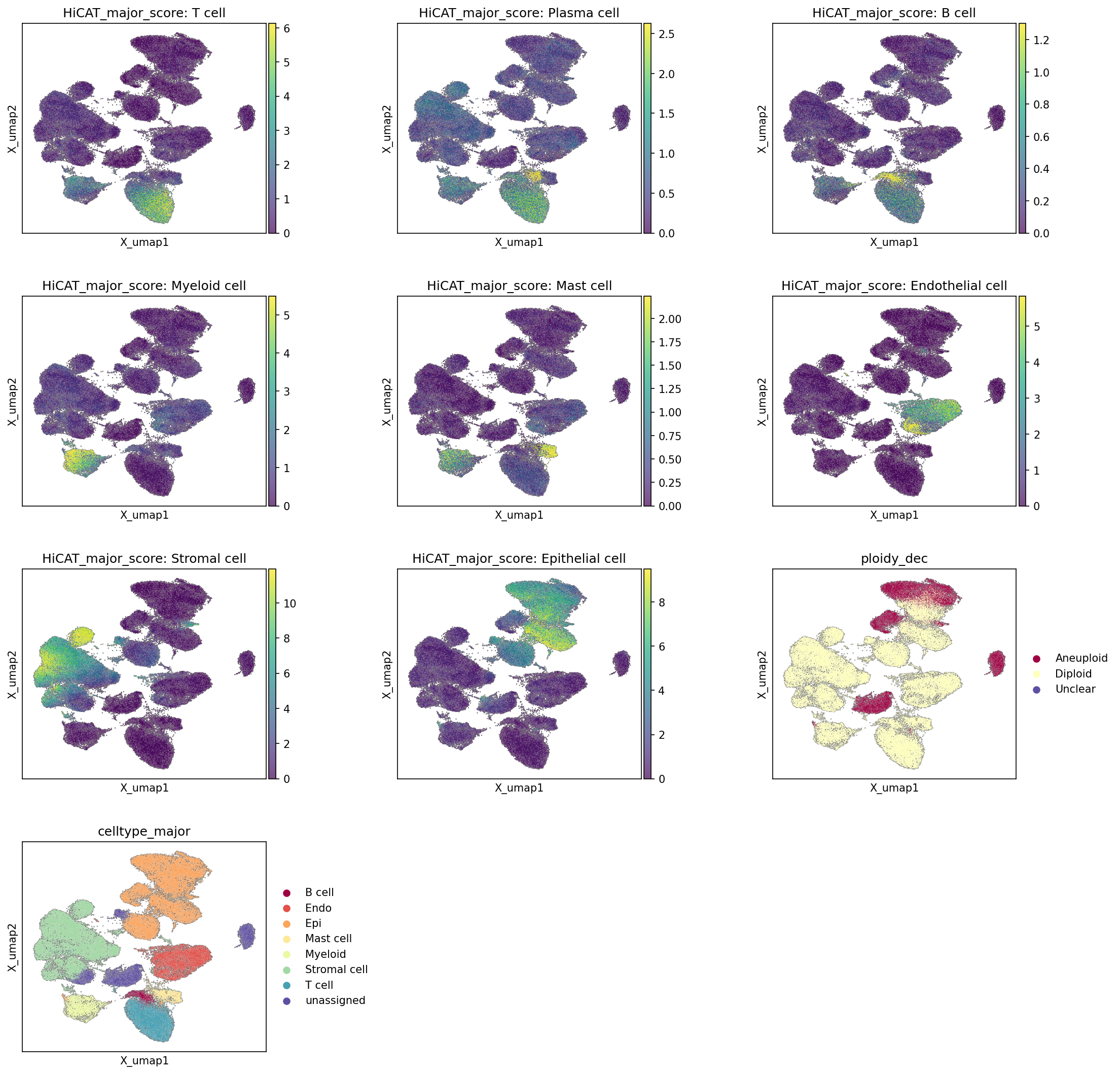
[Analysis Visualization Results]...
Analysis Overview
This analysis presents UMAP (Uniform Manifold Approximation and Projection) visualizations to depict the distribution of major cell type scores across the single-cell RNA-seq dataset. Each plot illustrates the calculated "HiCAT_major_score" for a specific major cell type, indicating the likelihood or confidence of cells belonging to that type. Additionally, the UMAP embedding is shown annotated by the final celltype_major assignment and ploidy_dec status (Aneuploid, Diploid, or Unclear) to provide context for cell identity and genomic stability. This allows for an assessment of the consistency of cell type annotations and the spatial distribution of cells with different ploidy states within the overall cellular landscape.
Visual Summary
The UMAP embedding reveals a complex architecture with distinct clusters representing various cell populations.
Cell Type Score Distributions (HiCAT_major_score)
- T cell, Plasma cell, B cell, Myeloid cell, Mast cell, Endothelial cell, Stromal cell, Epithelial cell: For each of these major cell types, the high-score regions (indicated by warmer colors, typically yellow-green) largely localize to distinct, non-overlapping clusters or segments of the UMAP. This suggests that the HiCAT major cell type scoring effectively distinguishes between different cell populations based on their transcriptional profiles. For instance, T cell scores are high in a specific cluster, as are Stromal cell and Epithelial cell scores in their respective dominant clusters. This pattern is consistent across all major cell types shown.
- Ploidy Status (ploidy_dec): The ploidy_dec UMAP plot distinctly highlights two main populations:
- Diploid cells (light yellow): These cells constitute the vast majority of cells and are broadly distributed across most clusters, particularly in regions dominated by immune and stromal cells.
- Aneuploid cells (dark red): These cells are primarily concentrated in a few specific, well-defined clusters within the UMAP, appearing as distinct "islands" rather than being diffusely spread.
- Unclear cells (dark blue): Only a very small number of cells are classified as "Unclear" and are barely visible on the plot, consistent with their low count (3 cells).
- Major Cell Type Annotation (celltype_major): The celltype_major UMAP plot serves as the reference for the final cell type assignments. It shows well-separated clusters for each major cell type (e.g., T cell, B cell, Epithelial cell, Stromal cell). The regions with high HiCAT scores for each cell type generally correspond well with the clusters assigned to that specific cell type in the celltype_major plot, affirming the quality of the cell type identification.
Biological Interpretation
- Robust Cell Type Identification: The strong compartmentalization of high HiCAT_major_scores for each cell type on the UMAP, mirroring the celltype_major annotations, indicates a robust and consistent cell type identification. This suggests that the underlying transcriptional differences between these major cell types are well-captured by the UMAP embedding and score calculation. This is crucial for downstream analyses, as accurate cell typing underpins all subsequent biological interpretations.
- Aneuploidy in Epithelial Cells: A striking observation is the prominent localization of aneuploid cells (ploidy_dec = Aneuploid) to specific clusters. When cross-referencing this with the celltype_major plot, these aneuploid clusters strongly overlap with the Epithelial cell population. This finding aligns perfectly with the provided data context that "Epithelial cell" is the "Resolved tumor-origin cell type" and the inferred disease context of breast cancer. Aneuploidy, a hallmark of cancer, is frequently observed in tumor cells, which in breast cancer often originate from epithelial cells [1]. The distinct clustering of aneuploid epithelial cells further supports their neoplastic nature and separation from the surrounding diploid cells.
- Tumor Microenvironment Composition: The majority of the UMAP consists of diploid cells, distributed across various immune (T cell, B cell, Myeloid cell, Mast cell, Plasma cell) and stromal cell populations, as well as some normal epithelial cells and endothelial cells. These diploid cells represent the non-malignant components of the breast tissue, including the immune infiltrate and the stromal support cells, which together form the tumor microenvironment (TME). The distinct separation of aneuploid epithelial cells from these diploid cell types provides a clear visual distinction between tumor cells and surrounding healthy or immune cells.
- Homogeneity of Aneuploid Population: The relatively consolidated appearance of aneuploid cells in distinct clusters suggests a degree of homogeneity within the aneuploid epithelial cell population, or at least a clear transcriptional signature that distinguishes them from their diploid counterparts and other cell types.
Annotation Notes
The UMAP plots provide strong visual evidence for high-quality cell type annotation. The consistency between the HiCAT_major_scores and the celltype_major labels validates the cell type assignments. Furthermore, the clear segregation of aneuploid cells, predominantly within the epithelial compartment, strongly supports the biological context of breast cancer and the identification of tumor-origin cells. The embedding effectively separates major cell types and also distinguishes between cells based on their ploidy status, indicating a meaningful representation of cellular heterogeneity.
---
References:
- Aneuploidy in Cancer: PubMed search for "aneuploidy cancer hallmark epithelial cells" https://pubmed.ncbi.nlm.nih.gov/?term=aneuploidy+cancer+hallmark+epithelial+cells
3. Marker Gene Expression Dot Plot for Cell Type Subsets
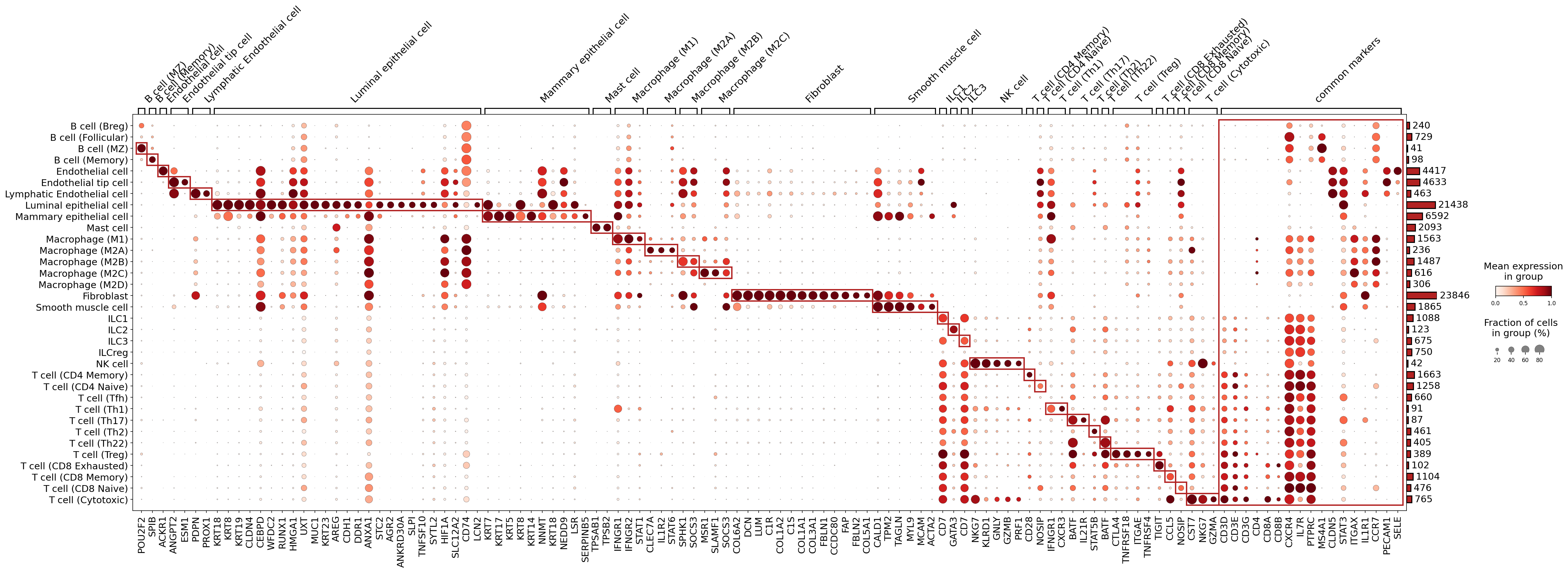
[Analysis Visualization Results]...
Analysis Overview
This analysis presents a marker gene expression dot plot, visualizing the mean expression level and the fraction of expressing cells for identified marker genes across various celltype_subset populations. The primary objective is to validate the assigned cell type annotations based on characteristic gene expression patterns, consistent with an identity-check result for single-cell RNA-seq data from breast tissue samples under normal and primary tumor conditions. The marker gene discovery process prioritized surfaceome genes and filtered out markers common to many groups to highlight cell-type-specific signatures.
Visual Summary
The dot plot effectively illustrates the distinct expression profiles of marker genes across 30 identified celltype_subset populations.
- Diagonal Pattern: A clear diagonal pattern of enriched expression (darker, larger dots) is observed, indicating that each celltype_subset generally expresses a unique set of marker genes specifically. This pattern suggests robust and well-defined cell type annotations.
Dot Size and Color:
- Dot size represents the fraction of cells within a given celltype_subset that express the marker gene. Larger dots signify higher prevalence of expression.
- Dot color intensity (ranging from light red to dark red) indicates the mean expression level of the marker gene in that cell type. Darker red denotes higher mean expression.
- Marker Grouping: Genes are grouped by the celltype_subset they are characteristic of, making it intuitive to assess the defining markers for each population. Red boxes around these groups further highlight their specificity.
- "Common markers" Column: The right-most column displays "common markers," which are genes expressed broadly across multiple cell types. These likely include pan-cell type markers or genes involved in general cellular processes, filtered from the specific marker sets.
Biological Interpretation and Annotation Notes
The marker expression patterns strongly support the assigned celltype_subset annotations:
B cells:
- B cell (Memory), B cell (MZ), B cell (Follicular), B cell (Breg) subsets are clearly identified by pan-B cell markers such as *CD19* and *MS4A1* (CD20) (visible in the "common markers" section, or for some B cell subsets in the specific marker section if less common). *POU2F2* is also a B cell lineage marker.
Endothelial cells:
- Endothelial cell, Endothelial tip cell, Lymphatic Endothelial cell subsets show strong expression of characteristic markers. *PECAM1* (CD31) is a classical endothelial marker, also appearing in "common markers". *ESM1* and *ANGPT2* are specific to general endothelial cells. *PROX1* is a defining marker for Lymphatic Endothelial cells, which is evident. *ACKR1* (DARC) is also expressed in endothelial cells.
Epithelial cells:
- Luminal epithelial cell and Mammary epithelial cell are well-defined by their expression of keratins (*KRT8, KRT18, KRT19, KRT7*) and other epithelial markers like *CDH1* (E-cadherin), *CLDN4*, *MUC1*, *AGR2*, *STC2*, *ANXA1*. The distinct expression profiles for luminal vs. mammary epithelial cells indicate a successful sub-typing. *KRT5* and *KRT17* for Mammary epithelial cells are also observed.
Macrophages:
- Various Macrophage (M1, M2A, M2B, M2C, M2D) subsets are distinguished by specific marker sets. *SPP1* (Osteopontin) is a prominent marker for Macrophage (M1). *MSR1* is a scavenger receptor found on macrophages. The clear separation of M1, M2A, M2B, M2C, M2D indicates a detailed characterization of macrophage polarization states.
Fibroblasts:
- Fibroblast cells exhibit strong expression of canonical fibroblast markers such as *COL1A1, COL3A1, DCN, LUM, FBLN1, FAP, CCDC80, FBLN2, COL5A1*.
Smooth muscle cells:
- Smooth muscle cell populations are characterized by markers like *ACTA2* (alpha-SMA), *MYL9, CALD1, TPM2, TAGLN*.
Innate Lymphoid Cells (ILCs) and Natural Killer (NK) cells:
- ILC1, ILC2, ILC3, ILCreg, and NK cell populations are identified by specific markers within the broader lymphoid context. *CD7* is characteristic of ILCs. *NKG7, KLRD1* (CD94), *GZMB*, and *PRF1* are prominent markers for NK cells and cytotoxic ILCs.
T cells:
- Numerous T cell subsets, including CD4 Memory, CD4 Naive, Tfh, Th1, Th17, Th2, Th22, Treg, and CD8 Naive, CD8 Memory, CD8 Exhausted, Cytotoxic, are well-demarcated. Pan-T cell markers like *CD3D, CD3E, CD3G* are present in the "common markers" section or broadly within T cell groups.
- Specific markers include: *CD4* for CD4+ T cells; *CD8A, CD8B* for CD8+ T cells; *FOXP3* for T cell (Treg); *GATA3* for T cell (Th2); *STAT3* for T cell (Tfh); *CTLA4* and *TIGIT* for T cell (Exhausted); *GZMB, PRF1* for T cell (Cytotoxic).
- The distinct markers for each T cell subtype, including naive, memory, and various helper T cell lineages, validate the fine-grained resolution of T cell annotation.
- Common Markers: The "common markers" column highlights genes like *PTPRC* (CD45, a pan-leukocyte marker) and *PECAM1* (CD31, pan-endothelial marker), *CD74*, *STAT1*, *SLC12A2*, which are broadly expressed in immune cells or structural cells, consistent with their known biological roles.
Overall, the marker expression dot plot provides strong evidence for the distinct identity and purity of the celltype_subset populations. The specificity and high expression levels of the displayed markers within their respective groups, coupled with minimal off-target expression, validate the accuracy of the cell type annotations derived from the single-cell RNA-seq data.
4. Copy Number Variation Analysis in Breast Epithelial Cells
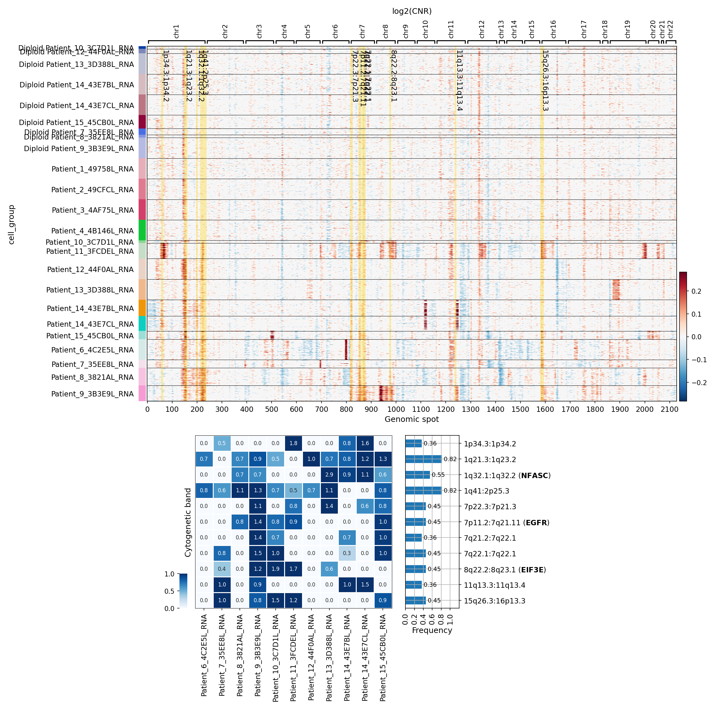
[Analysis Visualization Results]...
Analysis Overview
This analysis investigates copy number variations (CNVs) specifically within the Epithelial cell population from breast tissue samples. The dataset includes both 'normal' and 'primary_tumor' conditions, with Epithelial cells identified as the resolved tumor-origin cell type. The objective is to visualize CNV patterns across patient samples and summarize regions with significant copy number amplifications, which are hallmarks of genomic instability often associated with cancer.
Visual Summary (CNV Heatmap)
The heatmap displays log2(CNR) (Copy Number Ratio) values across the genome (Genomic spot) for Epithelial cells grouped by individual patient samples (cell_group).
- Distinct Genomic Profiles: The heatmap clearly segregates samples into two main groups based on their CNV profiles.
- Diploid Samples: The top rows, labeled with "Diploid Patient\_...", exhibit minimal deviation from a log2(CNR) of 0 (green/light colors). This indicates a predominantly diploid genomic state with very few or no detectable large-scale CNVs in their epithelial cell populations, consistent with a normal or non-cancerous profile. This observation also supports the accuracy of the ploidy_dec annotation.
- Aneuploid/Tumor Samples: The lower group of rows, labeled "Patient\_...", displays extensive and widespread regions of both amplifications (red, log2(CNR) > 0) and deletions (blue, log2(CNR) < 0) across multiple chromosomes. This pattern is highly characteristic of aneuploidy and genomic instability, strongly indicative of tumor cells.
- Inter-Patient Heterogeneity: Within the aneuploid group, significant variability in CNV patterns is observed among different patients. While some regions are frequently affected (e.g., Chr1, Chr7, Chr8, Chr11, Chr15), the precise combination and extent of amplifications and deletions differ between individual patients (e.g., Patient_2, Patient_4, Patient_12, Patient_8).
- Commonly Amplified Regions: Several chromosomal regions show recurrent amplifications across multiple tumor samples, including portions of chromosomes 1, 7, 8, 11, and 15.
Visual Summary (Amplification Frequency)
The accompanying summary provides a heatmap showing the frequency of amplification in specific cytogenetic bands per sample, alongside a bar chart illustrating the overall frequency of these amplifications across all tumor samples analyzed.
- High-Frequency Amplifications: The bar chart highlights the most frequently amplified cytogenetic bands across the tumor epithelial cells:
1p34.3:1p34.2 (0.82 frequency)
1q21.3:1q23.2 (0.82 frequency)
- 1q41:2p25.3 (0.82 frequency): Note: This label describing a region spanning two chromosomes (Chr1 and Chr2) is unusual for a single cytogenetic band in this type of CNV summary and might represent a complex event or a specific data processing artifact in the labeling.
1q32.1:1q32.2 (NFASC) (0.55 frequency)
7p22.3:7p21.3 (0.45 frequency)
7p11.2:7q21.11 (EGFR) (0.45 frequency)
7q21.2:7q22.1 (0.45 frequency)
8q22.2:8q23.1 (EIF3E) (0.45 frequency)
- 15q26.3:16p13.3 (0.45 frequency): Similar to 1q41:2p25.3, this label spanning two chromosomes (Chr15 and Chr16) is unusual.
11q13.3:11q13.4 (0.36 frequency)
- Sample-Specific Amplification Patterns: The summary heatmap shows that while some amplifications are widespread (e.g., 1q regions in Patient_6, Patient_7, Patient_8, Patient_9, Patient_10, Patient_11, Patient_12, Patient_13, Patient_14, Patient_15), others are more specific to certain patients. For example, EGFR amplification is observed in Patient_6, Patient_7, Patient_8, Patient_10, Patient_11, Patient_12, Patient_13, Patient_14, Patient_15.
Biological Interpretation
The observed CNV patterns in Epithelial cells strongly support the inference of Breast cancer in the 'primary_tumor' samples.
- Tumor-Specific Genomic Instability: The extensive aneuploidy (amplifications and deletions) in the epithelial cells of "Patient\_..." samples, contrasting sharply with the diploid "Diploid Patient\_..." samples, is a hallmark of malignancy. This genomic instability is a key driver of tumor evolution and heterogeneity. The explicit identification of "Epithelial cell" as the Resolved tumor-origin cell type for breast tissue further reinforces that these CNVs are indeed occurring within the cancerous cell population.
- Recurrent Oncogenic Amplifications: The frequently amplified regions harbor several known oncogenes:
- EGFR (Epidermal Growth Factor Receptor) on 7p11.2:7q21.11: Amplification and overexpression of EGFR are well-established drivers in various cancers, including subsets of breast cancer, promoting cell proliferation, survival, and metastasis. EGFR is a significant therapeutic target. GeneCards: EGFR
- NFASC (Neurofascin) on 1q32.1:1q32.2: NFASC, a cell adhesion molecule, has been implicated in cancer progression, with increased expression or amplification reported in several tumor types, contributing to cell motility and invasion. GeneCards: NFASC
- EIF3E (Eukaryotic translation initiation factor 3 subunit E) on 8q22.2:8q23.1: EIF3E is a component of the eukaryotic translation initiation factor 3 complex, involved in protein synthesis. Its overexpression or amplification can promote cell growth and proliferation, contributing to tumorigenesis. GeneCards: EIF3E
- 11q13.3:11q13.4 amplification: This region is a well-known amplification hotspot in breast cancer, often containing crucial oncogenes such as CCND1 (Cyclin D1), which regulates cell cycle progression, as well as FGF3 and FGF4. Amplification of this region is associated with aggressive tumor phenotypes. PubMed search: 11q13 amplification breast cancer
- Chromosomal Arm Amplifications: Broader amplifications on 1q, 7q, and 8q are frequently observed in breast cancer and other solid tumors, indicating widespread genomic aberrations that likely contain multiple oncogenes and regulatory elements contributing to cancer development and progression.
Clinical or Translational Implications
- Identification of Tumor Cells: The clear distinction in CNV profiles between "Diploid Patient\_..." and "Patient\_..." samples provides strong evidence for the successful identification and isolation of tumor-derived epithelial cells, validating the ploidy_dec and Resolved tumor-origin cell type annotations. This is critical for downstream analyses focusing on tumor-specific biology.
- Potential Therapeutic Targets: The recurrent amplification of oncogenes like EGFR highlights potential therapeutic vulnerabilities. Patients with EGFR amplification might respond to targeted therapies against EGFR, which are already available in the clinic for other cancer types.
- Biomarker Discovery: The identified high-frequency amplified regions and specific genes (e.g., NFASC, EIF3E, genes within 11q13) could serve as candidate biomarkers for disease progression, prognosis, or response to therapy in breast cancer. Further research would be needed to validate their clinical utility.
- Understanding Tumor Heterogeneity: The inter-patient variability in CNV patterns underscores the genomic heterogeneity of breast cancer, suggesting that patient-specific molecular profiling is crucial for personalized treatment strategies.
5. UMAP Analysis of CNV Patterns Across Cell Types, Ploidy, and Conditions in Breast Tissue
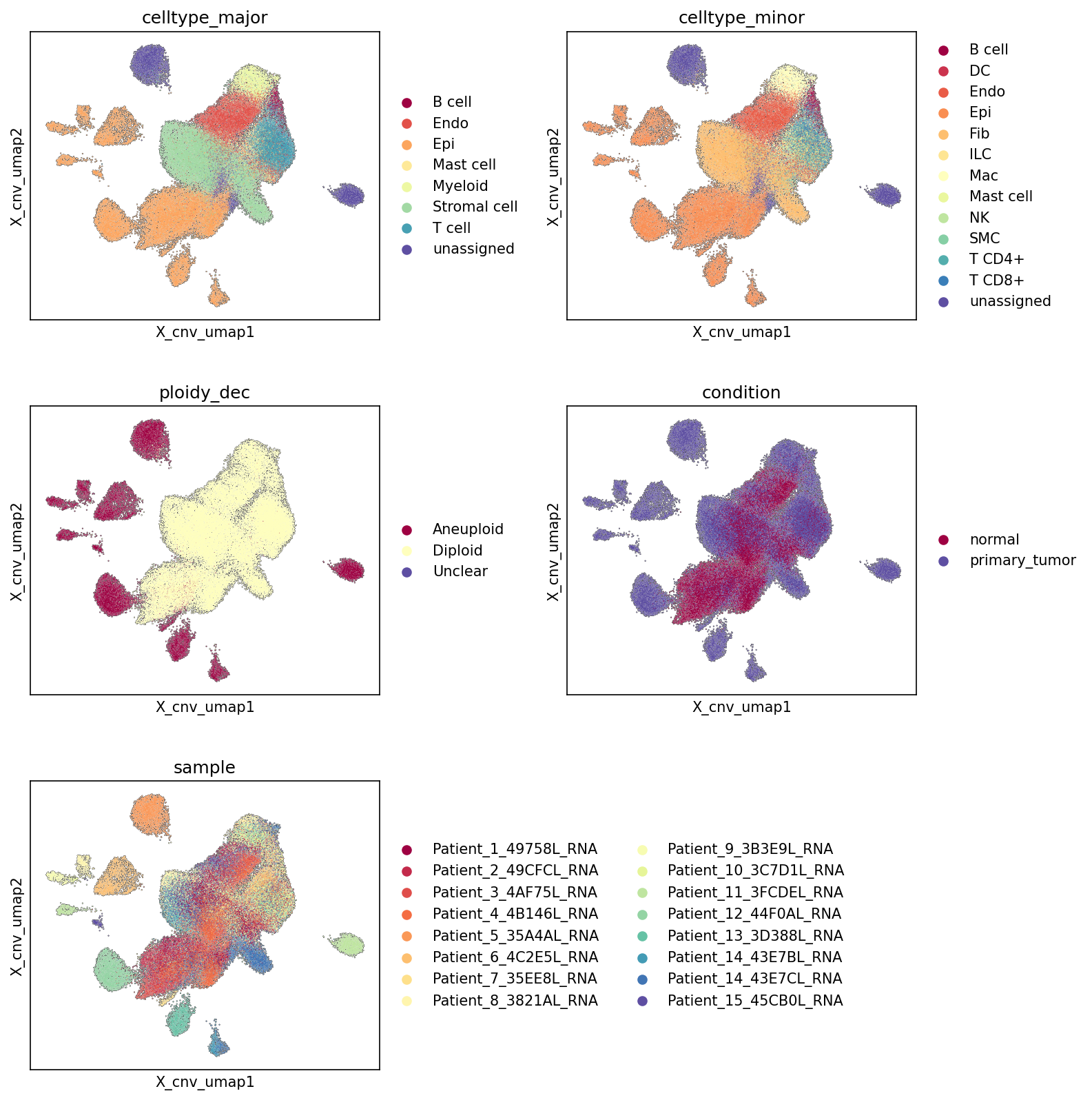
[Analysis Visualization Results]...
Analysis Overview
This analysis presents five UMAP (Uniform Manifold Approximation and Projection) plots, all generated using copy number variation (CNV) patterns, to visualize the distribution of cells colored by celltype_major, celltype_minor, ploidy_dec (ploidy declaration), condition (normal vs. primary_tumor), and individual sample. The primary goal is to assess cell type identity, ploidy status, and sample/condition-level heterogeneity based on their genomic CNV profiles, especially in the context of breast cancer. The UMAP embedding is specifically configured to leverage CNV information, emphasizing differences in genomic copy number alterations.
Visual Summary
Cell Type and Ploidy Distribution
- celltype_major and celltype_minor UMAPs: These plots reveal clear clustering of cells by both major and minor cell types based on their CNV profiles. Epithelial cell (orange in major, also prominent in minor) forms a large, central cluster. Other major cell types like Stromal cell (green) and Endothelial cell (red) also form distinct, albeit sometimes partially overlapping, clusters. Immune cell populations (B cell, T cell, Myeloid cell, Mast cell, NK cell, ILC) tend to form more dispersed or peripheral clusters. The fine-grained celltype_minor UMAP further refines these distinctions, showing sub-populations within major categories. The presence of unassigned cells in various regions suggests populations that are less clearly defined by current annotations or exhibit ambiguous CNV signatures.
- ploidy_dec UMAP: This UMAP demonstrates a striking separation between Aneuploid (dark red) and Diploid (light yellow) cell populations. A large portion of the central UMAP space, notably overlapping with the Epithelial cell cluster, is predominantly Aneuploid. Diploid cells tend to occupy distinct, often surrounding or separate, clusters. The Unclear category is minimally represented, consistent with the provided data context (3 cells). This strong separation indicates that CNV patterns are highly effective at distinguishing cells based on their ploidy status.
Condition and Sample Heterogeneity
- condition UMAP: This plot shows a clear segregation of cells based on condition. The primary_tumor cells (dark blue) strongly co-localize with the Aneuploid regions identified in the ploidy_dec UMAP and the dominant Epithelial cell cluster. Conversely, normal cells (dark red) are primarily found in the regions corresponding to Diploid populations, often occupying distinct clusters on the periphery. While there is a general separation, some degree of mixing or adjacency is observed, particularly at the interfaces of tumor and normal regions, reflecting the complex microenvironment.
- sample UMAP: The sample UMAP highlights significant inter-patient heterogeneity. While the central primary_tumor and Aneuploid regions show contributions from multiple patients, distinct clusters and sub-regions are often dominated by cells from specific patients (represented by different colors). This indicates that each patient contributes a unique CNV landscape, leading to patient-specific clustering patterns, even within broadly defined primary_tumor or normal conditions. This variability is expected in cancer genomics.
Biological Interpretation
The UMAPs provide compelling evidence that CNV profiles are highly informative for dissecting the cellular composition and disease state in breast tissue.
- Tumor Cell Identification and Origin: The strong overlap of the Epithelial cell cluster with Aneuploid cells and cells from the primary_tumor condition unequivocally supports the designation of Epithelial cell as the 'Resolved tumor-origin cell type'. This is a hallmark of breast cancer, which often originates from epithelial cells undergoing chromosomal instability leading to aneuploidy.
- CNV as a Determinant of Cell Identity and State: The ability of CNV patterns to distinctly cluster major and minor cell types, and more importantly, to separate aneuploid from diploid populations, underscores the profound impact of genomic alterations on cellular identity and biological function within this tissue. The UMAP algorithm, configured with CNV data, effectively captures these underlying genomic differences.
- Tumor Microenvironment Composition: While tumor cells (Epithelial cell, Aneuploid, primary_tumor) form a central and dominant mass, the presence of Stromal cell, Endothelial cell, and various Immune cells in distinct but sometimes adjacent clusters suggests a complex tumor microenvironment. These non-tumor cells, predominantly Diploid and often associated with normal regions, represent the host's response and structural components interacting with the evolving tumor.
- Patient-Specific Genomic Signatures: The sample UMAP reveals that despite a common disease (breast cancer), individual patients exhibit unique CNV landscapes. This inter-patient heterogeneity is crucial for understanding disease progression and therapeutic responses, implying that 'primary_tumor' can manifest with varied genomic alterations across individuals.
- Robustness of Ploidy and Condition Annotations: The clear separation in the ploidy_dec and condition UMAPs validates the quality and biological relevance of these annotations. It confirms that the bioinformatic assignment of ploidy and the experimental classification of samples as 'normal' or 'primary_tumor' are strongly reflected in the underlying CNV profiles.
Annotation Notes
- The unassigned cell populations observed in both celltype_major and celltype_minor UMAPs warrant further investigation. These could represent novel cell states, transitional cells, or cells with ambiguous CNV signatures that don't fit well into current annotations.
- The ploidy_dec UMAP shows excellent separation, with very few Unclear cells, indicating high confidence in the ploidy assignments based on CNVs.
- The clear separation of normal and primary_tumor conditions in the CNV-based UMAP provides strong evidence for distinct genomic profiles associated with disease state.
- The observed sample-specific clustering within the UMAP, particularly within the 'primary_tumor' region, highlights the intrinsic heterogeneity of cancer and the need to consider individual patient genomic profiles.
6. Minor Cell Type Population Analysis in Breast Tissue
[Analysis Visualization Results]...
Analysis Overview
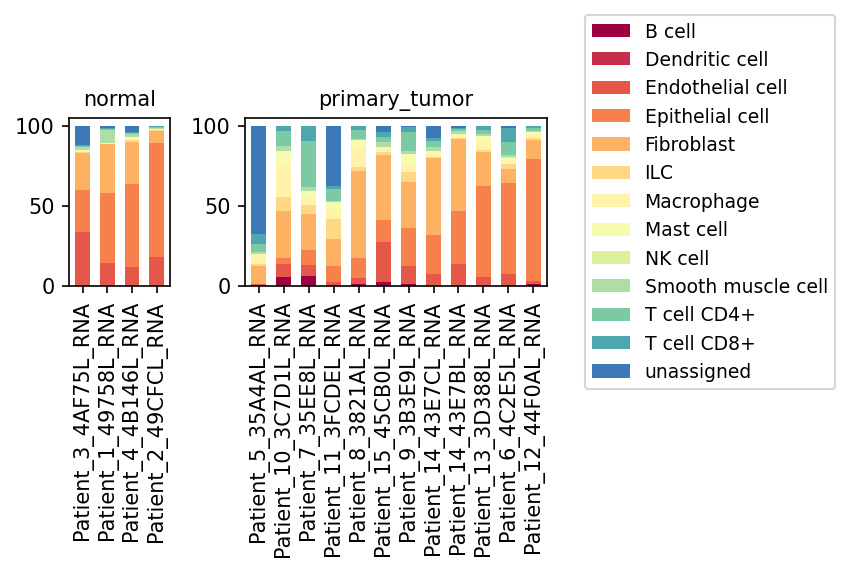
This analysis presents a population bar plot visualizing the relative proportions of minor cell types across individual samples, stratified by 'normal' and 'primary_tumor' conditions. The plot provides a high-level overview of cellular composition differences, highlighting the cellular heterogeneity within breast tissue in healthy and cancerous states. This type of analysis is crucial for understanding the basic architecture of the tumor microenvironment (TME) and how it differs from normal tissue.
Visual Summary
The visualization displays two main panels: one for 'normal' samples and one for 'primary_tumor' samples. Each bar represents an individual sample, with colored segments indicating the proportion of each minor cell type.
- Normal Samples: The four 'normal' samples show a relatively consistent cell type composition. Epithelial cells (orange) constitute a significant proportion, typically over 50%, followed by Fibroblasts (light orange) and Endothelial cells (red-orange). Immune cell populations like T cells (CD4+ and CD8+) and Macrophages are present but in smaller, relatively stable proportions. The 'unassigned' category (dark blue) is minimal in normal samples.
- Primary Tumor Samples: The 'primary_tumor' samples exhibit significantly higher heterogeneity in cell type composition compared to normal samples.
- Epithelial cells remain a major component in most tumor samples, consistent with their designation as the 'tumor-origin cell type'. However, their proportion varies widely across different tumor samples, ranging from dominant (e.g., Patient_6_4C2E5L_RNA, Patient_12_44F0AL_RNA) to considerably reduced (e.g., Patient_5_35A4AL_RNA, Patient_11_3FCDEL_RNA).
- Fibroblasts show a noticeable increase in many primary tumor samples (e.g., Patient_10_3C7D1L_RNA, Patient_7_35EE8L_RNA) compared to normal tissue, suggesting an expansion of the stromal compartment.
- Macrophages (yellow) also appear to be more prominent in several tumor samples (e.g., Patient_5_35A4AL_RNA, Patient_10_3C7D1L_RNA, Patient_11_3FCDEL_RNA), indicating potential immune cell infiltration.
- T cells (CD4+ and CD8+) and B cells (maroon) are also more variable in their proportions within tumors, with some samples showing higher immune cell content than others.
- The 'unassigned' cell population is notably higher in several primary tumor samples (e.g., Patient_5_35A4AL_RNA, Patient_10_3C7D1L_RNA, Patient_11_3FCDEL_RNA) compared to normal samples. This could represent cells difficult to classify, novel cell states, or artifacts.
Biological Interpretation
The observed shifts in cell type proportions between normal and primary tumor samples provide crucial insights into the biological changes occurring in breast cancer.
- Tumor Microenvironment (TME) Remodeling: The increased heterogeneity and altered proportions of stromal cells (Fibroblasts, Endothelial cells) and immune cells (Macrophages, T cells) in 'primary_tumor' samples strongly indicate significant remodeling of the TME. This is a hallmark of cancer progression, where non-malignant cells contribute to tumor growth, invasion, and metastasis.
- Epithelial Cell Dynamics: While epithelial cells are the tumor-origin cell type, their variable proportion in tumors suggests different tumor cellularities or varying degrees of stromal infiltration. Tumors with a higher proportion of epithelial cells might be more homogeneous or less infiltrated by stromal and immune components, while those with lower epithelial content could be highly desmoplastic or have extensive immune infiltration.
- Cancer-Associated Fibroblasts (CAFs): The apparent increase in Fibroblast populations in several tumor samples aligns with the known role of Cancer-Associated Fibroblasts (CAFs) in breast cancer. CAFs secrete extracellular matrix components, growth factors, and cytokines that promote tumor growth, angiogenesis, and immune suppression PMID: 32669614.
- Tumor-Associated Macrophages (TAMs): The elevated proportion of Macrophages in some tumor samples is consistent with the presence of Tumor-Associated Macrophages (TAMs). TAMs are often polarized towards an M2-like phenotype, fostering angiogenesis, immune evasion, and metastasis in breast cancer PMID: 35165971.
- Immune Cell Infiltration: The variable presence of T cells (CD4+, CD8+) and B cells indicates diverse immune responses within different tumors. Some tumors might be "immune-hot" with significant infiltration, while others could be "immune-cold" or show distinct immune cell compositions that influence prognosis and response to immunotherapy.
- "Unassigned" Cell Population: The higher proportion of 'unassigned' cells in some tumor samples warrants further investigation. These could represent novel cell states unique to the tumor microenvironment, cells undergoing dedifferentiation, or cells whose transcriptional profiles make them challenging to categorize with existing reference annotations. Understanding these cells could reveal new aspects of tumor biology.
Clinical or Translational Implications
The observed differences in minor cell type populations between normal and primary breast tumors have several potential clinical implications:
- Biomarker Discovery: The distinct cellular compositions could serve as prognostic or predictive biomarkers. For instance, a higher proportion of CAFs or TAMs might correlate with more aggressive disease or resistance to certain therapies.
- Therapeutic Targeting: Understanding the dominant stromal and immune cell types in specific tumors could guide the development of targeted therapies. For example, strategies to deplete CAFs, repolarize TAMs, or enhance T-cell infiltration are under investigation in breast cancer PMID: 33716345.
- Personalized Medicine: The significant inter-tumor heterogeneity suggests that a "one-size-fits-all" approach to breast cancer treatment may be suboptimal. Characterizing the specific cellular makeup of each patient's tumor could facilitate more personalized treatment strategies, including optimizing combinations of conventional therapies with immunotherapies or stromal-targeting agents.
- Improved Diagnosis and Staging: Detailed cellular composition analysis could potentially augment current pathological assessments, providing a deeper understanding of tumor biology beyond histological grading.
7. T Cell Subtype Population Analysis in Breast Tissue: Normal vs. Primary Tumor
[Analysis Visualization Results]...
Analysis Overview
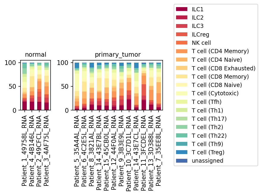
This analysis presents a population bar plot illustrating the relative proportions of various T cell subsets, including Innate Lymphoid Cells (ILCs) and NK cells, within different samples from 'normal' and 'primary_tumor' breast tissue. Each bar represents a single sample, with stacked segments indicating the percentage contribution of each T cell or ILC/NK subset. The goal is to identify potential shifts in immune cell composition associated with the primary tumor microenvironment.
Visual Summary
The visualization displays the T cell subset composition for four 'normal' samples and ten 'primary_tumor' samples.
- Overall Composition: Both normal and primary tumor samples contain a diverse array of T cell subsets, including CD4+ T cells (Memory, Naive), CD8+ T cells (Memory, Naive, Cytotoxic, Exhausted), various helper T cell types (Th1, Th2, Th9, Th17, Th22, Tfh), T regulatory cells (Treg), ILCs (ILC1, ILC2, ILC3, ILCreg), and NK cells. An 'unassigned' category is also present, albeit in small proportions.
- Normal Samples: Show a relatively consistent distribution of T cell subsets across the four analyzed samples. Key populations observed include CD4 Memory, CD8 Memory, CD8 Naive, and Cytotoxic T cells, along with a mix of Th cells. T cell (CD8 Exhausted) and T cell (Treg) populations appear to be present at lower proportions.
- Primary Tumor Samples: Exhibit greater heterogeneity in T cell subset distribution across individual patient samples compared to normal controls.
- Enrichment of T cell (CD8 Exhausted): A notable increase in the proportion of T cell (CD8 Exhausted) is observed in several primary tumor samples compared to normal tissue. This population, represented by the light yellow segment, is visibly more prominent in the tumor microenvironment.
- Increased T cell (Treg): The proportion of T cell (Treg), represented by the dark blue segment at the top of the stack, appears generally higher in many primary tumor samples compared to normal samples.
- Variability in ILC3: The ILC3 population (dark red at the bottom) shows variable but sometimes increased proportions in primary tumor samples.
- Shifts in other populations: While less uniformly striking, there appear to be subtle shifts in other T cell subsets, with some naive and memory populations potentially reduced proportionally in certain tumor samples.
Biological Interpretation
The observed shifts in T cell subset populations provide significant biological insights into the immune landscape of breast cancer.
- T Cell Exhaustion in Tumors: The increased proportion of T cell (CD8 Exhausted) in primary tumor samples is a critical finding. CD8+ T cells are crucial for anti-tumor immunity by directly killing cancer cells. However, in the chronic antigen-rich and immunosuppressive tumor microenvironment (TME), these cells can become "exhausted," losing their effector functions (e.g., cytokine production, cytolytic activity) and upregulating inhibitory receptors (e.g., PD-1, TIM-3). This exhaustion represents a key mechanism by which tumors evade immune surveillance [PubMed Search: T cell exhaustion cancer PubMed Search].
- Regulatory T Cells (Tregs) and Immunosuppression: The apparent enrichment of T cell (Treg) in primary tumor samples is highly indicative of an immunosuppressive TME. Tregs play a pivotal role in maintaining immune tolerance by suppressing the activity of effector T cells and other immune cells. Their increased presence in cancer is a well-established mechanism contributing to tumor immune evasion and progression in breast cancer [PubMed Search: regulatory T cells breast cancer immunosuppression PubMed Search].
- ILC3 Dynamics: The variable but sometimes increased presence of ILC3 in tumor samples suggests potential alterations in innate immune responses. ILC3s are known to respond to cytokines and contribute to tissue homeostasis and inflammation, with complex roles in cancer that can be either pro- or anti-tumorigenic depending on the context and specific TME signals [PubMed Search: ILC3 cancer PubMed Search].
- Heterogeneity in Tumor Immunity: The significant inter-sample variability among primary tumor samples underscores the heterogeneous nature of immune responses in cancer patients. This suggests that individual patients may mount different immune responses or have distinct immune evasion mechanisms, which could influence treatment outcomes.
Clinical or Translational Implications
The alterations in T cell subset populations observed in breast primary tumors have direct clinical and translational implications:
- Immunotherapy Response: The enrichment of exhausted CD8 T cells and Tregs strongly suggests an immunosuppressive tumor microenvironment. This makes breast cancer a prime candidate for immunotherapeutic strategies aiming to reverse T cell exhaustion (e.g., immune checkpoint blockade targeting PD-1/PD-L1 or CTLA-4) or to deplete/inhibit Tregs.
- Biomarkers for Prognosis and Treatment Selection: The proportions of exhausted CD8 T cells and Tregs could potentially serve as prognostic biomarkers, predicting disease progression or recurrence. Furthermore, these immune cell compositions might help in stratifying patients who are more likely to respond to specific immunotherapies.
- Combination Therapies: The complex interplay between different immune cell subsets suggests that combination therapies targeting multiple immunosuppressive pathways (e.g., combining checkpoint inhibitors with Treg-depleting agents) might be more effective in a subset of breast cancer patients.
- Monitoring Immune Landscape: Serial monitoring of T cell subsets could provide insights into treatment response or resistance mechanisms during anti-cancer therapy.
8. Differential T Cell Subset Proportions in Breast Cancer
[Analysis Visualization Results]...
Analysis Overview
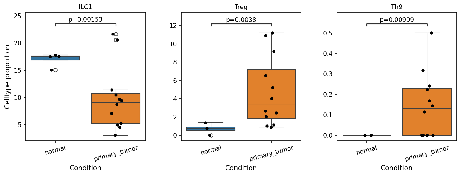
This analysis investigates the proportional changes of T cell subsets between 'normal' and 'primary_tumor' conditions within breast tissue single-cell RNA-seq data. The tool plot_box_for_celltype_population_with_signif_difference was used to identify and visualize cell types with statistically significant differences in their proportions. The analysis focused on subsets within the broader T cell compartment, aiming to highlight shifts in immune cell composition within the tumor microenvironment.
Visual Summary
The box plots display the celltype proportion for three distinct T cell subsets: ILC1, Treg, and Th9, comparing 'normal' and 'primary_tumor' conditions. Each plot includes individual data points (black dots for normal, orange dots for primary_tumor) and p-values indicating statistical significance.
- ILC1 (Type 1 Innate Lymphoid Cells): The proportion of ILC1s is significantly lower in the 'primary_tumor' condition compared to 'normal' tissue (p=0.00153). In normal tissue, ILC1s constitute approximately 17-18% of the cell population, whereas in primary tumors, their proportion drops to around 9-10%. This suggests a substantial decrease in this cell type in the tumor microenvironment.
- Treg (Regulatory T cells): Conversely, the proportion of Tregs is significantly higher in the 'primary_tumor' condition compared to 'normal' tissue (p=0.0038). Normal tissue shows a very low Treg proportion (approximately 0.5-1%), which increases notably to about 3-4% in primary tumors. This indicates an enrichment of Tregs in the tumor microenvironment.
- Th9 (T helper 9 cells): The proportion of Th9 cells also shows a statistically significant increase in the 'primary_tumor' condition compared to 'normal' tissue (p=0.00999). While their overall proportion remains relatively low, normal tissue exhibits nearly undetectable levels, which rise to a median of approximately 0.15% in primary tumors.
Biological Interpretation
The observed shifts in T cell subset proportions between normal and primary tumor breast tissue provide critical insights into the immune landscape of breast cancer.
- Decrease in ILC1s in Tumors: ILC1s are known to play a role in anti-tumor immunity, primarily through the production of IFN-γ, which can promote cytotoxic responses and tumor suppression. A significant decrease in ILC1s within the primary tumor microenvironment suggests a potential weakening of type 1 anti-tumor immune responses, which could contribute to immune evasion and tumor progression.
PubMed search: ILC1 cancer immunity
- Increase in Tregs in Tumors: The substantial increase in Regulatory T cells (Tregs) in primary tumors is a hallmark of an immunosuppressive tumor microenvironment in many cancers, including breast cancer. Tregs are critical for maintaining immune tolerance by suppressing the activity of effector T cells (like CD8+ cytotoxic T cells) and other immune cells. Their enrichment in tumors allows cancer cells to evade immune surveillance, contributing to tumor growth and metastasis. This finding aligns with the inferred context of breast cancer, where increased Tregs are often associated with poor prognosis.
UniProt: FOXP3 (key Treg marker)
PubMed search: Treg breast cancer immunosuppression
- Increase in Th9 cells in Tumors: The increase in Th9 cells in primary tumors presents a more complex picture. Th9 cells produce cytokines like IL-9 and IL-10. While IL-9 has been implicated in both pro- and anti-tumor responses depending on the cancer type and context (e.g., sometimes promoting mast cell activation for anti-tumor effects, sometimes supporting tumor growth), the overall context of decreased ILC1s and increased Tregs suggests a predominantly immunosuppressive environment. In breast cancer, the role of Th9 cells is still being elucidated, but some studies indicate they might contribute to tumor progression or immune evasion in certain contexts.
PubMed search: Th9 breast cancer
Collectively, these findings paint a picture of an immune microenvironment in breast cancer characterized by dampened anti-tumor immunity (decreased ILC1s) and enhanced immune suppression (increased Tregs).
Clinical or Translational Implications
These findings have several potential clinical and translational implications for breast cancer:
- Prognostic Biomarkers: The proportions of ILC1s, Tregs, and Th9 cells, particularly the ILC1/Treg ratio, could serve as prognostic biomarkers, potentially indicating disease progression or response to therapy. A lower ILC1 proportion and higher Treg proportion might correlate with more aggressive disease or poorer outcomes.
- Therapeutic Targets: The observed shifts highlight specific T cell subsets as potential targets for immunotherapy.
- Targeting Tregs: Strategies to deplete or inhibit Tregs within the tumor microenvironment could unleash anti-tumor immune responses, potentially enhancing the efficacy of existing immunotherapies (e.g., checkpoint inhibitors).
- Enhancing ILC1s: Approaches to boost ILC1 numbers or their function could help re-establish a more effective anti-tumor immune response.
- Monitoring Immunotherapy Response: Changes in these T cell subset proportions could be monitored during immunotherapy to assess treatment efficacy and predict patient response. For example, a successful immunotherapy might lead to a decrease in Tregs or an increase in ILC1s.
9. Macrophage Subset Population Analysis in Breast Tissue
[Analysis Visualization Results]...
Analysis Overview
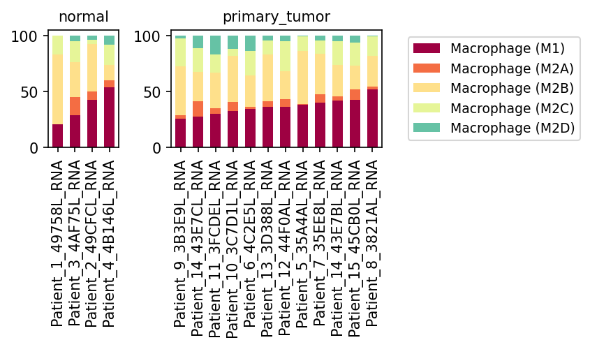
This analysis presents the population distribution of macrophage subsets (M1, M2A, M2B, M2C, M2D) across individual samples derived from breast tissue. The data are categorized into 'normal' and 'primary_tumor' conditions. The primary objective is to evaluate how the composition of macrophage subsets shifts between healthy and cancerous breast microenvironments, providing insight into macrophage polarization associated with breast cancer.
Visual Summary
The population bar plots illustrate the relative proportions of five macrophage subsets for each individual sample, grouped by 'normal' and 'primary_tumor' conditions.
- Normal Condition: In the 'normal' samples, Macrophage (M1) cells (dark red) represent a substantial proportion, often co-existing with significant proportions of Macrophage (M2B) cells (light yellow). Macrophage (M2A) (orange), Macrophage (M2C) (light green), and Macrophage (M2D) (teal) subsets are generally present in smaller proportions.
- Primary Tumor Condition: In contrast, 'primary_tumor' samples show a distinct shift in macrophage composition. While Macrophage (M1) cells are still present, their relative proportion appears reduced compared to many normal samples. There is a noticeable increase in the collective representation of M2-like macrophage subsets, particularly Macrophage (M2B) which consistently forms a large component across most tumor samples. Macrophage (M2C) and Macrophage (M2D) also appear to contribute more significantly to the overall macrophage population in the tumor microenvironment compared to normal samples. This suggests an expansion of specific M2 subtypes within the tumor.
Biological Interpretation
Macrophages are highly plastic immune cells that polarize into distinct functional phenotypes, primarily M1 (classically activated) and M2 (alternatively activated), in response to microenvironmental cues.
- M1 Macrophages: Characterized by pro-inflammatory functions, M1 macrophages are typically involved in pathogen clearance and anti-tumor immunity 1. The observed relative decrease in M1 macrophages in the primary tumor context suggests a suppression of these anti-tumor functions within the breast cancer microenvironment.
- M2 Macrophages: M2 macrophages, encompassing various subtypes (M2A, M2B, M2C, M2D), are generally associated with anti-inflammatory responses, tissue repair, angiogenesis, and immunosuppression, often promoting tumor growth and metastasis 2.
- The consistent prevalence of Macrophage (M2B) in tumor samples, alongside increased contributions from Macrophage (M2C) and Macrophage (M2D), is a key finding. This suggests a significant shift towards an M2-polarized phenotype, which is characteristic of tumor-associated macrophages (TAMs) in many cancers, including breast cancer 3.
- M2B macrophages can exhibit mixed M1/M2 characteristics but are often linked to immune complex-mediated responses. M2C macrophages are typically immunosuppressive, promoting wound healing and tissue remodeling. M2D macrophages are often associated with angiogenesis and tumor progression, induced by factors like adenosine in the tumor microenvironment 2.
This shift towards M2-like macrophage subsets indicates an immune environment that is likely conducive to tumor progression, facilitating processes such as angiogenesis, tissue remodeling, and suppression of anti-tumor immune responses.
Clinical or Translational Implications
The observed shift from an M1-predominant macrophage population in normal tissue to an M2-predominant population in primary breast tumors has significant clinical implications:
- Therapeutic Targeting: The prevalence of M2-like macrophages (TAMs) in breast tumors suggests that these cells could be promising therapeutic targets. Strategies aimed at inhibiting M2 macrophage polarization, depleting TAMs, or reprogramming them towards an M1-like anti-tumor phenotype could enhance the efficacy of existing cancer therapies, including immunotherapies 3.
- Biomarker Potential: Specific M2 macrophage subsets (e.g., M2B, M2C, M2D) or their characteristic gene expression signatures could serve as prognostic biomarkers for disease progression or as predictive markers for response to immunomodulatory treatments in breast cancer patients.
- Heterogeneity and Precision Medicine: The variability in macrophage subset composition across individual tumor samples highlights the importance of personalized approaches. Characterizing the specific macrophage polarization patterns in each patient's tumor could help tailor therapeutic strategies for more effective disease management.
---
References:
- Locati, M., Mantovani, A. Macrophage Plasticity and Polarization. *Annu. Rev. Pathol. Mech. Dis.* 2017. 12:12.1–12.26. https://pubmed.ncbi.nlm.nih.gov/27918751/
- Qian, B. Z., & Pollard, J. W. Macrophage Diversity Enhances Tumor Progression and Metastasis. *Cell*. 2010. 141(1), 39–51. https://pubmed.ncbi.nlm.nih.gov/20346754/
- Pan, Y., Yu, D., Wang, X., et al. Tumor-Associated Macrophages in Breast Cancer. *Frontiers in Immunology*. 2020. 11:582522. https://pubmed.ncbi.nlm.nih.gov/33133036/
10. Ploidy Analysis of Epithelial Cells in Breast Tissue Samples
[Analysis Visualization Results]...
Analysis Overview
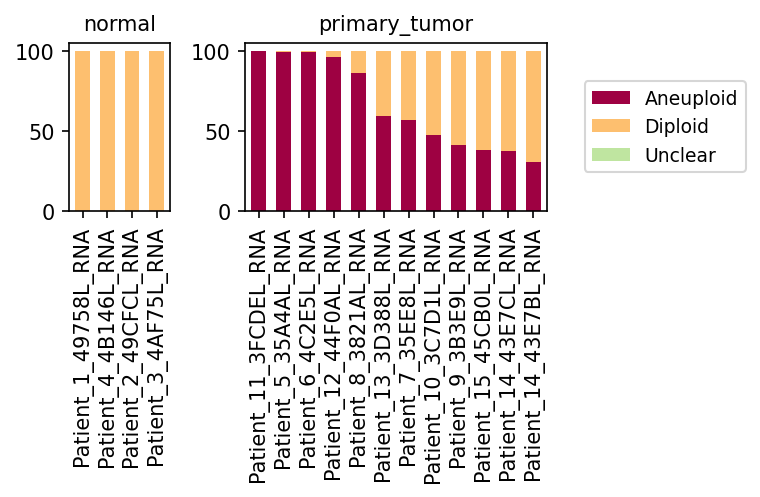
This analysis investigates the ploidy status (diploid vs. aneuploid) of Epithelial cells across various samples, categorized by 'normal' and 'primary_tumor' conditions. Epithelial cells were selected as the focus, given their identification as the 'Resolved tumor-origin cell type' in this breast tissue dataset. The results are presented as a stacked bar plot, showing the percentage of aneuploid and diploid epithelial cells for each individual sample.
Visual Summary
The visualization clearly segregates samples based on their condition: 'normal' and 'primary_tumor'.
- Normal Samples: The left panel, displaying results for four distinct normal samples (e.g., Patient_1_49758L_RNA), shows a uniform pattern where 100% of the Epithelial cells are classified as Diploid (peach bars). There is no detectable aneuploidy in the epithelial cell population of these normal samples.
- Primary Tumor Samples: The right panel, representing primary tumor samples, presents a striking contrast. All primary tumor samples exhibit a substantial proportion of Aneuploid Epithelial cells (burgundy bars).
- Several tumor samples (e.g., Patient_11_3FCDEL_RNA, Patient_5_35A4AL_RNA, Patient_6_4C2E5L_RNA) are almost entirely composed of Aneuploid Epithelial cells, nearing 100%.
- Other primary tumor samples (e.g., Patient_7_35EE8L_RNA, Patient_10_3C7D1L_RNA, Patient_9_3B3E9L_RNA) show a mixed population, with varying proportions of Aneuploid cells ranging from approximately 30% to 80%, alongside a remaining fraction of Diploid cells.
- The 'Unclear' ploidy category (light green in legend) is not visibly represented in any of the bars, indicating a negligible count of such cells in the Epithelial cell population across these samples.
Biological Interpretation
The findings from this ploidy analysis strongly corroborate the inferred biological context of breast cancer, where epithelial cells are the primary site of tumor initiation.
- Aneuploidy as a Hallmark of Cancer: The presence of a high proportion of aneuploid epithelial cells in all primary tumor samples, and their complete absence in normal samples, is a classic genomic hallmark of malignancy. Aneuploidy, an abnormal number of chromosomes, is a common feature of cancer cells resulting from genomic instability, which drives tumor evolution and heterogeneity [PubMed Search].
- Tumor Cell Identification: This analysis effectively distinguishes neoplastic epithelial cells (aneuploid) from normal epithelial cells (diploid). The 'Resolved tumor-origin cell type' (Epithelial cell) is confirmed to harbor the expected genomic instability characteristic of cancer.
- Inter-tumor Heterogeneity: The variability in the percentage of aneuploid cells among different primary tumor samples suggests underlying inter-patient heterogeneity. Some tumors may be more clonally advanced, or have a higher proportion of malignant cells, while others might contain a significant admixture of non-neoplastic stromal cells or quiescent epithelial cells. The presence of diploid epithelial cells within tumor samples could represent non-malignant bystander cells or residual normal ducts entrapped within the tumor microenvironment.
Clinical or Translational Implications
- Diagnostic Potential: The clear distinction in epithelial cell ploidy between normal and primary tumor tissues highlights aneuploidy as a potent diagnostic marker for breast cancer. This genomic feature could aid in confirming malignancy, especially in cases with equivocal histological findings.
- Prognostic and Therapeutic Relevance: The proportion of aneuploid cells within the tumor's epithelial compartment could potentially serve as a prognostic indicator. Tumors with a higher burden of aneuploid cells might represent a more aggressive phenotype, which could influence treatment strategies. Monitoring ploidy could also offer insights into disease progression or response to therapies targeting genomic stability.
- Tumor Microenvironment Analysis: Recognizing the presence of diploid epithelial cells within tumor samples is crucial for understanding the tumor microenvironment. These diploid cells might represent normal epithelial components, or even potentially early-stage tumor cells that have not yet fully acquired aneuploidy, offering targets for preventative or early intervention strategies.
11. Condition-Specific Cell-Cell Interaction Patterns in Breast Tissue Reveal Tumor Microenvironment Dynamics
[Analysis Visualization Results]...
Analysis Overview
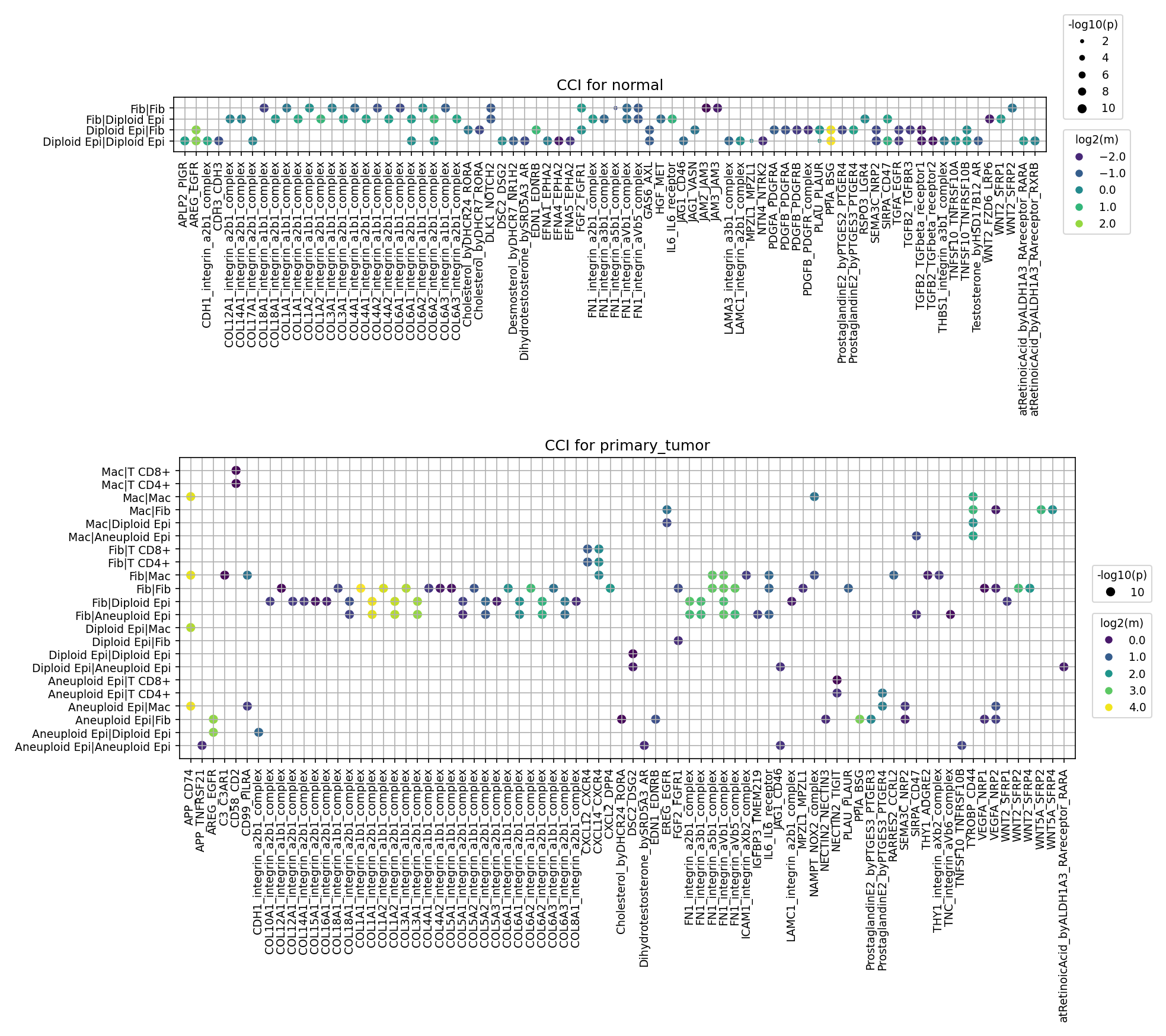
This analysis investigates cell-cell interaction (CCI) patterns in human breast tissue, comparing normal and primary_tumor conditions using single-cell RNA sequencing data. The objective was to identify significant ligand-receptor interactions between key cell types, including Epithelial cells, Fibroblasts, Macrophages, and T cells (CD4+ and CD8+), with a focus on how these interactions change in the tumor microenvironment. The results incorporate ploidy information, specifically highlighting aneuploid epithelial cells identified as the tumor-origin cell type. The visualization employs dot plots, where dot size reflects the significance of the interaction (-log10(p-value)) and color intensity represents the interaction strength (log2(mean) expression of the ligand-receptor pair), for up to 80 top interactions per condition.
Visual Summary
Normal Condition:
The cell-cell interaction landscape in normal breast tissue is characterized by a less complex and diverse set of interacting cell pairs. The primary interactions observed involve diploid Fibroblasts and Epithelial cells, both interacting with themselves and each other. Key interactions are dominated by extracellular matrix (ECM) components such as various Collagen (e.g., COL12A1, COL1A1, COL3A1, COL4A1, COL4A2, COL6A1, COL6A2, COL6A3) and Fibronectin (FN1) complexes interacting with Integrins (e.g., integrin_a2b1_complex, integrin_a1b1_complex, integrin_avb3_complex). Growth factors such as PDGF (PDGFA, PDGFB) and TGFB (TGFB2, TGFB3) also show significant interactions. These patterns largely reflect the fundamental communications necessary for maintaining tissue structure and homeostasis.
Primary Tumor Condition:
In stark contrast, the primary tumor condition exhibits a significantly more complex and altered cell-cell interaction profile. A crucial feature is the prominent involvement of Aneuploid Epi (Aneuploid Epithelial) cells, which are central to many novel and intensified interactions. The diversity of interacting cell pairs expands significantly, now including interactions between tumor epithelial cells and immune cells (Macrophages, T cells), within tumor epithelial cells, and with fibroblasts.
- Emergence of Novel and Intensified Interactions: Several interactions that are absent or weak in normal tissue become highly prominent and strong in the tumor. These include:
- APP_integrin_a2b1_complex (ubiquitous across various tumor-associated pairs including Macrophages, T cells, and Aneuploid Epi).
- Growth factor signaling: EREG_EGFR, LIF_LIFR, NAMPT_NMPR (predominantly within Aneuploid Epi cells).
- ECM-related: SPP1_CD44, SPP1_integrin_avb1_complex, and TNC_integrin complexes (involving Aneuploid Epi and Fibroblasts).
- Angiogenic signaling: VEGFA_FLT1 and VEGFA_KDR (within Aneuploid Epi and with Macrophages).
- Chemokine signaling: CXCL12_CXCR4 (between Aneuploid Epi and Fibroblasts).
- Immune-related: CD58_CD2, NECTIN2_TIGIT, MIF_CD74, ICAM1_integrin_aLb2_complex (involving Macrophages, T cells, and Aneuploid Epi).
- Altered ECM and Growth Factor Signaling: While some FN1-integrin interactions persist or are amplified, particularly involving aneuploid epithelial cells, many COL-integrin interactions appear less dominant compared to the normal state. WNT signaling patterns also appear qualitatively different.
Biological Interpretation
The stark differences in cell-cell interaction patterns between normal and primary tumor conditions reveal critical biological processes underlying breast cancer development and progression, primarily driven by the tumor-origin aneuploid epithelial cells and their interactions within the evolving tumor microenvironment (TME).
- ECM Remodeling and Integrin-Mediated Signaling: The shift from homeostatic collagen-integrin interactions in normal tissue to the prominent involvement of SPP1 (Osteopontin) with CD44 and various integrin complexes, alongside Tenascin-C (TNC)-integrin interactions in the tumor, signifies profound ECM remodeling. SPP1 and TNC are well-established components of the tumor-promoting ECM that facilitate cancer cell adhesion, migration, invasion, and angiogenesis, thus driving metastatic potential PubMed search: SPP1 cancer progression integrin CD44, GeneCards: TNC.
- Oncogenic Growth Factor and Cytokine Signaling Activation: The tumor microenvironment exhibits highly activated signaling pathways crucial for cancer cell proliferation and survival.
- EGFR Signaling: Strong EREG_EGFR and CD74_EGFR interactions, especially autocrine/paracrine signaling within aneuploid epithelial cells, indicate activated EGFR signaling. This pathway is a known driver of cell growth and survival in breast cancer PubMed search: EREG EGFR breast cancer.
- LIF and NAMPT Pathways: The prominence of LIF_LIFR and NAMPT_NMPR interactions, primarily within aneuploid epithelial cells, suggests roles in maintaining tumor cell stemness, promoting survival, and driving metabolic reprogramming to support rapid tumor growth GeneCards: LIF, GeneCards: NAMPT.
- Angiogenesis Promotion: The robust VEGFA_FLT1 and VEGFA_KDR interactions, involving both aneuploid epithelial cells and macrophages, highlight active angiogenesis, a critical process for tumor growth and metastasis by ensuring nutrient and oxygen supply PubMed search: VEGFA angiogenesis cancer.
- Immune Landscape Modulation: Interactions involving immune cells (Macrophages and T cells) and aneuploid epithelial cells point to complex immune modulation within the TME.
- Adhesion and co-stimulation: CD58_CD2 and ICAM1_integrin_aLb2_complex mediate crucial cell adhesion and communication among immune cells and with tumor cells.
- Immune Checkpoint Interactions: The presence of NECTIN2_TIGIT interactions, particularly between macrophages and T cells, suggests the activation of immune checkpoint mechanisms where TIGIT on T cells can inhibit their anti-tumor activity upon binding to NECTIN2 PubMed search: TIGIT NECTIN2 cancer immunology.
- Chemokine-Mediated Tumor-Stromal Crosstalk: The robust CXCL12_CXCR4 interactions between aneuploid epithelial cells and fibroblasts represent a well-characterized signaling axis. This pathway is pivotal in promoting tumor growth, metastasis, and the recruitment of pro-tumorigenic stromal cells and immune cells into the TME in breast cancer PubMed search: CXCL12 CXCR4 breast cancer.
Clinical or Translational Implications
The identified condition-specific cell-cell interactions offer a valuable roadmap for understanding breast cancer pathobiology and pinpointing potential therapeutic targets:
- Targeting Growth Factor Pathways: The hyperactivation of EGFR (via EREG and CD74) and LIFR signaling on aneuploid epithelial cells presents these receptors as attractive therapeutic targets. Strategies involving small molecule inhibitors or blocking antibodies, similar to existing EGFR-targeted therapies, could be explored to suppress tumor proliferation and survival.
- Modulating the Tumor Microenvironment (TME):
- ECM-Targeted Therapies: The increased SPP1-CD44/integrin and TNC-integrin interactions highlight the critical role of ECM remodeling in tumor progression. Disrupting these interactions, for instance, by targeting SPP1 or its receptors, or interfering with TNC-integrin binding, could impede tumor cell invasion and metastasis.
- Anti-Angiogenic Strategies: The robust VEGFA-FLT1/KDR signaling reinforces the continued relevance of anti-angiogenic therapies to inhibit blood vessel formation, thereby starving tumors and limiting their growth and spread.
- Stromal-Tumor Axis Disruption: Targeting the CXCL12-CXCR4 axis could disrupt crucial communication between tumor cells and fibroblasts, thereby inhibiting tumor cell migration, reducing metastasis, and preventing the recruitment of pro-tumorigenic stromal cells.
- Immune Checkpoint Modulation: The presence of NECTIN2_TIGIT interactions suggests TIGIT as a potential immune checkpoint. Blocking TIGIT could re-invigorate anti-tumor T cell responses, offering a novel immunotherapeutic strategy for breast cancer, potentially in combination with other immune checkpoint inhibitors.
These insights provide a prioritized list of ligand-receptor pairs for further experimental validation in preclinical models and for consideration in the development of targeted therapies or companion diagnostics in breast cancer.
12. Condition-Specific Cell-Cell Interaction Patterns in Breast Cancer
[Analysis Visualization Results]...
Analysis Overview
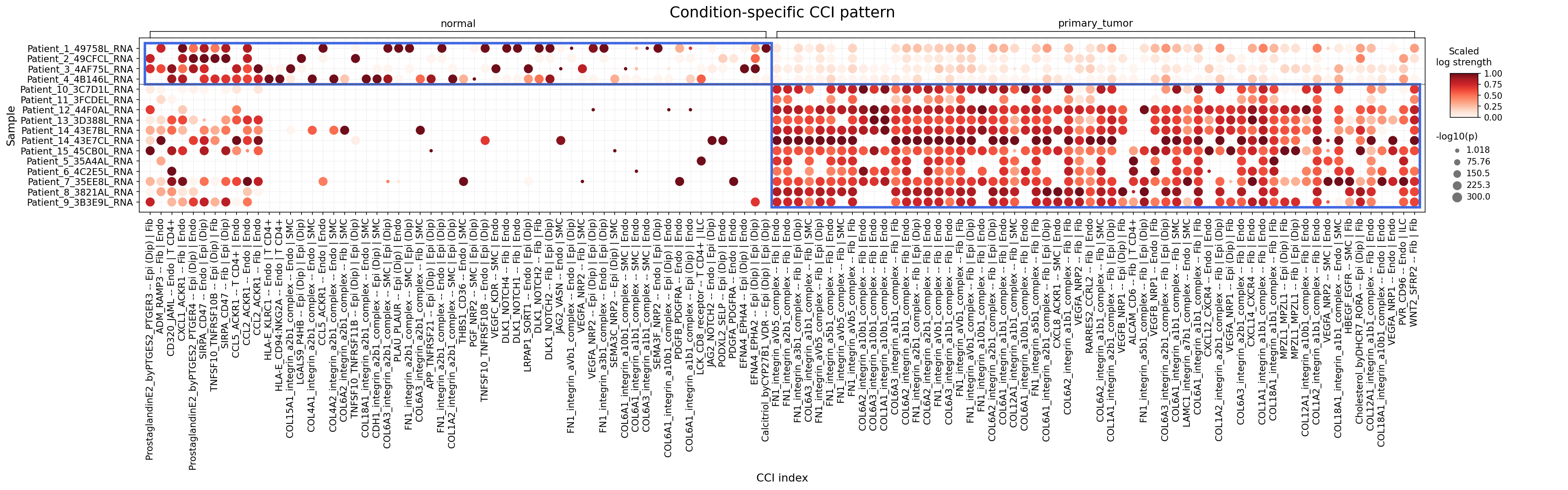
This analysis investigates cell-cell interaction (CCI) patterns involving Epithelial cells (the identified tumor-origin cell type), major immune cells (B cells, Myeloid cells, T cells, Mast cells), and stromal cells (Fibroblasts, Endothelial cells, Smooth muscle cells) across normal and primary_tumor breast tissue conditions. The plot_dot_for_cci_with_signif_difference tool was used to identify and visualize interactions that significantly differ between conditions, with a focus on highlighting interactions more pronounced in the tumor microenvironment. The visualization displays the scaled log strength of interactions (color intensity) and their statistical significance (-log10(p-value) as dot size) for individual patient samples.
Visual Summary
The dot plot provides a clear visual distinction in cell-cell interaction patterns between normal and primary_tumor conditions.
- Normal Condition: Interactions are generally sparse, with fewer and smaller dots, indicating lower interaction strength and statistical significance across samples. While some interactions are present (e.g., involving ProstaglandinE2, ACKR1, and specific integrin complexes), they are less prevalent and potent compared to the tumor condition.
- Primary Tumor Condition: There is a dramatic increase in the number, strength (darker red color), and significance (larger dot size) of cell-cell interactions. A large cluster of highly active and significant interactions is evident in the primary_tumor samples (highlighted by the blue box on the right). This indicates extensive remodeling of the tumor microenvironment and heightened intercellular communication.
- Key Differences: The most striking observation is the pervasive upregulation of interactions involving integrins and various collagen/fibronectin complexes, particularly between Epithelial (Dip) cells (diploid epithelial cells within the tumor context) and stromal components such as Fibroblast, Endothelial cell, and Smooth muscle cell. Angiogenic and chemokine signaling pathways also show significant activity.
Biological Interpretation
The observed shifts in CCI patterns in primary_tumor samples point to several critical biological processes driving breast cancer progression, largely orchestrated through enhanced communication between tumor-associated epithelial cells and their surrounding stromal and immune microenvironment. The Epithelial cell has been identified as the tumor-origin cell type, and Epi (Dip) refers to diploid epithelial cells found within the tumor microenvironment, which are actively involved in these pathological interactions.
1. Extracellular Matrix (ECM) Remodeling and Integrin Signaling
A dominant feature in the primary_tumor microenvironment is the widespread and highly significant activity of integrin-mediated interactions, primarily involving Epithelial (Dip) cells and Fibroblasts, Endothelial cells, or Smooth muscle cells. These interactions are nearly absent or very weak in normal tissue.
Specific Interactions
- Many collagen (e.g., COL1A1, COL1A2, COL6A1, COL6A2) and fibronectin (FN1) complexes interacting with various integrin subunits (e.g., integrin_a1b1_complex, integrin_a2b1_complex, integrin_a5b1_complex, integrin_avb5_complex, integrin_a10b1_complex, integrin_a11b1_complex, integrin_a12b1_complex) are highly activated.
- Example: COL6A1_integrin_a1b1_complex -- Fib | Epi (Dip) indicates Fibroblast-secreted Collagen VI interacts with integrin_a1b1 on Epithelial (Diploid) cells.
- Mechanism: Integrins are transmembrane receptors that link the ECM to the intracellular cytoskeleton, modulating cell adhesion, migration, proliferation, and survival. In cancer, altered ECM composition and integrin signaling promote tumor cell invasion, metastasis, and angiogenesis by providing migratory cues and survival signals. Collagen VI, for instance, is increasingly recognized for its role in promoting breast cancer stemness and metastasis.
- Therapeutic Potential: Targeting specific integrin subunits or their ECM ligands could disrupt critical tumor-stroma communication.
- Relevant Agents: Integrin-blocking antibodies (e.g., targeting αVβ3, α5β1) or small molecule inhibitors are being developed for various cancers [1]. For example, Cilengitide targets αVβ3 and αVβ5 integrins.
2. Angiogenesis Signaling
Several interactions related to vascular development and remodeling are significantly upregulated in primary_tumor samples, particularly between Epithelial (Dip) cells and Endothelial cells.
Specific Interactions
- VEGFA_NRP1 -- Fib | Endo, VEGFA_NRP2 -- Epi (Dip) | Endo, VEGFB_NRP1 -- Epi (Dip) | Endo, VEGFC_KDR -- SMC | Endo.
- Mechanism: Vascular Endothelial Growth Factors (VEGFA, VEGFB, VEGFC) and their receptors (KDR/VEGFR2, Neuropilin-1 (NRP1), Neuropilin-2 (NRP2)) are master regulators of angiogenesis, the formation of new blood vessels. Uncontrolled angiogenesis is essential for tumor growth, nutrient supply, and metastasis. NRP1 and NRP2 act as co-receptors that enhance VEGFR signaling.
- Therapeutic Potential: Anti-angiogenic therapies aim to starve tumors by inhibiting new blood vessel formation.
- Relevant Agents: Bevacizumab (anti-VEGFA antibody), Ramucirumab (anti-VEGFR2 antibody) are established treatments in various cancers, including breast cancer.
3. Chemokine Signaling
The CXCL12-CXCR4 axis shows enhanced activity in the tumor microenvironment.
- Specific Interactions: CXCL12_CXCR4 -- Epi (Dip) | Endo.
- Mechanism: C-X-C motif chemokine ligand 12 (CXCL12) and its receptor CXCR4 are critical for cell migration, homing, and retention. In cancer, this axis promotes tumor cell migration, invasion, metastasis to distant organs, and recruitment of pro-tumorigenic immune cells and stromal cells.
- Therapeutic Potential: Blocking the CXCL12-CXCR4 axis can inhibit metastasis and modulate the immune microenvironment.
- Relevant Agents: Plerixafor (a CXCR4 antagonist) is approved for stem cell mobilization in multiple myeloma and non-Hodgkin lymphoma and is being investigated in solid tumors [2].
4. Immune Evasion and Modulation
An important interaction related to immune suppression is activated in the tumor setting.
- Specific Interactions: SIRPA_CD47 -- Fib | Epi (Dip).
- Mechanism: CD47, expressed on cancer cells, binds to SIRPA (signal regulatory protein alpha) on macrophages, delivering a "don't eat me" signal that prevents phagocytosis. This interaction is a critical mechanism for immune evasion by cancer cells. Fibroblasts can also contribute to this by interacting with tumor epithelial cells.
- Therapeutic Potential: Blocking CD47 can unleash anti-tumor immunity by promoting macrophage-mediated phagocytosis.
- Relevant Agents: Anti-CD47 antibodies like Magrolimab are in clinical development for various malignancies [3].
5. Notch Signaling
Evidence of Notch pathway activation is also present in the primary_tumor environment.
- Specific Interactions: DLK1_NOTCH1 -- Fib | Endo, DLK1_NOTCH2 -- Fib | Endo.
- Mechanism: Notch signaling, activated by ligand (e.g., Delta-like ligand, DLK1) binding to Notch receptors, is crucial for cell fate determination, development, and stem cell maintenance. Aberrant Notch signaling contributes to cancer development, epithelial-mesenchymal transition (EMT), angiogenesis, and drug resistance.
- Therapeutic Potential: Inhibiting Notch signaling can target cancer stem cells and reduce tumor progression.
- Relevant Agents: Gamma-secretase inhibitors (GSIs) and anti-Notch antibodies are under investigation in clinical trials.
6. Inflammation and Other Interactions
- Prostaglandin E2 (PGE2) Signaling: ProstaglandinE2_byPTGES2_PTGER3 -- Epi (Dip) | Fib is observed in both normal and tumor conditions, but its specific role and modulation in the tumor require further investigation. PGE2 often promotes inflammation, angiogenesis, and immune suppression in cancer. COX inhibitors could indirectly affect this.
- CD36 Interactions: THBS1_CD36 -- Fib | Epi (Dip) and CD36 -- Endo | SMC show activity. CD36 is a scavenger receptor involved in lipid metabolism, angiogenesis, and immune responses. Its interaction with Thrombospondin-1 (THBS1) can modulate angiogenesis and inflammation.
Clinical or Translational Implications
The comprehensive upregulation of diverse and significant cell-cell interactions in breast primary_tumor samples highlights the dynamic and complex nature of the tumor microenvironment as a central driver of disease.
- Multi-Targeted Therapies: Given the multifaceted nature of these interactions, combination therapies targeting multiple pathways (e.g., angiogenesis, integrin signaling, and immune evasion) could be more effective than single-agent approaches.
- Biomarker Discovery: Highly active and specific CCI pairs identified here could serve as novel biomarkers for breast cancer prognosis or response to therapy.
- Targeting ECM-Integrin Axis: The strong involvement of various integrins suggests that targeting specific integrin receptors (e.g., using monoclonal antibodies or small molecule inhibitors) could impede tumor growth, invasion, and metastasis by disrupting critical tumor-stroma connections.
- Enhancing Immunotherapy: Blocking immune checkpoints like the CD47-SIRPA axis presents a promising strategy to activate anti-tumor immune responses, potentially synergistic with other therapies.
- Anti-angiogenic Strategies: The confirmed role of VEGF-related pathways further supports the continued use and refinement of anti-angiogenic agents in breast cancer, possibly in combination with agents disrupting ECM or immune interactions.
- Stromal Targeting: The prominent role of Fibroblasts, Endothelial cells, and Smooth muscle cells in mediating these interactions underscores the importance of targeting not only the cancer cells but also the supportive stromal elements of the tumor microenvironment.
This analysis provides a rich landscape of specific cell-cell communication hubs that are pathologically activated in breast cancer, offering a foundation for developing novel targeted therapies and combination strategies.
---
References:
- Integrin Inhibitors in Cancer: PubMed search for "integrin inhibitors cancer" (PubMed Search)
- CXCR4 Antagonists in Cancer: PubMed search for "CXCR4 antagonist cancer" (PubMed Search)
- CD47 Blockade in Cancer: PubMed search for "CD47 blockade cancer" (PubMed Search)
13. Condition-Specific Cell-Cell Interaction Patterns in Breast Tumor Microenvironment
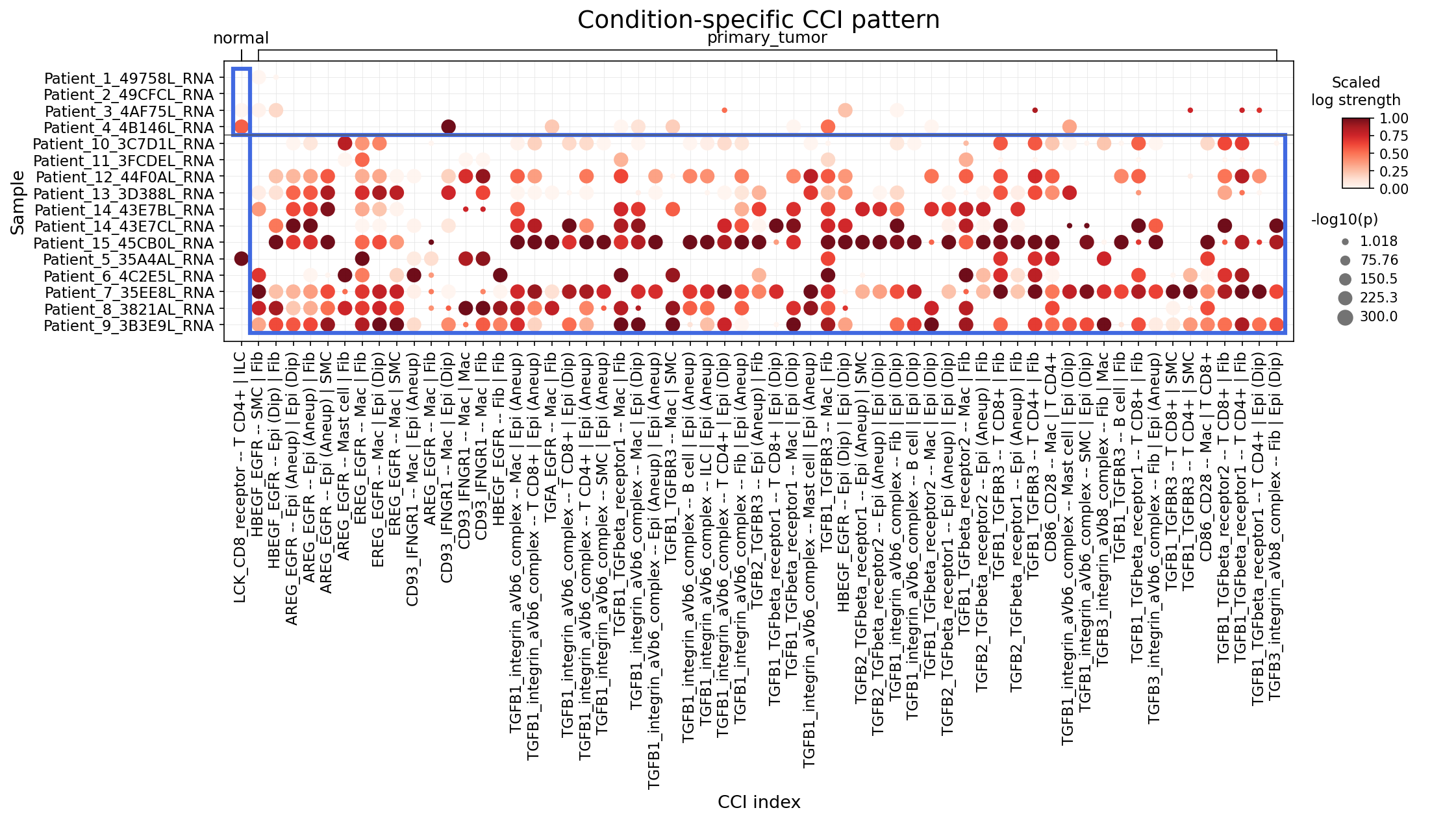
[Analysis Visualization Results]...
Analysis Overview
This analysis investigates condition-specific cell-cell interactions (CCIs) within human breast tissue, comparing primary tumor samples against normal tissue samples. The focus is on interactions involving a curated set of immune checkpoint and cell cycle pathway-related genes, and key cell types including Epithelial cells (the resolved tumor-origin cell type), major immune cell populations (B cell, Myeloid cell, T cell, Mast cell), and Stromal cells. The plot_dot_for_cci_with_signif_difference tool was utilized to visualize interactions that significantly differ between conditions, with a maximum of 60 top differentiating interactions displayed per group.
Visual Summary
The dot plot visualizes the strength and statistical significance of specific cell-cell interactions across individual patient samples under "normal" and "primary_tumor" conditions.
- X-axis: Represents individual cell-cell interaction pairs, indicating the ligand-receptor complex, the interacting cell types (e.g., Fibroblast -- Epithelial cell), and the ploidy status of the epithelial cell (Diploid or Aneuploid).
- Y-axis: Lists individual patient samples.
- Dot Color (Scaled log strength): The intensity of the red color indicates the scaled log strength of the interaction, with darker red corresponding to stronger interactions.
- Dot Size (-log10(p-value)): The size of the dot reflects the statistical significance of the interaction, with larger dots representing more significant interactions (smaller p-value).
A striking visual pattern emerges:
- Normal Condition: Samples from the normal condition (left side, enclosed by the blue box) exhibit very few and generally weaker/less significant cell-cell interactions. This suggests a relatively quiescent state with limited active signaling through these specific immune checkpoint and cell cycle-related pathways.
- Primary Tumor Condition: In contrast, primary tumor samples (right side) display a highly dense and vibrant pattern of strong and significant cell-cell interactions. This indicates a dramatically altered and highly active communicative microenvironment within the primary tumor, characterized by widespread engagement of the selected ligand-receptor pairs.
Biological Interpretation
The observed increase in cell-cell interactions within the primary tumor microenvironment highlights several critical biological processes contributing to breast cancer progression. The analysis predominantly reveals interactions related to the EGFR and TGF-beta signaling pathways, involving various cell types, prominently Fibroblasts, Macrophages, and Epithelial cells.
EGFR Signaling Pathway
Multiple interactions involving various EGFR ligands and the EGFR receptor are highly active in primary tumors:
- Ligands & Cell Types: Interactions such such as AREG_EGFR, HBEGF_EGFR, EGF_EGFR, and TGFA_EGFR are frequently observed. These ligands are secreted by different cells within the tumor microenvironment, including Fibroblasts and Macrophages, and primarily signal to Epithelial cells.
- Ploidy Context: These interactions target both diploid (Epi (Dip)) and aneuploid (Epi (Aneu)) epithelial cells, with particularly strong and significant interactions involving aneuploid epithelial cells (e.g., AREG_EGFR -- Epi (Aneu) | Fib), suggesting that tumor cells with genomic instability are heavily reliant on or highly responsive to these paracrine signals.
- Mechanisms: EGFR signaling is a well-established driver of cell proliferation, survival, invasion, and angiogenesis. Activation of EGFR by its ligands triggers downstream cascades like the RAS/RAF/MAPK and PI3K/AKT/mTOR pathways, which directly promote cell cycle progression, inhibit apoptosis, and facilitate epithelial-mesenchymal transition (EMT) in cancer cells [GeneCards].
TGF-beta Signaling Pathway
The TGF-beta pathway is another dominant feature of the tumor microenvironment:
- Ligands & Receptors: Interactions such as TGFB1_TGFbeta_receptor1, TGFB2_TGFbeta_receptor2, TGFB1_TGFBR3, TGFB1_integrin_avb6_complex, and TGFB3_integrin_avb6_complex are highly active.
- Cell Types & Activators: These interactions primarily involve Fibroblasts, Epithelial cells (both diploid and aneuploid), and T cells. Notably, the involvement of integrin_avb6_complex (alpha-v beta-6 integrin) is crucial. Integrin αvβ6 is known to activate latent TGF-beta, suggesting that Fibroblasts and Epithelial cells contribute to creating a high TGF-beta environment [GeneCards].
- Mechanisms: TGF-beta has a complex and context-dependent role in cancer. While it can act as a tumor suppressor in early stages by inhibiting cell proliferation and inducing apoptosis, in established tumors, it often switches to a pro-tumorigenic role. It promotes EMT, metastasis, angiogenesis, and significantly contributes to immune suppression by inhibiting anti-tumor immune responses and promoting regulatory T cell differentiation [GeneCards]. Its activation by integrins further amplifies this immunosuppressive and pro-metastatic environment.
Immune Cell Interactions
While not as numerous as EGFR and TGF-beta interactions, some immune cell interactions are also notable:
- CD86_CD28 -- Mac | T CD4+: This interaction represents a co-stimulatory signal between Macrophages and CD4+ T cells, crucial for T cell activation and subsequent immune responses. In the tumor context, this could represent attempts at immune activation or, conversely, support for regulatory T cells.
- LCK_CD8_receptor -- T CD4+ | ILC: LCK is a tyrosine kinase essential for T cell receptor signaling. This interaction suggests potential communication between T cells and innate lymphoid cells (ILCs), which play diverse roles in immune surveillance and inflammation.
- CD93_IFNGR1 -- Mac | Fib: This interaction suggests cross-talk between Macrophages and Fibroblasts involving the IFN-gamma receptor pathway, potentially indicating inflammatory responses or immune modulation within the stromal compartment.
Clinical or Translational Implications
The widespread and significant activation of EGFR and TGF-beta signaling pathways in primary breast tumor samples offers clear therapeutic avenues.
- EGFR Inhibition:
- Therapeutic Potential: Given the central role of EGFR in driving epithelial cell proliferation and survival, targeting this pathway is a well-established strategy in various cancers. The prominence of EGFR interactions with both diploid and aneuploid epithelial cells suggests its critical role in sustaining tumor growth.
- Therapeutic Agents: Several EGFR inhibitors are clinically available. While their efficacy in breast cancer can be subtype-dependent, agents such as Erlotinib, Lapatinib, or Cetuximab (in specific contexts) could be considered. New generation EGFR inhibitors or combination therapies might prove beneficial, especially for patients exhibiting high EGFR signaling in their tumor microenvironment [PubMed Search].
- TGF-beta Pathway Modulation:
- Therapeutic Potential: The high activity of TGF-beta signaling, particularly involving integrin-mediated activation, points to a heavily immunosuppressive and pro-metastatic tumor microenvironment. Blocking TGF-beta could alleviate immune suppression, inhibit EMT, and potentially enhance the efficacy of other anti-cancer treatments.
- Therapeutic Agents: Several TGF-beta inhibitors are under clinical investigation, including galunisertib (a TGFBR1 kinase inhibitor) and anti-TGF-beta antibodies like fresolimumab. Targeting integrin αvβ6 (e.g., with specific antibodies) could also be a strategy to block TGF-beta activation within the tumor [PubMed Search].
- Combination Therapies:
- The concurrent activation of both EGFR and TGF-beta pathways suggests that a combination therapy approach might be more effective. For example, combining an EGFR inhibitor with a TGF-beta pathway antagonist could simultaneously suppress tumor cell proliferation and reverse immune suppression in the tumor microenvironment.
- Furthermore, given the involvement of immune cells, integrating these targeted therapies with immunotherapies (e.g., PD-1/PD-L1 inhibitors) could be explored to achieve synergistic anti-tumor effects by disarming tumor-promoting signals and unleashing anti-tumor immunity.
These findings suggest that a detailed assessment of EGFR and TGF-beta pathway activity, possibly through analysis of these specific CCI patterns, could serve as prognostic or predictive biomarkers to stratify patients who would benefit most from targeted or combination therapeutic strategies.
14. Epithelial Cell Condition-Specific Surfaceome Markers in Breast Cancer
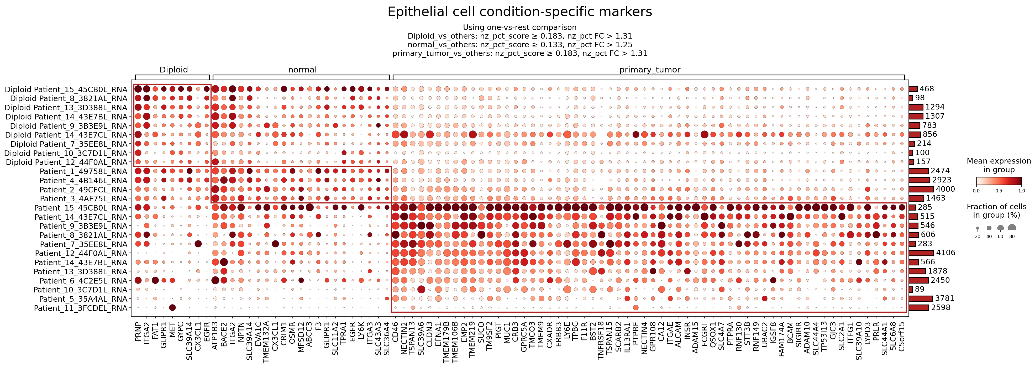
[Analysis Visualization Results]...
Analysis Overview
This analysis identifies and visualizes surfaceome marker expression in Epithelial cells across different patient samples, categorized by 'ploidy_init_group' (Diploid) and 'condition' (normal, primary_tumor). The goal is to highlight markers that are differentially expressed in breast cancer epithelial cells compared to normal counterparts, distinguishing between established therapeutic targets and less well-characterized markers with potential. The results are presented in a dot plot where dot size represents the fraction of cells expressing a gene (non-zero percentage), and color intensity indicates the mean expression level within each group. The dataset context indicates that breast cancer is the inferred disease, with Epithelial cells identified as the tumor-origin cell type and CNV estimates available, reinforcing the relevance of identifying surface markers in this context.
Visual Summary
The dot plot, titled "Epithelial cell condition-specific markers," effectively illustrates distinct expression patterns for surfaceome genes across different conditions and samples.
- Primary Tumor-Specific Upregulation: A prominent cluster of genes (highlighted by the red rectangle in the plot) exhibits high mean expression (dark red color) and a large fraction of expressing cells (large dot size) specifically within the primary_tumor samples. These markers are largely absent or expressed at very low levels in normal and Diploid samples. This pattern strongly indicates their association with the malignant phenotype of breast cancer epithelial cells.
- Normal/Diploid-Specific Expression: Conversely, a set of markers on the left side of the plot (e.g., PRNP, GJA1, GJB1, ITGA2, FAT1, GLIPR1, MET, GYPC, EGFR, ATP1B3) shows higher expression and prevalence in the normal and Diploid epithelial cell populations. This suggests their roles in normal breast epithelial cell function or their downregulation during tumor progression. Notably, some genes like EGFR appear on both sides, but their prevalence and mean expression often differ, with a more pronounced presence in certain normal/diploid samples compared to the general primary_tumor context for the depicted markers.
- Heterogeneity Across Samples: Within both normal and primary_tumor conditions, there is some variability in marker expression across individual patient samples, which is expected in complex diseases like cancer. However, the overall condition-specific patterns are robust.
- Surfaceome Focus: The analysis exclusively focused on surfaceome markers, making the identified genes highly relevant for therapeutic strategies involving antibodies, CAR T-cells, or other cell-surface targeting approaches.
Biological Interpretation
The distinct expression profiles observed highlight key biological differences between normal and malignant breast epithelial cells, offering insights into cancer progression and potential vulnerabilities. The robust upregulation of specific surfaceome markers in primary_tumor epithelial cells points towards altered cell adhesion, signaling, metabolism, and immune evasion mechanisms characteristic of cancer.
Established Therapeutic Targets
Several identified surface markers are either well-established targets in breast cancer or belong to families that are actively pursued therapeutically.
ERBB3 (HER3)
- Expression Pattern: Strongly upregulated in primary_tumor epithelial cells, largely absent in normal/diploid.
- Signaling Pathways/Mechanisms: ERBB3 is a member of the epidermal growth factor receptor (EGFR/ERBB) family. Unlike other family members, ERBB3 has impaired kinase activity but is a potent activator of the PI3K/Akt pathway upon heterodimerization, primarily with ERBB2 (HER2) or EGFR (ERBB1). This activation promotes cell proliferation, survival, and resistance to therapy. GeneCards: ERBB3
- Relevant Therapeutic Agents: Monoclonal antibodies targeting ERBB3 (e.g., patritumab deruxtecan, seribantumab) are in clinical trials for various cancers, including breast cancer, particularly in HER2-low or HR+/HER2- metastatic settings. PubMed: ERBB3 breast cancer therapeutics
- Supporting Evidence: ERBB3 overexpression is observed in various cancers, including breast cancer, and is associated with tumor growth, metastasis, and resistance to endocrine therapy and chemotherapy.
MUC1
- Expression Pattern: Highly and broadly expressed across primary_tumor epithelial cells, with minimal expression in normal/diploid.
- Signaling Pathways/Mechanisms: MUC1 (Mucin 1) is a large transmembrane glycoprotein that, when aberrantly expressed in cancer, loses its apical localization and becomes uniformly distributed across the cell surface. It functions as an oncoprotein, modulating cell signaling pathways (e.g., Wnt/β-catenin, MAPK, PI3K/Akt) to promote cell proliferation, survival, invasion, and immune evasion. GeneCards: MUC1
- Relevant Therapeutic Agents: MUC1 has been a long-standing target for cancer immunotherapy, including therapeutic vaccines (e.g., GO-203, previously in trials for various cancers) and antibody-drug conjugates (ADCs). PubMed: MUC1 breast cancer therapeutics
- Supporting Evidence: MUC1 is overexpressed and aberrantly glycosylated in over 90% of breast cancers and is associated with aggressive disease and poor prognosis. It also serves as a serum biomarker (CA15-3).
INSR (Insulin Receptor)
- Expression Pattern: Elevated in primary_tumor epithelial cells, low in normal/diploid.
- Signaling Pathways/Mechanisms: The Insulin Receptor (INSR) is a receptor tyrosine kinase that binds insulin and insulin-like growth factors (IGFs), activating downstream pathways like PI3K/Akt/mTOR and MAPK/ERK. In cancer, INSR overexpression and/or activation can promote cell proliferation, survival, and metabolic reprogramming, contributing to tumor growth and resistance. GeneCards: INSR
- Relevant Therapeutic Agents: Inhibitors targeting INSR and IGF-1R (which often heterodimerizes with INSR) are under investigation as potential anticancer agents, though specific success in breast cancer has been mixed. Dual IGF-1R/INSR inhibitors aim to overcome compensatory mechanisms. PubMed: INSR breast cancer therapy
- Supporting Evidence: High INSR expression is found in various cancers, including breast cancer, and is linked to metabolic alterations in tumor cells and potentially resistance to standard therapies.
Less Well-Characterized Markers with Potential as Therapeutic Targets
These markers show significant upregulation in primary_tumor epithelial cells and represent promising avenues for future research and therapeutic development.
SLC2A1 (GLUT1)
- Expression Pattern: Highly expressed in primary_tumor epithelial cells, largely absent in normal/diploid.
- Signaling Pathways/Mechanisms: SLC2A1, also known as Glucose Transporter 1 (GLUT1), is a facilitative glucose transporter. Its overexpression is a hallmark of the Warburg effect in cancer, where tumor cells rely heavily on glycolysis for energy production, even in the presence of oxygen. This metabolic shift supports rapid proliferation and biomass accumulation. GeneCards: SLC2A1
- Relevant Therapeutic Agents: Given its critical role in cancer metabolism, GLUT1 is a target for metabolism-based therapies. Inhibitors of GLUT1 (e.g., WZB117) are being explored to starve cancer cells of glucose. PubMed: GLUT1 inhibitors breast cancer
- Supporting Evidence: High GLUT1 expression is associated with aggressive phenotypes, poor prognosis, and resistance to chemotherapy in breast cancer. It is often induced by hypoxia.
ALCAM (CD166)
- Expression Pattern: Elevated in primary_tumor epithelial cells, with minimal expression in normal/diploid.
- Signaling Pathways/Mechanisms: Activated Leukocyte Cell Adhesion Molecule (ALCAM/CD166) is a cell adhesion molecule involved in cell-cell interactions, migration, and differentiation. In cancer, ALCAM can promote tumor progression, metastasis, and stemness, possibly through interactions with CD6 on immune cells or other receptors on cancer cells, modulating signaling pathways related to cell survival and motility. GeneCards: ALCAM
- Relevant Therapeutic Agents: ALCAM has been investigated as a potential target for antibody-based therapies, particularly in contexts where it marks cancer stem cells. PubMed: ALCAM antibody breast cancer
- Supporting Evidence: ALCAM expression is often increased in various cancers, including breast cancer, and has been identified as a marker for cancer stem-like cells, contributing to resistance and recurrence.
CLDN3 (Claudin-3)
- Expression Pattern: Strongly expressed in a subset of primary_tumor samples, generally low/absent elsewhere.
- Signaling Pathways/Mechanisms: Claudin-3 (CLDN3) is a component of tight junctions, which regulate paracellular permeability. In cancer, CLDN3 expression is often dysregulated. While tight junctions typically restrict cell migration, aberrant CLDN3 can promote invasion and metastasis through mechanisms involving altered cell adhesion, epithelial-mesenchymal transition (EMT), and interaction with growth factor receptors. GeneCards: CLDN3
- Relevant Therapeutic Agents: CLDN3 overexpression makes it a potential target for therapeutic antibodies or drug conjugates designed to deliver cytotoxic agents specifically to CLDN3-expressing cancer cells. PubMed: Claudin-3 therapeutics cancer
- Supporting Evidence: CLDN3 is frequently overexpressed in breast, ovarian, and other epithelial cancers, correlating with aggressive disease and metastatic potential.
CD46
- Expression Pattern: Highly expressed across most primary_tumor epithelial cells, with some presence in normal/diploid but generally lower.
- Signaling Pathways/Mechanisms: CD46 is a complement regulatory protein that protects host cells from complement-mediated lysis. In cancer, CD46 overexpression allows tumor cells to evade immune surveillance by inhibiting complement activation on their surface. It can also modulate cellular signaling, contributing to cell proliferation and survival. GeneCards: CD46
- Relevant Therapeutic Agents: Antibodies targeting CD46 are being explored as a means to sensitize cancer cells to complement-dependent cytotoxicity or as vehicles for drug delivery. PubMed: CD46 antibody cancer therapy
- Supporting Evidence: CD46 is often upregulated in various cancers, including breast cancer, contributing to immune evasion and tumor survival.
TNFRSF18 (GITR)
- Expression Pattern: Elevated in a subset of primary_tumor epithelial cells, low in normal/diploid.
- Signaling Pathways/Mechanisms: TNFRSF18, also known as Glucocorticoid-Induced TNFR-Related Protein (GITR), is a costimulatory receptor expressed primarily on immune cells (T cells, Tregs, NK cells). While primarily known for its role in immune cell activation and regulation, its expression on cancer cells, as observed here, could potentially modulate cell survival or interaction with the immune microenvironment. It can activate NF-κB and MAPK pathways. GeneCards: TNFRSF18
- Relevant Therapeutic Agents: Agonistic antibodies targeting GITR are under investigation in clinical trials to enhance anti-tumor immunity by activating effector T cells and inhibiting regulatory T cells. If expressed on tumor cells, it might also be a direct target. PubMed: GITR agonist breast cancer
- Supporting Evidence: While mainly an immune checkpoint target, some studies suggest GITR expression can occur on certain cancer cells, impacting tumor-immune interactions.
CA12 (Carbonic Anhydrase XII)
- Expression Pattern: Expressed in a subset of primary_tumor epithelial cells, minimal in normal/diploid.
- Signaling Pathways/Mechanisms: CA12 is a transmembrane enzyme involved in regulating pH homeostasis by catalyzing the reversible hydration of carbon dioxide. In the hypoxic and acidic tumor microenvironment, CA12 contributes to maintaining intracellular pH, thereby promoting tumor cell survival, proliferation, and invasion. GeneCards: CA12
- Relevant Therapeutic Agents: Inhibitors of carbonic anhydrases (CAIs) are being developed as anti-cancer agents, with specific inhibitors for tumor-associated CAs like CA12. PubMed: CA12 inhibitors cancer
- Supporting Evidence: CA12 is often overexpressed in various solid tumors, including breast cancer, particularly in regions of hypoxia, and is associated with unfavorable prognosis.
Clinical or Translational Implications
The identification of these epithelial cell-specific surfaceome markers in primary breast tumors carries significant clinical and translational implications:
- Biomarker Development: Markers like MUC1, ERBB3, ALCAM, CLDN3, SLC2A1, and CA12, which are highly specific to primary_tumor epithelial cells, represent excellent candidates for diagnostic and prognostic biomarkers. Their expression could be assessed via immunohistochemistry or liquid biopsy to monitor disease status, predict recurrence, or guide treatment selection.
- Targeted Therapies: The surface localization of these markers makes them ideal candidates for targeted therapeutic interventions.
- Antibody-Drug Conjugates (ADCs): MUC1, ERBB3, ALCAM, CLDN3, CD46, and NECTIN2 are attractive targets for ADCs, which can deliver cytotoxic payloads specifically to tumor cells, minimizing systemic toxicity.
- Monoclonal Antibodies: Neutralizing or agonistic antibodies (e.g., anti-ERBB3, anti-GITR) could directly modulate cancer cell signaling or enhance anti-tumor immunity.
- CAR T-cell Therapy: Highly specific surface markers could be utilized to design Chimeric Antigen Receptor (CAR) T-cell therapies, directing immune cells specifically against breast cancer epithelial cells.
- Metabolic Inhibitors: The strong upregulation of SLC2A1 (GLUT1) highlights the metabolic vulnerability of breast cancer cells, suggesting that GLUT1 inhibitors could be effective in combination with standard therapies.
- pH Regulation Targets: CA12 presents an opportunity to target the unique metabolic adaptations of cancer cells in hypoxic microenvironments.
- Understanding Tumor Biology: The discovery of these markers provides deeper insights into the underlying molecular mechanisms driving breast cancer progression, including altered adhesion, immune evasion, and metabolic reprogramming. Further research into their precise roles and downstream signaling networks can unveil novel pathways for therapeutic intervention.
- Patient Stratification: Differential expression of these markers across various primary_tumor samples suggests potential for stratifying patients into subgroups that might benefit from specific targeted therapies, moving towards more personalized treatment approaches. For instance, patients with high ERBB3 expression might be candidates for ERBB3-targeting agents.
In summary, this analysis identifies a rich set of surfaceome markers distinguishing primary breast tumor epithelial cells from normal counterparts. These markers represent critical biological features of the disease and offer promising avenues for the development of novel diagnostic tools and targeted therapies.
15. Macrophage Condition-Specific Surfaceome Markers in Breast Cancer
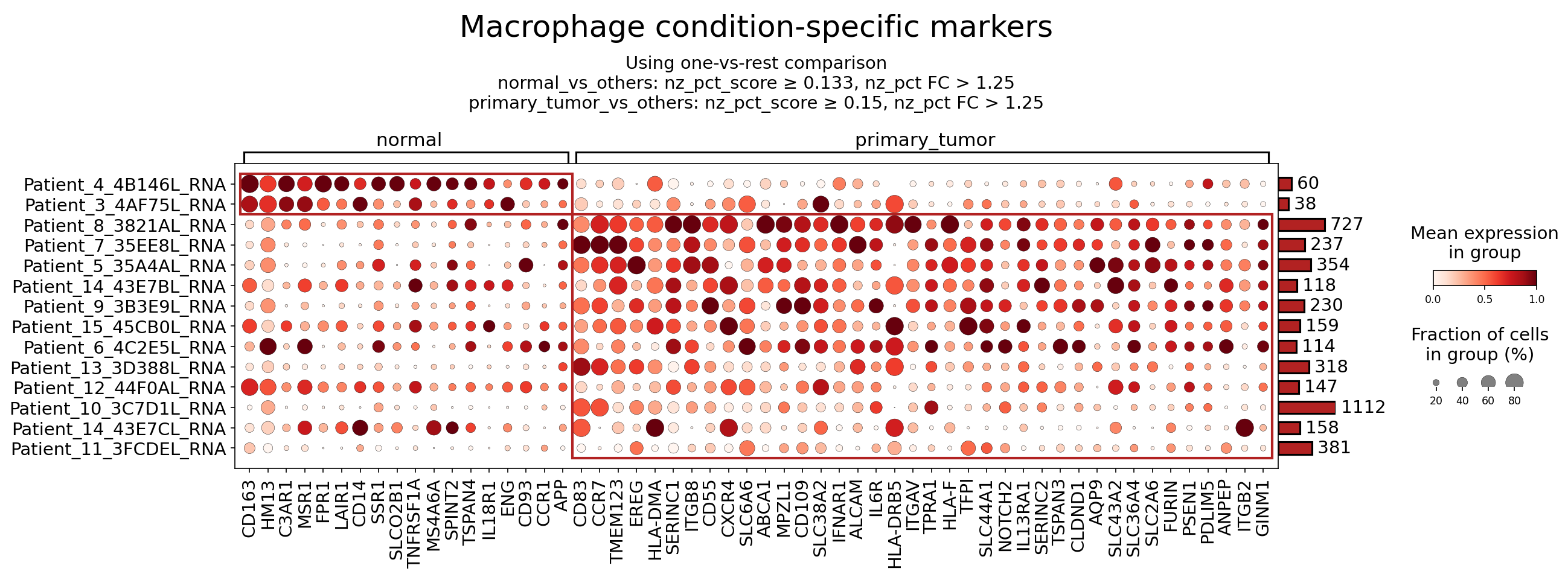
[Analysis Visualization Results]...
Analysis Overview
This analysis identifies and visualizes condition-specific surfaceome markers for Macrophages, comparing 'normal' breast tissue with 'primary_tumor' breast tissue, derived from single-cell RNA-seq data. The objective is to highlight genes encoding cell surface proteins that are differentially expressed in macrophages under these two conditions, offering insights into macrophage plasticity and function within the tumor microenvironment (TME) of breast cancer. The dot plot displays mean expression levels (color intensity) and the fraction of cells expressing the marker (dot size) for each gene across individual patient samples within the 'normal' and 'primary_tumor' groups. Markers were selected based on specific differential expression criteria (nz_pct_score and nz_pct_FC thresholds) and restricted to surfaceome markers to emphasize their potential as therapeutic targets or diagnostic biomarkers.
Visual Summary
The dot plot clearly differentiates macrophage populations in 'normal' versus 'primary_tumor' conditions based on their surfaceome marker expression profiles.
- Condition-Specific Expression Patterns: A distinct set of markers is highly expressed in macrophages from 'normal' samples, while a largely different set of markers characterizes macrophages from 'primary_tumor' samples. This indicates a significant phenotypic shift in macrophages in the context of breast cancer.
- Normal Macrophage Markers: Macrophages in 'normal' breast tissue consistently express markers such as CD163, HM13 (CLEC10A), C3AR1, MSR1, CD14, and ENG (CD105). These markers generally show high mean expression and are present in a large fraction of cells within the normal samples.
- Primary Tumor Macrophage Markers: Macrophages in 'primary_tumor' samples exhibit a much broader and often higher expression of markers, including CD68, ITGB8, CXCR4, CD83, EREG, IL6R, ITGAV, HLA-DRB5, and various other genes. These markers are highly expressed and prevalent across most tumor samples, suggesting a robust and consistent reprogramming of macrophages in the TME.
- Heterogeneity Across Patients: While overall patterns are clear, there is some patient-to-patient variability in expression levels and prevalence for certain markers, particularly within the 'primary_tumor' group, reflecting potential inter-patient heterogeneity in macrophage phenotypes.
- Prevalence and Expression: For 'primary_tumor' markers, many genes show large dot sizes (high fraction of expressing cells) and dark red colors (high mean expression), indicating their widespread and strong upregulation in tumor-associated macrophages (TAMs).
Biological Interpretation
Macrophages are pivotal components of the breast cancer tumor microenvironment (TME), exhibiting significant plasticity. Their phenotype can range from anti-tumor (M1-like) to pro-tumor (M2-like, commonly referred to as Tumor-Associated Macrophages or TAMs). The surfaceome markers identified here provide crucial insights into the functional state and potential roles of macrophages in both normal and cancerous breast tissue.
Macrophage Markers in Normal Breast Tissue
Macrophages in normal tissue predominantly exhibit markers associated with tissue homeostasis, anti-inflammatory responses, and M2-like phenotypes, which are essential for tissue repair and immune regulation in a non-diseased state.
- CD163: A scavenger receptor expressed by tissue-resident macrophages, often associated with an M2-like, anti-inflammatory, and immunosuppressive phenotype. It is involved in clearing hemoglobin-haptoglobin complexes.
- Signaling Pathways: Interacts with components of the innate immune system. Its shedding can generate a soluble form (sCD163) that acts as a biomarker for inflammation and macrophage activation.
- Role in TME: While often associated with pro-tumorigenic TAMs, in normal tissue, it indicates a homeostatic, tissue-resident macrophage population.
- Therapeutic Target Potential: In cancer, high CD163 expression on TAMs correlates with poor prognosis, making it a potential target for depleting pro-tumorigenic macrophages or delivering immunotoxins.
- Relevant Therapeutic Agents: Antibody-drug conjugates (ADCs) targeting CD163 are under investigation for specific cancer types.
- Reference: GeneCards: CD163
- HM13 (CLEC10A): C-type lectin domain family 10 member A, a macrophage galactose-type C-type lectin (MGL) expressed by tolerogenic dendritic cells and M2 macrophages. Involved in antigen capture and presentation.
- Signaling Pathways: Engages in glycan recognition, influencing immune responses.
- Role in TME: In normal tissue, may contribute to immune tolerance. Its upregulation in some cancers suggests a role in immune evasion.
- Therapeutic Target Potential: Its restricted expression makes it an attractive target for selective drug delivery to certain immune cell subsets or tumor cells expressing specific glycans.
- Reference: UniProt: CLEC10A
- C3AR1: Complement C3a receptor 1, a G-protein coupled receptor for the anaphylatoxin C3a. Activation promotes inflammatory responses.
- Signaling Pathways: Initiates intracellular signaling cascades upon C3a binding, mediating chemotaxis and inflammatory mediator release.
- Role in TME: In normal contexts, part of the innate immune response. Dysregulation in cancer can contribute to chronic inflammation and immune suppression.
- MSR1: Macrophage scavenger receptor 1 (CD204), involved in the uptake of modified lipoproteins and host defense. Expressed by macrophages and plays a role in innate immunity.
- Signaling Pathways: Involved in endocytosis and cellular adhesion.
- Role in TME: In normal tissue, contributes to waste clearance. In cancer, can be associated with M2-like TAMs and phagocytosis of apoptotic cells.
- Reference: GeneCards: MSR1
- FPR1: Formyl peptide receptor 1, a G-protein coupled receptor that binds bacterial N-formylated peptides, initiating inflammatory and chemotactic responses.
- Signaling Pathways: Mediates chemotaxis and activation of phagocytes.
- Role in TME: Important for pathogen sensing and inflammation in normal conditions. Can be hijacked by tumors to recruit immune cells that support tumor growth.
- LAIR1: Leukocyte-associated immunoglobulin-like receptor 1, an inhibitory receptor expressed on various immune cells, including macrophages.
- Signaling Pathways: Upon ligand binding, it recruits phosphatases (SHP-1, SHP-2) to inhibit immune cell activation.
- Role in TME: In normal tissue, maintains immune homeostasis. In cancer, can contribute to immune evasion by suppressing anti-tumor immune responses.
- Therapeutic Target Potential: Blocking LAIR1 could enhance anti-tumor immunity.
- Reference: GeneCards: LAIR1
- CD14: A co-receptor for Toll-like receptors (TLRs), particularly TLR4, involved in detecting bacterial lipopolysaccharide (LPS). Highly expressed on monocytes and macrophages.
- Signaling Pathways: Key component of TLR signaling, leading to NF-κB activation and inflammatory cytokine production.
- Role in TME: Essential for innate immune responses. In the TME, can mediate responses to bacterial components or DAMPs, influencing macrophage polarization.
- Reference: UniProt: CD14
- SSR1: Signal sequence receptor subunit 1, part of the translocon complex in the endoplasmic reticulum, involved in protein translocation and glycosylation. While primarily an ER component, it can be detected on the cell surface.
- Signaling Pathways: Not a classical signaling receptor, but critical for protein trafficking and folding.
- ENG (CD105): Endoglin, a co-receptor for TGF-β, primarily expressed on endothelial cells but also on activated macrophages and other cells. Involved in angiogenesis and TGF-β signaling modulation.
- Signaling Pathways: Modulates TGF-β/BMP signaling, influencing cell proliferation, differentiation, and angiogenesis.
- Role in TME: In normal tissue, important for vascular integrity. Its expression on macrophages in normal tissue might reflect a quiescent state or early activation.
- Therapeutic Target Potential: Highly expressed on tumor vasculature and some TAMs, making it a target for anti-angiogenic therapies or drug delivery.
- Reference: GeneCards: ENG
- CD93: A C-type lectin-like domain containing receptor expressed on endothelial cells and myeloid cells. Involved in cell adhesion and angiogenesis.
- Signaling Pathways: Primarily involved in cell-cell interactions and modulating vascular development.
- Role in TME: In normal tissue, contributes to tissue maintenance. In tumor, can promote angiogenesis and macrophage survival.
- CCR1: C-C chemokine receptor type 1, a receptor for chemokines such as CCL3, CCL5, and CCL7. Involved in leukocyte recruitment to inflammatory sites.
- Signaling Pathways: G-protein coupled receptor signaling, leading to cell migration.
- Role in TME: Essential for recruiting monocytes and other immune cells to sites of inflammation or injury. Can be involved in recruiting both protective and pro-tumorigenic immune cells.
- APP: Amyloid beta A4 protein precursor, a transmembrane protein extensively studied in Alzheimer's disease, but also has roles in cell adhesion, neurogenesis, and immune regulation.
- Signaling Pathways: Cleavage products can have signaling functions. Involved in cell surface interactions.
Macrophage Markers in Primary Tumor Tissue
Macrophages in primary tumor tissue (TAMs) display markers reflecting their altered, often pro-tumorigenic functions, including immune suppression, angiogenesis promotion, tissue remodeling, and metastasis support.
- CD68: A lysosomal membrane protein highly expressed by macrophages and monocytes, often used as a pan-macrophage marker. Its upregulation in tumors can reflect increased phagocytic activity and lysosomal content.
- Signaling Pathways: Primarily intracellular, but its high expression indicates active macrophage states.
- Role in TME: A general marker for TAMs, indicating a significant macrophage presence in the tumor.
- Reference: UniProt: CD68
- ITGB8: Integrin beta 8, forms a heterodimer with ITGAV (alphaV integrin). This integrin pair is known to activate latent TGF-β, a critical cytokine in cancer progression.
- Signaling Pathways: Integrin signaling, specifically activating latent TGF-β, which then signals through TGF-β receptors, promoting tumor growth, immunosuppression, and fibrosis.
- Role in TME: Key in converting latent TGF-β into its active form, thus enhancing the immunosuppressive and pro-fibrotic environment within the TME. Highly relevant to breast cancer progression.
- Therapeutic Target Potential: Targeting ITGB8 or the ITGAV/ITGB8 complex could inhibit TGF-β activation in the TME, thereby reducing immunosuppression and tumor growth.
- Reference: GeneCards: ITGB8
- CXCR4: C-X-C chemokine receptor type 4, a receptor for CXCL12. Crucial for cell trafficking, development, and a known driver of cancer metastasis.
- Signaling Pathways: G-protein coupled receptor signaling, leading to cell migration, proliferation, and survival. The CXCL12-CXCR4 axis is highly implicated in cancer.
- Role in TME: Macrophage expression of CXCR4 can facilitate their recruitment to the TME and interaction with CXCL12-expressing tumor cells or stromal cells, promoting tumor progression and metastasis.
- Therapeutic Target Potential: CXCR4 antagonists (e.g., Plerixafor) are clinically used in other contexts and are being explored in cancer to block tumor cell migration and TAM recruitment.
Relevant Therapeutic Agents: Plerixafor (Mozobil)
- Reference: PubMed Search: CXCR4 cancer therapy
- CD83: A glycoprotein expressed on activated dendritic cells, B cells, and T cells, but also found on some activated macrophages. Involved in immune regulation.
- Signaling Pathways: Modulates immune cell activation and differentiation.
- Role in TME: Its expression on TAMs suggests an activated state, potentially linked to immune modulation or antigen presentation (though sometimes compromised in TAMs).
- EREG: Epiregulin, a ligand for EGFR (epidermal growth factor receptor). Promotes cell proliferation, survival, and angiogenesis.
- Signaling Pathways: Activates EGFR, leading to downstream MAPK/ERK and PI3K/Akt signaling, crucial for cell growth and survival.
- Role in TME: Macrophage-derived EREG can act in a paracrine fashion to stimulate tumor cell growth and angiogenesis, contributing to the pro-tumorigenic TME.
- Therapeutic Target Potential: Targeting EGFR signaling or EREG itself could inhibit tumor growth and angiogenesis.
- IL6R: Interleukin-6 receptor, the receptor for the pro-inflammatory cytokine IL-6. IL-6 signaling is a major driver of chronic inflammation and cancer progression.
- Signaling Pathways: Upon IL-6 binding, activates JAK/STAT3 pathway, promoting cell survival, proliferation, and immune suppression.
- Role in TME: Macrophage-expressed IL6R can mediate responses to tumor-derived or stromal-derived IL-6, promoting TAM survival and pro-tumorigenic functions. High IL-6 signaling is associated with poor prognosis in breast cancer.
- Therapeutic Target Potential: Blocking IL6R (e.g., Tocilizumab) is used in autoimmune diseases and is being investigated in cancer to inhibit chronic inflammation and STAT3 signaling.
Relevant Therapeutic Agents: Tocilizumab
- Reference: GeneCards: IL6R
- ITGAV: Integrin alpha V, forms heterodimers with various beta integrins (e.g., ITGB1, ITGB3, ITGB5, ITGB6, ITGB8), mediating cell adhesion and signaling.
- Signaling Pathways: Integrin signaling, connecting ECM to cytoskeleton, involved in cell migration, proliferation, and survival. As mentioned with ITGB8, it activates latent TGF-β.
- Role in TME: ITGAV-containing integrins on TAMs facilitate their migration within the TME, adhesion to tumor cells or ECM, and critically, activation of TGF-β.
- Therapeutic Target Potential: Pan-alphaV integrin inhibitors are in development for cancer therapy, aiming to disrupt cell-ECM interactions and TGF-β activation.
- Reference: GeneCards: ITGAV
- HLA-DRB5 & HLA-DMA & HLA-F: These are components of the Major Histocompatibility Complex (MHC) class II, involved in presenting antigens to CD4+ T cells. HLA-DRB5 is an MHC class II beta chain, HLA-DMA is involved in peptide loading onto MHC class II molecules, and HLA-F is a non-classical MHC class I molecule.
- Signaling Pathways: Antigen presentation pathway, crucial for adaptive immune responses.
- Role in TME: Upregulation of MHC II components on TAMs could suggest an attempt at antigen presentation. However, TAMs often present antigens inefficiently or in a way that tolerizes T cells, contributing to immune evasion. HLA-F can also have immunoregulatory roles.
- Reference: PubMed Search: HLA-DRB5 cancer immunology
- CD55: Complement decay-accelerating factor (DAF), a GPI-anchored protein that protects cells from complement-mediated lysis by inhibiting C3 convertase.
- Signaling Pathways: Inhibits the complement cascade.
- Role in TME: Upregulation on TAMs (and tumor cells) helps them evade complement-mediated destruction, contributing to immune evasion.
- Therapeutic Target Potential: Inhibiting CD55 could sensitize tumor cells and TAMs to complement-dependent cytotoxicity.
- IFNAR1: Interferon alpha/beta receptor 1, a subunit of the receptor for type I interferons (IFN-α/β).
- Signaling Pathways: Mediates type I interferon signaling, leading to activation of JAK/STAT pathway and induction of antiviral and anti-proliferative genes.
- Role in TME: Macrophage response to IFNs can be complex; while IFNs can have anti-tumor effects, chronic IFN signaling can also promote immune suppression or tumor cell survival in some contexts. Upregulation on TAMs could indicate a response to the inflammatory TME.
- ALCAM: Activated leukocyte cell adhesion molecule (CD166), involved in cell-cell adhesion, especially between immune cells and epithelial cells.
- Signaling Pathways: Mediates homophilic and heterophilic adhesion.
- Role in TME: Can promote tumor cell invasion and metastasis by facilitating interactions between tumor cells and stromal cells, including macrophages.
- TPRA1 (TLR8AP): TLR8 accessory protein 1, potentially involved in TLR8 signaling.
- Signaling Pathways: May modulate innate immune signaling via TLR8.
- TFPI: Tissue factor pathway inhibitor, a natural anticoagulant that inhibits the initiation of the extrinsic coagulation pathway.
- Signaling Pathways: Regulates coagulation.
- Role in TME: Coagulation pathways are often dysregulated in cancer and can contribute to tumor growth, angiogenesis, and metastasis. Macrophage-derived TFPI might modulate these processes.
- NOTCH2: Notch receptor 2, a transmembrane receptor involved in cell-cell communication, development, and cell fate decisions.
- Signaling Pathways: Notch signaling pathway, critical for cell proliferation, differentiation, and apoptosis.
- Role in TME: Notch signaling in TAMs can influence their polarization and functions, potentially promoting pro-tumorigenic activities.
- Therapeutic Target Potential: Notch inhibitors are being explored in cancer therapy.
- IL13RA1: Interleukin 13 receptor subunit alpha 1, a component of the receptor for IL-13 (and partially IL-4). Involved in type 2 immune responses and M2 macrophage polarization.
- Signaling Pathways: Mediates IL-13 and IL-4 signaling, activating JAK/STAT6 pathway, leading to M2 macrophage differentiation.
- Role in TME: Strong indicator of M2-like polarization in TAMs, which are pro-tumorigenic and immunosuppressive in breast cancer.
- Therapeutic Target Potential: Targeting IL-13/IL-13RA1 axis could reprogram TAMs or inhibit their pro-tumor functions.
- Reference: GeneCards: IL13RA1
- SLC2A6 (GLUT6): Solute carrier family 2 member 6, a glucose transporter.
- Signaling Pathways: Involved in glucose metabolism.
- Role in TME: Altered metabolism is a hallmark of cancer. Changes in glucose transporters on TAMs may reflect their metabolic adaptation to the TME, which supports their pro-tumor functions.
- FURIN: A proprotein convertase that cleaves various proproteins, including growth factors, receptors, and bacterial toxins, into their active forms.
- Signaling Pathways: Activates precursor proteins.
- Role in TME: Can activate pro-tumorigenic factors such as TGF-β, promoting tumor growth and metastasis. Its upregulation on TAMs suggests a role in activating key molecules in the TME.
- Therapeutic Target Potential: Furin inhibitors are being investigated as anti-cancer agents.
- ANPEP (CD13): Aminopeptidase N, a transmembrane metalloprotease expressed on myeloid cells, endothelial cells, and epithelial cells. Involved in peptide metabolism and cell adhesion.
- Signaling Pathways: Cleaves various peptides, influencing local signaling.
- Role in TME: Involved in angiogenesis, tumor invasion, and metastasis. Macrophage-expressed ANPEP can contribute to matrix remodeling and pro-angiogenic processes.
- Therapeutic Target Potential: CD13 inhibitors are being explored in cancer for their anti-angiogenic and anti-metastatic properties.
- Reference: GeneCards: ANPEP
- ITGB2: Integrin beta 2 (CD18), forms heterodimers with various alpha integrins (e.g., ITGAL, ITGAM, ITGAX) to form leukocyte-specific integrins. Critical for leukocyte adhesion and extravasation.
- Signaling Pathways: Integrin signaling, mediating cell adhesion to endothelial cells and ECM, crucial for immune cell trafficking.
- Role in TME: In TAMs, ITGB2-containing integrins are essential for their recruitment to the tumor site and interactions with other cells, influencing the inflammatory and immunosuppressive milieu.
Clinical or Translational Implications
The distinct surfaceome marker profiles observed for macrophages in normal vs. primary_tumor breast tissue have significant clinical and translational implications:
- Diagnostic and Prognostic Biomarkers:
- Markers highly expressed on TAMs (e.g., ITGB8, CXCR4, IL6R, IL13RA1, CD68, ANPEP) could serve as diagnostic markers for identifying pro-tumorigenic macrophage populations in breast cancer biopsies.
- The expression levels of these markers, or the ratios between normal-associated and tumor-associated markers, could potentially be developed into prognostic indicators for disease progression, recurrence, or response to therapy.
- Soluble forms of some of these markers (e.g., sCD163) are already being explored as non-invasive biomarkers.
- Therapeutic Targets:
- The identified surfaceome markers on TAMs offer promising avenues for targeted therapeutic interventions in breast cancer. Surface proteins are accessible to antibody-based therapies, antibody-drug conjugates (ADCs), or engineered cell therapies (e.g., CAR-macrophages, though still in early stages).
- Immune Reprogramming: Targeting markers like ITGB8 (TGF-β activation), CXCR4 (recruitment/metastasis), or IL6R/IL13RA1 (M2 polarization) could help reprogram pro-tumorigenic TAMs into anti-tumor phenotypes or deplete them, thereby enhancing anti-tumor immunity.
Specific Examples:
- ITGB8 and ITGAV: Given their role in activating latent TGF-β, blocking ITGB8/ITGAV could reduce immunosuppression and fibrosis in the TME, potentially synergizing with immunotherapies.
- CXCR4: Antagonists could prevent TAM recruitment and hinder metastatic processes.
- IL6R and IL13RA1: Inhibiting these receptors could disrupt key pro-tumorigenic signaling pathways driving M2-like macrophage polarization.
- ANPEP (CD13): Targeting this metalloprotease could impede angiogenesis and tumor invasion.
- LAIR1 (in normal but potentially inhibitory in tumor): Blocking LAIR1 on TAMs might unleash anti-tumor immune responses.
- Combination Therapies:
- Therapies targeting TAMs via these surface markers could be particularly effective when combined with existing treatments like chemotherapy, radiation, or checkpoint inhibitors, to overcome immune resistance mechanisms driven by TAMs.
- Drug Development:
- The detailed understanding of these macrophage-specific surface markers provides a rational basis for the development of novel drugs tailored to target the pro-tumorigenic functions of TAMs in breast cancer.
16. Fibroblast Condition-Specific Surfaceome Markers in Breast Tissue
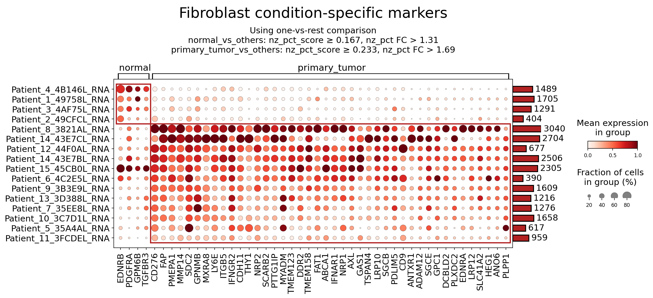
[Analysis Visualization Results]...
Analysis Overview
This analysis identifies surfaceome markers differentially expressed in Fibroblasts across normal and primary tumor conditions in breast tissue, derived from single-cell RNA-seq data. The goal is to highlight potential markers for distinguishing cancer-associated fibroblasts (CAFs) from normal fibroblasts and to assess their utility as therapeutic targets, especially given the context of breast cancer with epithelial cell origin and aneuploidy. The dot plot visualizes the mean expression level (color intensity) and the fraction of cells expressing each marker (dot size) per patient sample. Only surfaceome markers, with at most 40 markers per condition, were considered for this analysis.
Visual Summary
The dot plot clearly differentiates two main groups of fibroblast markers based on their expression patterns across normal and primary tumor samples:
- Normal-enriched Markers: A distinct cluster of markers (e.g., EDNRB, PDGFRA, GPM6B, TGFBR3) shows high mean expression and prevalence (larger, darker red dots) exclusively within the "normal" patient samples (Patient_4_4B146L_RNA, Patient_1_49758L_RNA, Patient_3_4AF75L_RNA, Patient_2_49CFCL_RNA). These markers are largely absent or very lowly expressed in the "primary_tumor" samples.
- Primary Tumor-enriched Markers: A much larger panel of markers exhibits high expression and prevalence within the "primary_tumor" patient samples, with minimal to no expression in the normal samples. Notable examples with high expression and widespread presence across most tumor samples include FAP, CD276, PMEPA1, MMP14, SDC2, GPNMB, MXRA8, LY6E, ITGB5, NRP1, NRP2, AXL, and ADAM12.
- Patient Heterogeneity: Within the primary tumor group, there's some heterogeneity in marker expression across different patient samples. For instance, markers like EDNRA and PLPP1 show strong expression in a subset of tumor samples but are less prominent in others. This suggests inter-patient variability in CAF profiles.
- Overall Trend: The overall pattern strongly indicates a significant transcriptional reprogramming of fibroblasts in the primary tumor microenvironment, leading to a distinct "CAF phenotype" characterized by the upregulation of numerous surfaceome proteins.
Biological Interpretation
The observed differential expression of surfaceome markers in fibroblasts between normal and primary tumor conditions underscores the critical role of these cells in breast cancer progression and the profound changes they undergo in the tumor microenvironment (TME).
Markers Enriched in Normal Fibroblasts
While the focus is often on tumor-associated changes, understanding markers in normal fibroblasts provides a baseline.
PDGFRA (Platelet-Derived Growth Factor Receptor Alpha):
- Signaling Pathways: PDGFRA is a receptor tyrosine kinase that binds PDGF ligands, activating downstream pathways such as PI3K/AKT, MAPK/ERK, and STAT pathways, promoting cell proliferation, survival, and migration.
- Role in TME: In normal breast tissue, PDGFRA signaling is crucial for fibroblast development, proliferation, and maintenance of tissue homeostasis. Its relatively lower expression in CAFs (compared to normal fibroblasts) might indicate a shift in growth factor dependency or receptor repertoire, although CAFs can still express PDGFRA variants or other PDGF receptors.
- Therapeutic Potential: While often targeted in certain cancers (e.g., GIST), its higher expression in normal fibroblasts suggests that systemic PDGFRA inhibition might have off-target effects on healthy stromal tissue.
- Therapeutic Agents: Tyrosine kinase inhibitors (TKIs) like imatinib can inhibit PDGFRA. GeneCards: PDGFRA
TGFBR3 (Transforming Growth Factor Beta Receptor Type III / Betaglycan):
- Signaling Pathways: TGFBR3 is a co-receptor that binds TGF-β ligands and presents them to the signaling receptors (TGFBR1 and TGFBR2). It can positively or negatively regulate TGF-β signaling, which is context-dependent and highly pleiotropic.
- Role in TME: In normal fibroblasts, TGFBR3 contributes to the regulated response to TGF-β, which can have anti-proliferative effects or regulate ECM production. In cancer, loss of TGFBR3 can sometimes promote progression, while its presence might modulate CAF activation or immune responses.
- Therapeutic Potential: Its role is complex. Modulating TGF-β signaling is a therapeutic strategy, but directly targeting TGFBR3 in CAFs might require a nuanced understanding of its specific function in breast cancer CAFs versus normal fibroblasts.
- Therapeutic Agents: Various agents target TGF-β signaling, including ligand traps, receptor kinase inhibitors, and antisense oligonucleotides. GeneCards: TGFBR3
Markers Enriched in Primary Tumor Fibroblasts (Cancer-Associated Fibroblasts, CAFs)
The high expression of these markers in primary tumor fibroblasts highlights their transformation into CAFs, which actively contribute to tumor progression.
FAP (Fibroblast Activation Protein):
- Signaling Pathways: FAP is a serine protease expressed on the surface of CAFs. It cleaves specific ECM components (e.g., collagen, gelatin) and can activate other signaling molecules, impacting ECM remodeling, tissue stiffness, and growth factor bioavailability.
- Role in TME: FAP is a canonical marker of CAFs and is highly expressed in breast cancer stroma. It promotes tumor growth, invasion, metastasis, and immune evasion by remodeling the ECM and creating an immunosuppressive environment.
- Therapeutic Potential: FAP is an attractive therapeutic target due to its restricted expression in adult normal tissues (except during wound healing) and high expression in CAFs across many cancers. Targeting FAP can reduce tumor growth, enhance drug delivery, and improve immunotherapy efficacy.
- Therapeutic Agents: FAP-targeting antibodies (e.g., sibrotuzumab, although it showed limited efficacy as a monotherapy), FAP-activated prodrugs, FAP-directed CAR T-cells, and FAP-targeted radionuclide therapies are under investigation. PubMed Search: FAP breast cancer therapy
CD276 (B7-H3):
- Signaling Pathways: CD276 is an immune checkpoint molecule of the B7 family. It primarily acts as an inhibitory co-stimulatory molecule, suppressing T-cell activation and proliferation, thereby promoting immune evasion.
- Role in TME: Upregulated on various cell types, including CAFs, in numerous cancers including breast cancer. Its expression on CAFs contributes to the immunosuppressive TME, hindering anti-tumor immune responses.
- Therapeutic Potential: CD276 is a promising immunotherapy target. Its widespread expression in tumor stroma and on tumor cells, combined with its immune-suppressive function, makes it an attractive candidate for antibody-based therapies or CAR T-cell approaches.
- Therapeutic Agents: Monoclonal antibodies targeting CD276 (e.g., enoblituzumab) are in clinical trials for various solid tumors. GeneCards: CD276
MMP14 (Matrix Metalloproteinase 14 / MT1-MMP):
- Signaling Pathways: MMP14 is a membrane-anchored matrix metalloproteinase that degrades various ECM components (e.g., collagen, fibronectin). It also activates other MMPs (like MMP2) and growth factors, facilitating cell invasion and angiogenesis.
- Role in TME: CAFs express high levels of MMP14, contributing significantly to ECM remodeling, which enables cancer cell invasion and metastasis. It also creates space for angiogenesis and immune cell infiltration, but can also limit immune cell access to tumor cells.
- Therapeutic Potential: Inhibiting MMP14 on CAFs could impede tumor invasion and metastasis. However, broad MMP inhibitors have faced challenges in clinical trials due to side effects. More specific targeting strategies or combination therapies might be effective.
- Therapeutic Agents: Various MMP inhibitors have been developed, but none are FDA-approved for cancer due to lack of specificity and efficacy issues. Novel, more specific MMP14 inhibitors are being explored. GeneCards: MMP14
SDC2 (Syndecan-2):
- Signaling Pathways: SDC2 is a heparan sulfate proteoglycan involved in cell adhesion, migration, proliferation, and growth factor signaling by binding to ECM components and various ligands (e.g., FGF, VEGF, Wnt).
- Role in TME: Elevated SDC2 in CAFs can promote cancer cell proliferation and survival, and facilitate cell-matrix interactions that are crucial for invasion and metastasis in breast cancer.
- Therapeutic Potential: As a cell surface molecule involved in multiple pro-tumorigenic pathways, SDC2 represents a potential target for therapeutic intervention, possibly through antibodies or agents that disrupt its interactions.
- Therapeutic Agents: Currently, no specific SDC2-targeting therapies are in widespread clinical use for cancer, but research is ongoing. GeneCards: SDC2
GPNMB (Glycoprotein NMB):
- Signaling Pathways: GPNMB is a transmembrane glycoprotein involved in cell adhesion, migration, and resistance to apoptosis. It can activate downstream signaling pathways like PI3K/AKT.
- Role in TME: GPNMB is overexpressed in various aggressive cancers, including breast cancer, and on associated stromal cells. It promotes tumor cell survival, invasion, and metastatic potential. In CAFs, it might contribute to maintaining the pro-tumorigenic phenotype.
- Therapeutic Potential: GPNMB's surface localization and association with aggressive disease make it an attractive target, particularly for antibody-drug conjugates (ADCs).
- Therapeutic Agents: Glembatumumab vedotin (an ADC targeting GPNMB) has been investigated in clinical trials for metastatic breast cancer, particularly in triple-negative breast cancer (TNBC) patients. GeneCards: GPNMB
AXL:
- Signaling Pathways: AXL is a receptor tyrosine kinase that binds to GAS6 (Growth Arrest-Specific 6). AXL-GAS6 signaling promotes cell survival, proliferation, migration, invasion, and epithelial-mesenchymal transition (EMT) through activation of PI3K/AKT and MAPK/ERK pathways.
- Role in TME: Upregulated in CAFs in many cancers, AXL signaling contributes to tumor progression, angiogenesis, and drug resistance. It mediates crosstalk between cancer cells and CAFs, enhancing the malignant phenotype.
- Therapeutic Potential: AXL is a promising therapeutic target to inhibit tumor progression and overcome therapeutic resistance, especially given its role in CAFs and tumor cells.
- Therapeutic Agents: AXL inhibitors (e.g., bemcentinib, gilteritinib) are in various stages of clinical development, often evaluated in combination with chemotherapy or immunotherapy. PubMed Search: AXL inhibitor breast cancer
NRP1 (Neuropilin 1) & NRP2 (Neuropilin 2):
- Signaling Pathways: NRPs are co-receptors for various ligands, including VEGF (vascular endothelial growth factor) and semaphorins. They modulate signaling through VEGF receptors (VEGFRs) and plexins, influencing angiogenesis, cell migration, and immune cell function.
- Role in TME: CAFs often express NRPs, which contribute to angiogenesis (by enhancing VEGF signaling), immune suppression (by modulating T-cell responses), and tumor cell invasion/migration. NRP1/2 are key players in orchestrating the tumor microenvironment.
- Therapeutic Potential: Targeting NRP1/2 could inhibit angiogenesis, reduce tumor growth, and potentially reverse immune suppression.
- Therapeutic Agents: Strategies include antibodies blocking ligand binding, small molecule inhibitors, and CAR T-cells. GeneCards: NRP1 GeneCards: NRP2
Clinical or Translational Implications
The identification of condition-specific surfaceome markers on fibroblasts has significant clinical and translational implications for breast cancer:
- Diagnostic/Prognostic Biomarkers: The distinct expression profiles of FAP, CD276, MMP14, SDC2, GPNMB, AXL, NRP1, and NRP2 in primary tumor fibroblasts suggest their potential as diagnostic or prognostic biomarkers. Their presence or specific expression levels could indicate the presence of a pro-tumorigenic stroma, potentially predicting patient outcomes or response to therapy.
- Therapeutic Targets for CAF Reprogramming: The strong enrichment of specific surface markers on CAFs provides actionable targets for stroma-targeting therapies.
- Direct CAF Ablation/Inhibition: Molecules like FAP are ideal for direct targeting strategies (e.g., ADCs, FAP-specific CAR T-cells, FAP-activated prodrugs) to eliminate or inactivate CAFs, thereby reducing their pro-tumorigenic functions.
- Modulation of TME: Targeting pathways like AXL or NRPs on CAFs could disrupt critical signaling loops that support tumor growth, angiogenesis, and metastasis. This could also make the TME more permissive to other therapies.
- Immunotherapy Enhancement: CD276 is a particularly attractive target for improving immunotherapy. Blocking CD276 on CAFs could help release the "immune brake" imposed by the stromal compartment, allowing T-cells to more effectively attack tumor cells.
- Combination Therapies: Stroma-targeting agents are likely to be most effective when combined with conventional chemotherapy, radiation, or immune checkpoint inhibitors. By remodeling the TME or reducing immunosuppression, these agents could improve the efficacy and overcome resistance to existing cancer treatments.
- Targeting Drug Resistance: AXL, in particular, is known to be involved in mediating resistance to various anti-cancer therapies. Targeting AXL on CAFs could help overcome therapeutic resistance in breast cancer.
- Minimizing Off-target Effects: Focusing on surfaceome markers specifically upregulated in CAFs, and largely absent from normal fibroblasts (e.g., FAP), offers the potential for highly specific therapies with fewer systemic side effects, which is a major advantage in cancer treatment.
In summary, the differential surfaceome expression in fibroblasts provides a rich resource for understanding CAF biology in breast cancer and identifying novel therapeutic avenues that target the critical stromal component of the tumor microenvironment.
17. T cell CD4+ Condition-Specific Surfaceome Marker Analysis in Breast Cancer
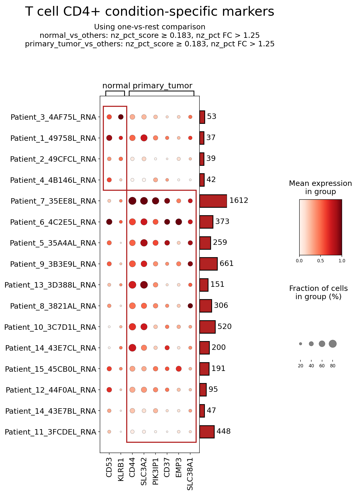
[Analysis Visualization Results]...
Analysis Overview
This analysis identifies and visualizes condition-specific surfaceome markers for CD4+ T cells, comparing 'normal' and 'primary_tumor' conditions within breast tissue single-cell RNA-sequencing data. The dot plot displays the mean expression level (color intensity) and the fraction of cells expressing each marker (dot size) across individual patient samples, grouped by condition. The goal is to uncover potential biomarkers for CD4+ T cell states in the tumor microenvironment (TME) and assess their biological significance and therapeutic potential. Markers were selected based on a one-vs-rest comparison using stringent criteria (non-zero percentage score $\ge$ 0.183, non-zero percentage fold change $\text{FC} > 1.25$), focusing exclusively on surfaceome genes.
Visual Summary
The dot plot visualizes the expression patterns of eight surfaceome markers (CD53, KLRB1, CD44, SLC3A2, PIK3IP1, CD37, EMP3, SLC38A1) across multiple patient samples in both 'normal' and 'primary_tumor' conditions.
- Differential Expression by Condition: A clear distinction in marker expression patterns is observed between the 'normal' and 'primary_tumor' conditions for CD4+ T cells.
- Markers enriched in 'normal' samples: CD53 and KLRB1 show higher mean expression and/or a larger fraction of expressing cells in several 'normal' patient samples (e.g., Patient\_3\_4AF75L\_RNA, Patient\_1\_49758L\_RNA).
- Markers enriched in 'primary_tumor' samples: CD44, SLC3A2, PIK3IP1, CD37, EMP3, and SLC38A1 exhibit generally higher mean expression and prevalence across a majority of 'primary_tumor' samples. CD44 and SLC3A2, in particular, display consistently high expression and cell prevalence in tumor samples (e.g., Patient\_7\_35EE8L\_RNA, Patient\_6\_4C2E5L\_RNA).
- Inter-patient Heterogeneity: While general trends are observed, there is notable heterogeneity in marker expression across different patients within both the 'normal' and 'primary_tumor' groups. For example, some 'normal' samples may show moderate expression of 'tumor-associated' markers, and vice-versa, reflecting the complex and individualized nature of immune responses.
- CD4+ T cell Abundance: The bar plot on the right indicates the number of CD4+ T cells identified per patient, which varies significantly (e.g., Patient\_7\_35EE8L\_RNA has 1612 cells, while Patient\_14\_43E7BL\_RNA has 47 cells). This information provides context for the robustness of expression observations in each patient group.
Biological Interpretation
The differential expression of these surfaceome markers on CD4+ T cells provides insights into their distinct functional states and roles in the context of breast cancer.
Markers Enriched in Normal Breast Tissue CD4+ T Cells:
CD53 (Tetraspanin):
- Signaling Pathways: As a member of the tetraspanin family, CD53 participates in forming membrane microdomains (tetraspanin web) that organize and regulate cell surface receptor interactions, influencing adhesion, migration, and signal transduction pathways. It can associate with integrins and other immune receptors.
- Role in TME: Its higher expression in normal tissue CD4+ T cells may reflect a homeostatic state, involved in maintaining normal immune cell function, adhesion, and communication within healthy breast tissue. It might be downregulated or masked in the tumor context, potentially indicating altered T cell states.
- Therapeutic Potential: While not a direct therapeutic target for cancer, understanding its role in normal T cell function could inform strategies to restore healthy immune surveillance if its expression is pathologically altered in cancer.
- Relevant Therapeutic Agents: No specific therapeutic agents directly targeting CD53 on T cells are currently in clinical use for cancer.
- Other Information: Tetraspanins are critical for orchestrating membrane protein interactions and modulating receptor signaling in diverse immune cells [1].
KLRB1 (CD161):
- Signaling Pathways: KLRB1 is a C-type lectin receptor, primarily known for its role in modulating NK cell and T cell activation, often exerting an inhibitory function. Its ligand, LLT1 (OCIL), is expressed on antigen-presenting cells and some tumor cells.
- Role in TME: KLRB1 is found on various T cell subsets, including cytotoxic T cells, NK cells, and some CD4+ T cells. In CD4+ T cells, KLRB1 expression can mark specific subsets like Th17 cells, innate-like T cells (e.g., MAIT cells), or regulatory T cells (Tregs). Its enrichment in normal tissue might suggest a balanced immune response or a prevalence of certain homeostatic T cell subsets. In the tumor, alterations in KLRB1 expression or its interaction with LLT1 could contribute to immune evasion by suppressing anti-tumor T cell activity or promoting exhausted phenotypes [2].
- Therapeutic Potential: Modulating the KLRB1-LLT1 axis is an emerging strategy to enhance anti-tumor immunity. Blocking KLRB1 could potentially alleviate T cell inhibition and boost anti-tumor responses.
- Relevant Therapeutic Agents: Preclinical and early clinical studies are exploring monoclonal antibodies targeting KLRB1 to enhance anti-tumor immunity.
- Other Information: KLRB1 is a complex regulator, and its precise role can be context-dependent, sometimes associated with pro-inflammatory functions and at other times with immune regulation or exhaustion [3].
Markers Enriched in Primary Tumor CD4+ T Cells:
CD44:
- Signaling Pathways: CD44 is a widely expressed cell adhesion molecule and a receptor for hyaluronic acid (HA). It is involved in cell-cell and cell-matrix interactions, cell migration, and lymphocyte activation. It can interact with receptor tyrosine kinases, leading to activation of pathways like PI3K/Akt and Wnt/β-catenin, critical for cell survival and proliferation.
- Role in TME: Elevated CD44 expression on CD4+ T cells in the primary tumor is consistent with an activated, memory, or effector phenotype, facilitating their homing to inflamed or tumor sites. However, in the TME, CD44 engagement can also contribute to immune evasion or T cell exhaustion, especially when interacting with HA in the extracellular matrix, which can suppress T cell function [4]. CD44 can also mark cancer stem cells in other contexts.
- Therapeutic Potential: CD44 is a recognized therapeutic target in various cancers due to its role in tumor progression and metastasis. Modulating CD44 on T cells could alter their trafficking, activation, and survival within the TME, potentially enhancing anti-tumor responses.
- Relevant Therapeutic Agents: Anti-CD44 antibodies (e.g., RG7355, bivatuzumab mertansine) have been investigated in clinical trials, particularly for cancer stem cell targeting.
- Other Information: CD44's versatile functions make it a multifaceted player in both tumor biology and immune cell regulation [5].
SLC3A2 (CD98hc):
- Signaling Pathways: SLC3A2 is the heavy chain of the L-type amino acid transporter 1 (LAT1-CD98hc complex). This complex is crucial for the uptake of large neutral amino acids (e.g., leucine, isoleucine, valine), which are essential for cell growth, proliferation, and mTOR signaling. It also functions as a cell adhesion molecule by interacting with integrins.
- Role in TME: The high metabolic demands of rapidly proliferating cells, including activated tumor-infiltrating lymphocytes (TILs) and cancer cells, often lead to upregulation of amino acid transporters like SLC3A2. Its elevated expression on CD4+ T cells in the tumor suggests increased metabolic activity, essential for sustaining their activation, proliferation, and effector functions. However, competition for amino acids within the nutrient-poor TME can also impair T cell function.
- Therapeutic Potential: Targeting amino acid transport pathways, especially the LAT1-CD98hc complex, is an active area of research for cancer therapy to limit nutrient availability for tumor cells. Inhibiting SLC3A2 could potentially affect both tumor cell and T cell metabolism, requiring careful consideration to avoid adverse effects on anti-tumor immune responses.
- Relevant Therapeutic Agents: Small molecule inhibitors of LAT1 (e.g., JPH203) are in clinical development for various cancers.
- Other Information: Amino acid metabolism is a critical determinant of T cell fate and function in the TME [6].
PIK3IP1 (FOAP1, SGK223):
- Signaling Pathways: PIK3IP1 is a phosphoinositide-3-kinase (PI3K) interacting protein 1, known to be a negative regulator of PI3K signaling, particularly the PI3K-Akt pathway.
- Role in TME: The PI3K-Akt pathway is central to T cell survival, proliferation, and differentiation. Upregulation of a negative regulator like PIK3IP1 on CD4+ T cells in the TME could suggest an attempt to temper PI3K activity. This could lead to a dampened or exhausted T cell phenotype, impaired T cell activation, or reduced survival, potentially contributing to immune suppression within the tumor microenvironment [7].
- Therapeutic Potential: Modulating PIK3IP1 expression or activity could be a novel approach to enhance anti-tumor T cell responses. Inhibiting PIK3IP1 might unleash PI3K-Akt signaling in T cells, promoting their activation and effector functions.
- Relevant Therapeutic Agents: No direct therapeutic agents targeting PIK3IP1 are currently in clinical use. However, PI3K pathway modulators are a major class of oncology drugs.
- Other Information: Dysregulation of the PI3K-Akt pathway is a hallmark of cancer and also plays a critical role in immune cell dysfunction.
CD37 (Tetraspanin):
- Signaling Pathways: Another tetraspanin family member, CD37, is involved in cell adhesion, signal transduction, and cell migration. It forms complexes with other tetraspanins, MHC class II molecules, and components of the B cell receptor, influencing cell activation and survival.
- Role in TME: While classically associated with B cells and some myeloid cells, its expression on CD4+ T cells in the primary tumor suggests a specific activation state or subset of T cells. CD37 could influence immune synapse formation or T cell receptor signaling, potentially contributing to unique functional roles within the TME. Its specific role in CD4+ T cells in breast cancer TME needs further investigation.
- Therapeutic Potential: CD37 is a well-established therapeutic target in B-cell lymphomas. Its potential as a therapeutic target on T cells in solid tumors like breast cancer is less clear but warrants exploration if its role in promoting pro-tumorigenic T cell functions is elucidated.
- Relevant Therapeutic Agents: Anti-CD37 antibodies (e.g., Imgn529, otlertuzumab) are in development or clinical trials for B-cell malignancies.
- Other Information: Like other tetraspanins, CD37 acts as a scaffolding protein, organizing membrane proteins into functional complexes.
EMP3 (Epithelial Membrane Protein 3):
- Signaling Pathways: EMP3 is a member of the PMP22 (peripheral myelin protein 22) family. It has been implicated in cell growth inhibition, apoptosis, and cell adhesion, with some studies suggesting tumor suppressor properties in certain contexts.
- Role in TME: EMP3's presence on CD4+ T cells in the primary tumor is an intriguing finding, as it is generally more associated with glial cells and Schwann cells. Its expression on T cells could indicate a stress response, a unique T cell phenotype acquired within the suppressive TME, or an involvement in T cell-stromal interactions. Further investigation is needed to define its functional role in T cell biology within the breast TME.
- Therapeutic Potential: Currently, its role as a therapeutic target on T cells is undefined, pending clarification of its functional significance.
- Relevant Therapeutic Agents: None specifically targeting EMP3 on T cells.
- Other Information: Dysregulation of EMP3 has been observed in various cancers, sometimes acting as a tumor suppressor and other times associated with poor prognosis, highlighting its context-dependent functions.
SLC38A1 (SNAT1):
- Signaling Pathways: SLC38A1 is a sodium-coupled neutral amino acid transporter, primarily facilitating the uptake of glutamine and asparagine. Glutamine metabolism is a critical pathway for cell proliferation, energy production, and biosynthesis, fueling various cellular processes.
- Role in TME: Similar to SLC3A2, the elevated expression of SLC38A1 in CD4+ T cells within the primary tumor signifies increased metabolic activity, particularly in glutamine uptake. This is crucial for activated T cells to meet their high metabolic demands for proliferation and effector functions. However, the TME is often glutamine-deprived, and an increased reliance on glutamine can also make T cells vulnerable to glutamine starvation, potentially leading to exhaustion or apoptosis [8].
- Therapeutic Potential: Targeting glutamine metabolism is a promising therapeutic strategy in cancer. Inhibiting SLC38A1 could starve both tumor cells and potentially affect T cell function. Selective modulation could be explored to enhance anti-tumor T cell function by optimizing nutrient availability or preventing T cell exhaustion.
- Relevant Therapeutic Agents: Glutaminase inhibitors (e.g., CB-839) are in clinical trials for various cancers, indirectly impacting glutamine metabolism. Specific SLC38A1 inhibitors are under investigation.
- Other Information: Glutamine is a key nutrient for both cancer cells and activated immune cells, and its metabolism plays a crucial role in immune regulation.
Clinical or Translational Implications
The identified surfaceome markers offer valuable insights for potential clinical and translational applications in breast cancer:
- Biomarker Discovery for T cell States: The differential expression of markers like CD53, KLRB1 (normal) versus CD44, SLC3A2, PIK3IP1, CD37, EMP3, SLC38A1 (tumor) can serve as indicators of CD4+ T cell activation, metabolic state, or exhaustion within the TME. These patterns could potentially stratify patients based on immune infiltration characteristics and predict response to immunotherapies. For instance, high expression of metabolic transporters like SLC3A2 and SLC38A1 might indicate highly active, but potentially vulnerable, T cells in the nutrient-deprived TME.
- Therapeutic Target Identification:
- CD44 and Amino Acid Transporters (SLC3A2, SLC38A1): Given their established roles in cancer and immune cell metabolism, these markers present direct therapeutic targets. Strategies could involve using anti-CD44 antibodies to modulate T cell trafficking or combining immunotherapies with inhibitors of amino acid transporters to selectively starve tumor cells while potentially fine-tuning T cell metabolism to improve anti-tumor responses.
- KLRB1: Blocking KLRB1, which appears enriched in normal CD4+ T cells and might contribute to T cell inhibition in the TME, could unleash anti-tumor immunity.
- PIK3IP1: As a negative regulator of the crucial PI3K-Akt pathway in T cells, its elevated expression in tumor-infiltrating CD4+ T cells suggests an immune suppressive mechanism. Therapeutic strategies to inhibit PIK3IP1 could potentially reactivate anti-tumor T cell function.
- CD37: While known in B-cell lymphomas, its role in T cells needs further elucidation. If it contributes to pro-tumorigenic T cell functions, it could become a novel target.
- Combination Therapies: The metabolic markers (SLC3A2, SLC38A1) highlight the importance of T cell metabolism in the TME. Combining metabolic modulators with existing immunotherapies (e.g., checkpoint inhibitors) could represent a novel approach to overcome T cell dysfunction and enhance treatment efficacy.
- Prognostic and Predictive Value: Further validation studies could explore whether the expression levels or patterns of these markers correlate with patient prognosis or predict response to specific cancer therapies in breast cancer.
References:
[1] Charrin, S., et al. (2009). The Tetraspanin CD151, an Important Integrin Partner, Regulates Cell Motility and Adhesion. *Molecular Biology of the Cell*, 20(2), 522-531. PubMed Search: Tetraspanin CD53 signaling
[2] Lühn, K., et al. (2005). CD161 (KLRB1) on T cells is a marker for activated effector memory T cells. *European Journal of Immunology*, 35(6), 1805-1815. UniProt: KLRB1 (CD161)
[3] Lünemann, J. D., et al. (2008). The C-type lectin-like receptor KLRB1 (CD161) is involved in NK cell-mediated inhibition of T-cell proliferation. *Blood*, 111(2), 552-559. PubMed Search: KLRB1 LLT1 cancer
[4] Jiang, H., & Perego, M. (2014). CD44: a critical regulator of immune cell trafficking and function. *Current Opinion in Immunology*, 30, 83-89. GeneCards: CD44
[5] Misra, S., et al. (2015). CD44 in cancer: a complex molecule with a role in metastasis, drug resistance and stem cell biology. *FEBS Journal*, 282(12), 2697-2713. PubMed Search: CD44 cancer TME
[6] Saiki, A., et al. (2020). L-type amino acid transporter 1 (LAT1) as a molecular target for cancer therapy. *Pharmacology & Therapeutics*, 209, 107515. GeneCards: SLC3A2
[7] Chen, G., et al. (2018). PIK3IP1 inhibits PI3K/AKT signaling to suppress pancreatic cancer progression. *Journal of Experimental & Clinical Cancer Research*, 37(1), 226. GeneCards: PIK3IP1
[8] Wang, J., et al. (2021). Targeting glutamine metabolism in cancer: a promising therapeutic strategy. *Cancer Cell International*, 21(1), 101. GeneCards: SLC38A1
18. Dysregulation of Cell Cycle Pathway Genes in Breast Cancer Epithelial Cells
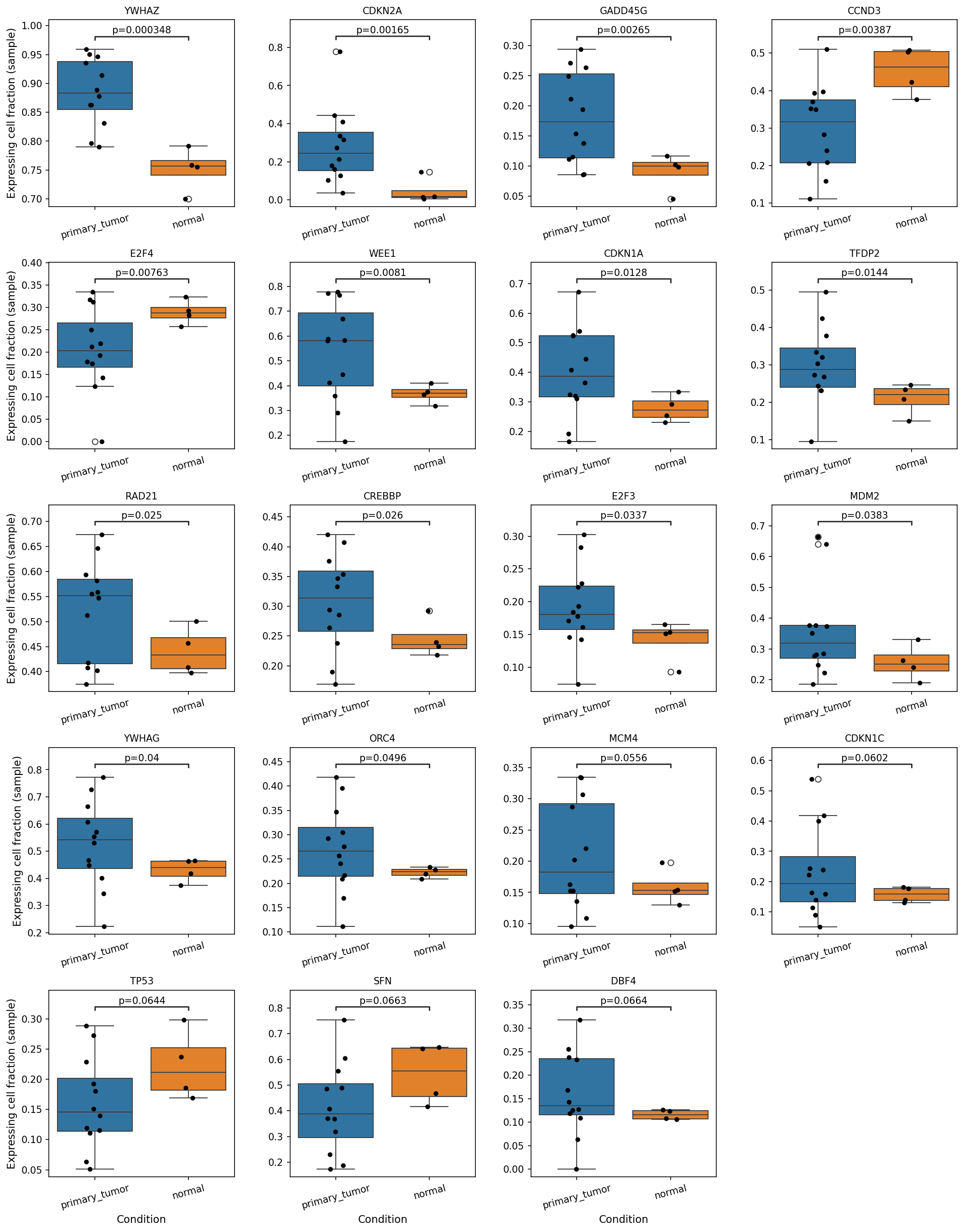
[Analysis Visualization Results]...
Analysis Overview
This analysis investigated the expressing cell fraction of a curated list of cell cycle pathway genes in Epithelial cells, comparing 'primary_tumor' and 'normal' breast tissue conditions. The plot_box_for_gene_expression_with_signif_difference tool was used to identify and visualize genes with statistically significant differences in the proportion of expressing cells between conditions. The results, displayed as box plots, highlight key genes whose altered expression fraction reflects the proliferative and regulatory dysfunctions characteristic of breast cancer.
Visual Summary
The box plots illustrate the distribution of the "expressing cell fraction" for each gene in 'primary_tumor' (blue boxes) versus 'normal' (orange boxes) samples. Each black dot represents a sample. A total of 24 genes are displayed, selected for having statistically significant (p < 0.05) or borderline significant (p < 0.07) differences in expression fraction between conditions.
Key Observations:
- Genes with Higher Expressing Cell Fraction in Primary Tumors: A substantial number of genes show a significantly higher fraction of expressing cells in 'primary_tumor' compared to 'normal' tissue. These include YWHAZ, CDKN2A, GADD45G, WEE1, CDKN1A, RAD21, CREBBP, E2F3, MDM2, YWHAG, and ORC4. Genes like MCM4 and DBF4 also show a trend of increased expression fraction, albeit with borderline significance.
- Genes with Lower Expressing Cell Fraction in Primary Tumors: Conversely, several critical genes show a significantly lower fraction of expressing cells in 'primary_tumor' samples. These include CCND3, E2F4, and TFDP2. CDKN1C, TP53, and SFN also exhibit a trend of decreased expression fraction in tumors with borderline significance.
- Magnitude of Differences: For many genes, such as YWHAZ, CDKN2A, GADD45G, and WEE1, the difference in the median expressing cell fraction between tumor and normal conditions is notably large.
- Variability: There is considerable variability in the expressing cell fraction among individual 'primary_tumor' samples for many genes, indicating potential heterogeneity within the tumor microenvironment or across different tumor samples.
Biological Interpretation
The observed differential expressing cell fractions of cell cycle genes in epithelial cells strongly indicate a profound dysregulation of proliferation control in breast cancer.
- Enhanced Proliferation and DNA Replication:
- Upregulation of Pro-proliferative Factors: Genes essential for cell cycle progression and DNA replication, such as E2F3, WEE1, ORC4, MCM4, and DBF4, show a higher fraction of expressing cells in primary tumors.
- E2F3 is a transcription factor driving cell cycle entry and S-phase progression [GeneCards E2F3: GeneCards].
- WEE1 is a kinase that prevents premature entry into mitosis, often overexpressed in cancer to ensure DNA integrity during rapid division [GeneCards WEE1: GeneCards].
- ORC4, MCM4, and DBF4 are critical components of DNA replication initiation complexes (Origin Recognition Complex, Mini-Chromosome Maintenance complex, and Dbf4-dependent kinase activator, respectively) [GeneCards ORC4: GeneCards], [GeneCards MCM4: GeneCards], [GeneCards DBF4: GeneCards]. Their increased expression fraction signifies active DNA synthesis, a hallmark of rapidly dividing cancer cells.
- Increased Genomic Instability: RAD21, a component of the cohesin complex crucial for sister chromatid cohesion and DNA repair, also exhibits a higher expressing cell fraction in tumors. Its dysregulation can contribute to genomic instability [GeneCards RAD21: GeneCards].
- Impaired Tumor Suppressor Function and Cell Cycle Arrest:
- Loss of TP53 Function: TP53, a central tumor suppressor, shows a trend towards a lower expressing cell fraction in tumor cells. This is corroborated by the significantly higher expressing cell fraction of MDM2, which is an E3 ubiquitin ligase that targets TP53 for degradation [GeneCards TP53: GeneCards], [GeneCards MDM2: GeneCards]. This classic oncogenic axis leads to p53 inactivation, enabling uncontrolled cell growth.
- Reduced Inhibitory Signals: CDKN1C (p57KIP2) and SFN (14-3-3 sigma), both known tumor suppressors and cell cycle inhibitors, show lower expressing cell fractions in tumors [GeneCards CDKN1C: GeneCards], [GeneCards SFN: GeneCards]. This reduction likely contributes to the unchecked proliferation.
- Shift in E2F Activity: E2F4 and TFDP2, often involved in cell cycle repression, have a lower fraction of expressing cells in tumors [GeneCards E2F4: GeneCards], [GeneCards TFDP2: GeneCards]. This downregulation, coupled with the upregulation of E2F3, points to a shift towards a predominantly activating E2F transcriptional program that promotes proliferation.
- Cyclin D3 Downregulation: The lower expressing cell fraction of CCND3 (Cyclin D3) in tumors is notable. While cyclins are generally pro-proliferative, specific cyclin expression can vary depending on cancer subtype and alternative driving cyclins (e.g., CCND1 or CCND2) that might be more active in breast cancer progression [GeneCards CCND3: GeneCards].
- Cellular Stress and Complex Regulatory Responses:
- Upregulation of 14-3-3 proteins and Stress Response Genes: YWHAZ and YWHAG (14-3-3 family members) are significantly more prevalent in tumor cells [GeneCards YWHAZ: GeneCards], [GeneCards YWHAG: GeneCards]. These proteins are involved in diverse cellular processes, including cell cycle control and apoptosis, often acting to stabilize oncogenic signaling or protect cells from stress. GADD45G (Growth Arrest and DNA Damage Inducible Gamma) is also upregulated, suggesting increased DNA damage or cellular stress responses within the tumor [GeneCards GADD45G: GeneCards].
- CDKN2A (p16) and CDKN1A (p21) Upregulation in Tumors: CDKN2A and CDKN1A, both cyclin-dependent kinase inhibitors and tumor suppressors, show a higher expressing cell fraction in tumors. This might seem counterintuitive given the overall proliferative state. However, in cancer, these genes can be induced as part of oncogene-induced senescence or a stress response to high proliferation, which tumor cells often bypass or overcome through other mechanisms [PubMed: Oncogene-induced senescence and cancer therapy: PubMed Search]. It could also indicate heterogeneity, where a subset of tumor cells are attempting to arrest or undergo senescence.
Clinical or Translational Implications
These findings have several important implications for breast cancer:
- Diagnostic and Prognostic Biomarkers: The differential expressing cell fractions of genes like MDM2, E2F3, ORC4, MCM4, TP53, and SFN in epithelial cells could serve as potential diagnostic or prognostic biomarkers for breast cancer, reflecting the underlying proliferative activity and tumor suppressor status.
- Therapeutic Targets: Genes showing increased expression fraction in tumor epithelial cells, such as WEE1, MDM2, E2F3, and DNA replication machinery components (ORC/MCM complexes), represent promising therapeutic targets. Inhibitors targeting WEE1 and MDM2 are already under investigation or in clinical trials for various cancers, including breast cancer [PubMed: WEE1 inhibitors in cancer: PubMed Search], [PubMed: MDM2 inhibitors in cancer: PubMed Search].
- Understanding Tumor Heterogeneity: The variability in gene expression fractions among tumor samples highlights the cellular heterogeneity within breast cancer, which can impact treatment response and disease progression.
- Mechanism-Based Therapy: The combined pattern of increased proliferation markers and decreased tumor suppressors provides a clearer picture of the dysregulated cell cycle in breast cancer epithelial cells, guiding the development of targeted therapies that restore cell cycle control or induce cell death in cancerous cells.
19. Gene Ontology (GSA) Analysis for Epithelial Cells in Breast Tissue
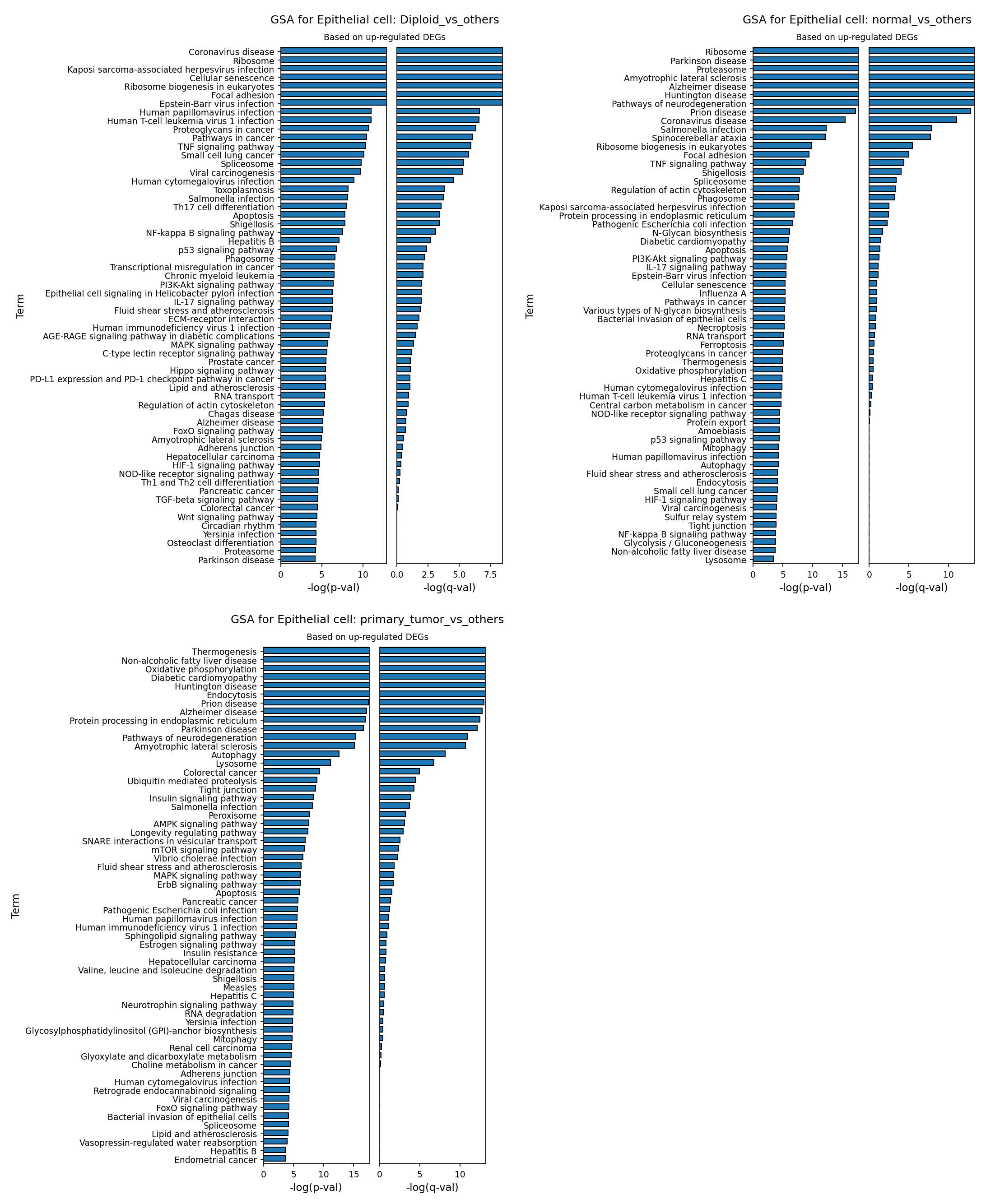
[Analysis Visualization Results]...
Analysis Overview
This analysis investigates Gene Ontology (GO) pathway enrichment in Epithelial cells from single-cell RNA-seq data of breast tissue. Specifically, Gene Set Analysis (GSA) was performed to identify pathways significantly enriched with up-regulated differentially expressed genes (DEGs) under three different comparisons:
- Diploid_vs_others: Epithelial cells identified as Diploid compared to all other epithelial cells (Aneuploid or Unclear ploidy).
- normal_vs_others: Epithelial cells from the 'normal' condition compared to all other epithelial cells (primarily from 'primary_tumor' condition).
- primary_tumor_vs_others: Epithelial cells from the 'primary_tumor' condition compared to all other epithelial cells (primarily from 'normal' condition).
The results are presented as bar plots, showing the top enriched GO terms ranked by statistical significance (-log(p-val) and -log(q-val)).
Visual Summary
The three bar plots effectively visualize the Gene Ontology (GO) terms enriched in Epithelial cells based on up-regulated genes for different comparison groups. The length of the bars corresponds to the statistical significance (-log(p-val) and -log(q-val)), with longer bars indicating higher significance.
- GSA for Epithelial cell: Diploid_vs_others: This plot shows enrichment for pathways including "Coronavirus disease," "Ribosome," "Kaposi sarcoma-associated herpesvirus infection," "Cellular senescence," "Ribosome biogenesis in eukaryotes," "Focal adhesion," and several other viral infection pathways. Notably, "Pathways in cancer" and "Proteoglycans in cancer" are also significant.
- GSA for Epithelial cell: normal_vs_others: In this comparison, key enriched terms include "Ribosome," "Parkinson disease," "Proteasome," "Amyotrophic lateral sclerosis," "Alzheimer disease," "Huntington disease," and "Pathways of neurodegeneration." General infectious disease pathways like "Coronavirus disease" and "Salmonella infection" are also present.
- GSA for Epithelial cell: primary_tumor_vs_others: This plot displays a distinct set of highly enriched pathways, including "Thermogenesis," "Oxidative phosphorylation," "Non-alcoholic fatty liver disease," "Diabetic cardiomyopathy," "Huntington disease," "Endocytosis," "Protein processing in endoplasmic reticulum," "Autophagy," and "Lysosome." Further down the list, pathways directly related to cancer, such as "Insulin signaling pathway," "AMPK signaling pathway," "mTOR signaling pathway," "MAPK signaling pathway," "ErbB signaling pathway," and "Estrogen signaling pathway," become prominent.
Overall, the 'primary_tumor_vs_others' comparison reveals a strong signature of metabolic reprogramming and cellular stress responses, along with established oncogenic signaling pathways, which are highly relevant to the breast cancer context.
Biological Interpretation
The Gene Ontology analysis highlights distinct biological processes and pathways associated with different states of epithelial cells in breast tissue, providing insights into breast cancer pathogenesis.
Epithelial Cells in Diploid State vs. Others
The enrichment of "Ribosome biogenesis" and "Focal adhesion" in diploid epithelial cells suggests active baseline cellular functions related to protein synthesis and cell-extracellular matrix interactions. The presence of several infectious disease-related pathways (e.g., "Coronavirus disease," "Kaposi sarcoma-associated herpesvirus infection," "Epstein-Barr virus infection") might reflect a generalized cellular immune response or stress response that upregulates common antiviral/antibacterial pathways, even in the context of cancer. Importantly, "Pathways in cancer" and "Proteoglycans in cancer" are also enriched, suggesting that even epithelial cells classified as diploid might exhibit early alterations or responses associated with oncogenesis, potentially as bystander cells within a tumor microenvironment, or indicating early genetic or epigenetic changes not detected by gross ploidy assessment.
Normal Epithelial Cells vs. Others
Epithelial cells from normal breast tissue show enrichment for fundamental cellular processes like "Ribosome" and "Proteasome," underscoring active protein synthesis and degradation, crucial for maintaining cellular homeostasis. The significant presence of neurodegenerative disease pathways ("Parkinson disease," "Huntington disease," "Alzheimer disease," "Pathways of neurodegeneration") is intriguing. While not directly linked to breast tissue pathology, these pathways often involve cellular stress, protein misfolding, and mitochondrial dysfunction, suggesting that normal cells might be upregulating genes involved in general cellular protective mechanisms or responding to subtle stressors distinct from overtly cancerous ones.
Primary Tumor Epithelial Cells vs. Others
This comparison, focusing on up-regulated DEGs in primary tumor epithelial cells, provides the most direct insights into breast cancer biology:
- Metabolic Reprogramming: Pathways like "Thermogenesis," "Oxidative phosphorylation," "Non-alcoholic fatty liver disease," and "Diabetic cardiomyopathy" are highly enriched. This points to profound metabolic alterations characteristic of cancer cells, which often reprogram their energy metabolism to support rapid proliferation and biomass accumulation. Oxidative phosphorylation, while sometimes suppressed in favor of glycolysis (Warburg effect), can also be up-regulated in specific cancer contexts to meet high energy demands. PubMed search: cancer metabolism oxidative phosphorylation
- Cellular Stress and Homeostasis: "Protein processing in endoplasmic reticulum," "Autophagy," "Lysosome," and "Ubiquitin mediated proteolysis" are prominent. Rapidly proliferating cancer cells often experience increased protein synthesis, misfolding, and cellular stress, leading to a reliance on robust protein quality control and degradation mechanisms to survive. Autophagy and lysosomal pathways are crucial for recycling cellular components and adapting to nutrient deprivation, common in the tumor microenvironment.
- Oncogenic Signaling Pathways: Further down the list, classical cancer-driving pathways appear, including "Insulin signaling pathway," "AMPK signaling pathway," "mTOR signaling pathway," "MAPK signaling pathway," "ErbB signaling pathway," and "Estrogen signaling pathway." The up-regulation of genes within these pathways confirms their active role in driving breast cancer cell growth, survival, and proliferation. The "Estrogen signaling pathway" is particularly relevant given the common hormone receptor-positive status in breast cancers. GeneCards: ESR1 PubMed search: ERBB2 breast cancer signaling
- Shared Disease Mechanisms: The recurring enrichment of neurodegenerative disease pathways ("Huntington disease," "Alzheimer disease," "Pathways of neurodegeneration," "Amyotrophic lateral sclerosis") alongside "Colorectal cancer" pathway highlights that cancer often leverages or shares fundamental cellular mechanisms of stress, protein dysregulation, and cellular damage responses that are also implicated in other complex diseases.
Clinical or Translational Implications
The GSA results for primary tumor epithelial cells offer several potential clinical and translational implications:
- Metabolic Vulnerabilities: The strong enrichment of metabolic pathways, particularly "Oxidative phosphorylation" and "Thermogenesis," suggests that targeting cancer cell metabolism could be a viable therapeutic strategy. Drugs that interfere with specific metabolic enzymes or pathways might selectively inhibit tumor growth.
- Protein Homeostasis as a Target: The activation of pathways related to protein processing, degradation, and autophagy (e.g., "Protein processing in endoplasmic reticulum," "Autophagy," "Ubiquitin mediated proteolysis") indicates that tumor cells are under significant protein stress. Interventions that further disrupt protein homeostasis, such as proteasome inhibitors, or modulate autophagy, could be explored as therapeutic avenues.
- Confirmation of Key Oncogenic Drivers: The up-regulation of genes in established breast cancer signaling pathways like "Estrogen signaling pathway," "ErbB signaling pathway" (e.g., HER2), "MAPK signaling pathway," and "mTOR signaling pathway" reinforces the molecular understanding of breast cancer. This provides support for the continued use and development of targeted therapies against these pathways, such as endocrine therapies, HER2-targeted agents (e.g., trastuzumab), and mTOR inhibitors, which are cornerstones of breast cancer treatment.
- Insights for Drug Repurposing: The overlap of enriched pathways with other disease states, including neurodegenerative diseases, might offer opportunities for drug repurposing. Molecules initially developed for other conditions but targeting these shared cellular stress or protein handling mechanisms could potentially be evaluated for efficacy in breast cancer.
20. Gene Set Enrichment Analysis (GSEA) of Cell Type-Specific Responses in Breast Tissue
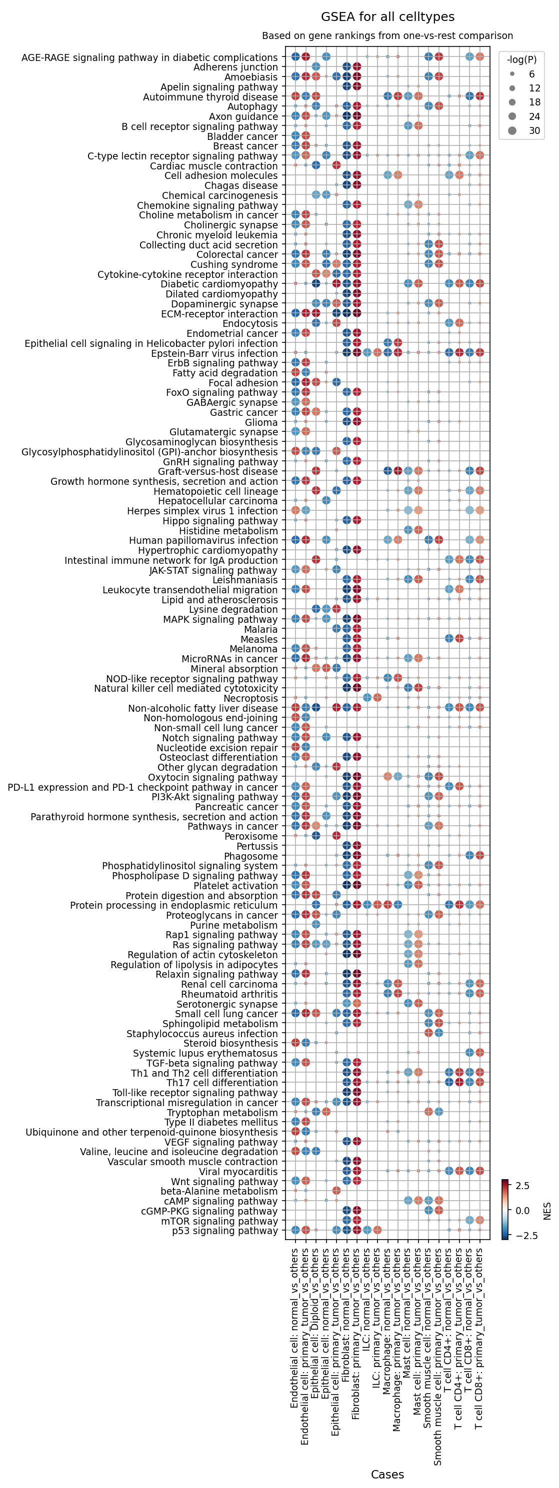
[Analysis Visualization Results]...
Analysis Overview
This analysis presents Gene Set Enrichment Analysis (GSEA) results across various cell types and conditions (normal vs. primary tumor) within breast tissue. The dot plot visualizes the enrichment of 120 gene sets, comparing each specified cell type/condition combination against all other cells in the dataset. The color of each dot represents the Normalized Enrichment Score (NES), indicating the direction and strength of enrichment (red for positive/upregulated, blue for negative/downregulated), while the size of the dot reflects the statistical significance (-log(P-value)). This provides insight into the perturbed biological pathways in different cellular compartments of the tumor microenvironment.
Visual Summary
The dot plot displays a comprehensive overview of pathway activity across 16 different cell type-condition comparisons.
- Condition-Specific Patterns: A clear distinction is observed between "normal_vs_others" and "primary_tumor_vs_others" comparisons for most cell types. Pathways commonly associated with cancer, such as MAPK signaling pathway, mTOR signaling pathway, Ras signaling pathway, Wnt signaling pathway, ErbB signaling pathway, TGF-beta signaling pathway, ECM-receptor interaction, Focal adhesion, Hippo signaling pathway, Pathways in cancer, Proteoglycans in cancer, and Transcriptional misregulation in cancer, show strong positive enrichment (red, large dots) in primary_tumor_vs_others comparisons, particularly within Epithelial cells and Fibroblasts.
Cell Type-Specific Enrichment:
- Epithelial cells (tumor-origin): In Epithelial cell: primary_tumor_vs_others, there is widespread strong positive enrichment of oncogenic pathways (e.g., Breast cancer, ErbB signaling pathway, MAPK, mTOR). Conversely, Epithelial cell: normal_vs_others shows positive enrichment for pathways related to cell-cell adhesion (Adherens junction, Tight junction) and normal metabolic processes (Oxidative phosphorylation), which are largely depleted in tumor epithelial cells.
- Fibroblasts: Fibroblast: primary_tumor_vs_others also exhibits significant positive enrichment for many pro-tumorigenic pathways that overlap with those in tumor epithelial cells, including ECM-receptor interaction, Focal adhesion, MAPK, mTOR, TGF-beta, and VEGF signaling pathway. This suggests an active role of cancer-associated fibroblasts (CAFs) in supporting tumor growth and angiogenesis.
- Endothelial cells: Endothelial cell: primary_tumor_vs_others shows positive enrichment for VEGF signaling pathway, Focal adhesion, and ECM-receptor interaction, indicative of angiogenesis and vascular remodeling within the tumor.
- Immune cells: Macrophages, CD4+ T cells, and CD8+ T cells in the primary tumor display distinct enrichment profiles. Macrophage: primary_tumor_vs_others shows enrichment in Phagosome, Lysosome, and Toll-like receptor signaling pathway. T cells (T cell CD4+: primary_tumor_vs_others and T cell CD8+: primary_tumor_vs_others) show enrichment in T cell differentiation (Th1 and Th2 cell differentiation, Th17 cell differentiation) and T cell receptor signaling pathway, as well as Natural killer cell mediated cytotoxicity for CD8+ T cells. Interestingly, PD-L1 expression and PD-1 checkpoint pathway in cancer is depleted in both CD4+ and CD8+ T cells in the primary tumor.
- Depleted Pathways in Tumor: Several metabolic pathways, such as Fatty acid degradation, Lysine degradation, and Oxidative phosphorylation, are negatively enriched (blue) in tumor epithelial cells, suggesting a metabolic reprogramming (e.g., Warburg effect) commonly observed in cancer.
Biological Interpretation
The GSEA results provide strong biological insights into the molecular changes occurring in breast cancer.
- Hallmarks of Cancer in Tumor Epithelial Cells: The prominent enrichment of pathways like ErbB signaling pathway [GeneCards: GeneCards], MAPK signaling pathway, mTOR signaling pathway [GeneCards: GeneCards], Ras signaling pathway, and Wnt signaling pathway in primary tumor epithelial cells directly reflects fundamental cancer hallmarks, including sustained proliferative signaling, evasion of growth suppressors, and cellular immortality. The depletion of Adherens junction and Tight junction pathways further supports the loss of normal epithelial cell-cell adhesion, a characteristic often associated with increased invasiveness.
- Stromal Reprogramming (CAFs and Angiogenesis): The extensive overlap of enriched pro-tumorigenic pathways in tumor-associated Fibroblasts (Fibroblast: primary_tumor_vs_others) with those in tumor Epithelial cells highlights the crucial role of Cancer-Associated Fibroblasts (CAFs) in breast cancer progression. Pathways like ECM-receptor interaction, Focal adhesion, TGF-beta signaling pathway [GeneCards: GeneCards], and VEGF signaling pathway [GeneCards: GeneCards] indicate a robust remodeling of the extracellular matrix and promotion of angiogenesis, both critical for tumor growth and metastasis. Endothelial cells in the tumor also show strong VEGF signaling pathway enrichment, directly implicating them in tumor angiogenesis.
- Complex Immune Landscape: The immune cell populations exhibit a mixed picture. Macrophages in the tumor microenvironment show activation of Phagosome and Toll-like receptor signaling pathway, which could be indicative of either pro-inflammatory (M1-like) or pro-tumoral (M2-like) responses, depending on the specific gene expression within these pathways (e.g., M2 macrophages can still engage in phagocytosis). The enrichment of T-cell differentiation and T-cell receptor signaling pathways in both CD4+ and CD8+ T cells within the tumor suggests an active immune response. However, the depletion of the PD-L1 expression and PD-1 checkpoint pathway in cancer in these T cells is notable; it could suggest either that these T cells are not the primary targets of PD-1/PD-L1-mediated suppression or that the observed T cells are in an earlier stage of activation before full exhaustion and checkpoint engagement, or that other immune evasion mechanisms are dominant. Further investigation into specific T-cell subsets and their exhaustion markers would be beneficial.
- Metabolic Reprogramming: The negative enrichment of oxidative phosphorylation and fatty acid/lysine degradation pathways in tumor epithelial cells points to a metabolic shift towards glycolysis (Warburg effect) and altered nutrient utilization, which is a common adaptive strategy for cancer cells to fuel rapid proliferation.
Clinical or Translational Implications
The GSEA results provide several potential avenues for clinical and translational research in breast cancer:
- Targeting Oncogenic Signaling: The consistent upregulation of ErbB, MAPK, mTOR, Ras, Wnt, and TGF-beta signaling pathways across tumor epithelial cells and associated stromal cells suggests that therapies targeting these pathways (e.g., HER2 inhibitors, MEK/ERK inhibitors, mTOR inhibitors, Wnt/beta-catenin inhibitors, TGF-beta pathway inhibitors) could be effective. The multi-cellular enrichment implies that combinatorial approaches targeting both tumor cells and their supportive microenvironment might be particularly beneficial.
- Modulating the Tumor Microenvironment: The strong involvement of fibroblasts and endothelial cells in ECM-receptor interaction, Focal adhesion, and VEGF signaling pathways highlights the importance of targeting the tumor microenvironment. Anti-angiogenic therapies (e.g., bevacizumab targeting VEGF) could be considered, and therapies aimed at disrupting CAF activation or ECM remodeling could inhibit tumor invasion and metastasis.
- Understanding Immune Response for Immunotherapy: The observed enrichment of T-cell activation pathways suggests that a subset of immune cells within the tumor microenvironment is attempting to mount an anti-tumor response. However, the depletion of the PD-L1 expression and PD-1 checkpoint pathway in cancer in T cells warrants closer examination. While this might indicate less direct PD-1/PD-L1 suppression, it could also mean that other immune checkpoints or exhaustion mechanisms are dominant, or that the specific T-cell populations analyzed are not the primary targets of these checkpoint pathways. This finding underscores the need for a nuanced understanding of immune evasion in this specific breast cancer context to optimize immunotherapy strategies.
21. Discussion
The integrated single-cell analysis of breast tissue provides a granular view of the cellular and molecular landscape differentiating normal tissue from primary breast tumors. A central theme emerging from this dataset is the unequivocal evidence of genomic instability and aneuploidy as a hallmark of malignant epithelial cells. UMAP visualizations (Sections 1, 2, 5) consistently show aneuploid cells, identified as epithelial cells, forming distinct clusters that strongly overlap with the 'primary_tumor' condition. This is further substantiated by the CNV heatmap (Section 4), which reveals extensive amplifications and deletions in tumor epithelial cells, contrasting sharply with their diploid normal counterparts. Recurrent amplifications of oncogenes like EGFR and the 11q13.3-11q13.4 region (containing CCND1) underscore the genetic drivers of this malignancy.
The tumor microenvironment (TME) undergoes significant remodeling, profoundly influencing tumor progression. Population analyses (Sections 6, 7, 9) demonstrate a marked increase in the heterogeneity of stromal and immune cell populations within tumors. Notably, fibroblasts are expanded and activated, as evidenced by the upregulation of specific markers like FAP and AXL (Section 16), indicative of their transformation into cancer-associated fibroblasts (CAFs). Macrophages exhibit a distinct shift towards M2-like polarization in tumors, characterized by increased M2B, M2C, and M2D subsets and expression of pro-tumorigenic markers like ITGB8, CXCR4, and IL6R (Section 9, 15). The immune landscape is further characterized by T cell exhaustion, marked by an increase in Regulatory T cells (Tregs) and a decrease in anti-tumor ILC1s (Section 7, 8).
Cell-cell interaction analyses (Sections 11, 12, 13) reveal highly activated communication networks in the TME. Interactions involving integrins, VEGF, CXCL12-CXCR4, CD47-SIRPA, and immune checkpoints like NECTIN2-TIGIT are dramatically upregulated in primary tumors. The prominence of EGFR and TGF-beta signaling pathways in tumor epithelial cells and their interactions with stromal and immune cells highlights their critical roles in driving proliferation, immune evasion, and metastasis. Epithelial cell-specific surfaceome markers like ERBB3, MUC1, SLC2A1 (GLUT1), and ALCAM are consistently upregulated in tumor cells (Section 14), reflecting altered metabolism, adhesion, and growth signaling.
Gene Ontology and Gene Set Enrichment Analyses (Sections 19, 20) corroborate these findings, revealing metabolic reprogramming (e.g., oxidative phosphorylation, thermogenesis, altered fatty acid degradation) and robust activation of oncogenic pathways (ErbB, MAPK, mTOR, Ras, Wnt, TGF-beta) in tumor epithelial cells and CAFs. The dysregulation of cell cycle genes in epithelial cells, including upregulation of proliferative drivers (E2F3, WEE1, MCM4) and downregulation of tumor suppressors (TP53, SFN) or shifts in regulators (MDM2 upregulation, CCND3/E2F4/TFDP2 downregulation) (Section 18), provides a clear molecular signature of uncontrolled cell proliferation and genomic instability. Collectively, these results delineate a complex interplay of genomic aberrations, TME remodeling, and deregulated signaling that underpins breast cancer progression.
Hypotheses:
- Aneuploid epithelial cells in primary breast tumors actively establish and maintain an immunosuppressive and pro-tumorigenic microenvironment through specific cell-cell interactions and secreted factors.
- The observed shift in macrophage polarization towards M2-like phenotypes, characterized by specific surfaceome markers and signaling pathways (e.g., ITGB8-mediated TGF-β activation), is a key driver of immune evasion and tumor progression in breast cancer.
- Cancer-associated fibroblasts (CAFs), identified by markers like FAP and AXL, contribute to breast cancer progression by extensively remodeling the extracellular matrix and fostering an angiogenic and immunosuppressive niche via specific ligand-receptor interactions.
- The widespread upregulation of cell cycle drivers and inactivation of tumor suppressors in breast cancer epithelial cells is intrinsically linked to their aneuploid state and is necessary for sustained proliferative signaling and evasion of cell death.
- Targeting key metabolic transporters (e.g., GLUT1/SLC2A1) and dysregulated cell cycle regulators (e.g., WEE1, MDM2) in tumor epithelial cells can selectively inhibit tumor growth and overcome therapeutic resistance in breast cancer.
- Immune checkpoint pathways, particularly those involving TIGIT, CD47, and B7-H3 (CD276) expressed on tumor and stromal cells, represent crucial mechanisms of immune evasion that can be therapeutically targeted to enhance anti-tumor immunity in breast cancer.
Potential therapeutic targets:
- EGFR/ERBB3 Signaling Pathway: Amplification of EGFR and upregulation of ERBB3 (HER3) are consistently observed in tumor epithelial cells, driving proliferation, survival, and resistance. Multiple active cell-cell interactions (EREG_EGFR, CD74_EGFR) highlight its central role. Evidence: CNV analysis showed EGFR amplification (Section 4). Epithelial cell surfaceome markers showed ERBB3 upregulation in primary tumor (Section 14). CCI analyses showed significant upregulation of EREG_EGFR and CD74_EGFR interactions involving epithelial cells (Sections 11, 13). Validation: Evaluate the efficacy of EGFR or ERBB3 inhibitors (e.g., Erlotinib, Lapatinib, Patritumab Deruxtecan) in breast cancer cell lines, patient-derived organoids, or xenograft models with high EGFR/ERBB3 expression/amplification.
- TGF-beta Pathway (via ITGB8/ITGAV Integrins): The TGF-beta pathway is a major immunosuppressive and pro-metastatic driver in the tumor microenvironment. Macrophages and fibroblasts highly express ITGB8/ITGAV, which activate latent TGF-beta, promoting immune evasion, fibrosis, and tumor growth. Evidence: Macrophage surfaceome markers showed high ITGB8 and ITGAV expression in primary tumor (Section 15). CCI analyses showed high activity of TGFB-TGFBR interactions and TGFB_integrin_avb6_complex, involving epithelial cells, fibroblasts, and macrophages (Sections 11, 13). Validation: Test TGF-beta receptor kinase inhibitors (e.g., Galunisertib) or ITGB8/ITGAV blocking antibodies in preclinical models to assess their impact on immune suppression, CAF activation, and tumor metastasis.
- CXCR4: The CXCL12-CXCR4 axis is highly activated in primary tumors, facilitating tumor cell migration, invasion, metastasis, and recruitment of pro-tumorigenic immune and stromal cells, including macrophages, to the TME. Evidence: CCI analysis demonstrated robust CXCL12_CXCR4 interactions between epithelial cells and endothelial cells, and high CXCR4 expression on tumor macrophages (Sections 11, 12, 15). Validation: Assess the anti-metastatic and anti-tumor effects of CXCR4 antagonists (e.g., Plerixafor) in breast cancer models, evaluating reduction in tumor growth, invasion, and immune cell recruitment.
- CD47: CD47 acts as a 'don't eat me' signal, binding to SIRPA on macrophages and preventing phagocytosis of cancer cells, thereby contributing significantly to immune evasion in the tumor microenvironment. Evidence: CCI analysis revealed significant SIRPA_CD47 interactions between fibroblasts and tumor epithelial cells in the primary tumor context (Section 12). Validation: Evaluate anti-CD47 antibodies (e.g., Magrolimab) in breast cancer models to promote macrophage-mediated phagocytosis of tumor cells and enhance anti-tumor immune responses.
- FAP (Fibroblast Activation Protein): FAP is a canonical marker of cancer-associated fibroblasts (CAFs), highly expressed in the breast cancer stroma. It actively remodels the ECM, promotes tumor growth, invasion, metastasis, and creates an immunosuppressive environment. Evidence: Fibroblast surfaceome markers showed strong upregulation of FAP in primary tumor fibroblasts (Section 16). Validation: Test FAP-targeting antibody-drug conjugates (ADCs) or FAP-directed CAR T-cells in breast cancer xenograft models to assess CAF depletion, ECM remodeling, and tumor growth inhibition.
- MUC1: MUC1 is aberrantly overexpressed and glycosylated on primary tumor epithelial cells, functioning as an oncoprotein that promotes cell proliferation, survival, invasion, and immune evasion. Evidence: Epithelial cell surfaceome markers showed high and broad MUC1 expression across primary tumor epithelial cells (Section 14). Validation: Investigate MUC1-targeting therapeutic vaccines or antibody-drug conjugates (e.g., GO-203) in MUC1-positive breast cancer cell lines or animal models, evaluating effects on tumor growth and immune response.
- WEE1 / MDM2: Dysregulation of cell cycle control is a hallmark of breast cancer. WEE1, which prevents premature mitosis, and MDM2, which degrades tumor suppressor TP53, are significantly upregulated in tumor epithelial cells, enabling uncontrolled proliferation. Evidence: Cell cycle gene expression analysis showed a significantly higher expressing cell fraction for WEE1 and MDM2 in primary tumor epithelial cells (Section 18). Validation: Test WEE1 inhibitors and MDM2 inhibitors in breast cancer cell lines or organoids, particularly those with high WEE1/MDM2 expression and p53 dysfunction, to assess cell cycle arrest and induction of apoptosis.
Follow-up validation ideas:
- Validate recurrent CNV amplifications (e.g., EGFR, 11q13.3) in primary tumor epithelial cells using Fluorescence In Situ Hybridization (FISH) on breast cancer tissue sections or targeted sequencing of single cells.
- Confirm the increased proportion of M2-like macrophages (e.g., M2B, M2C, M2D) and their specific markers (ITGB8, CXCR4, IL6R) in breast tumors via multiplexed immunohistochemistry (IHC) or flow cytometry on patient samples, correlating with clinical outcomes.
- Investigate the functional impact of CAF-specific markers (FAP, AXL, CD276) on tumor growth, invasion, and immune suppression using in vitro co-culture assays with breast cancer cell lines and primary CAFs, followed by in vivo xenograft models with genetically modified CAFs or specific inhibitor treatments.
- Perform spatial transcriptomics or multiplexed imaging (e.g., CyTOF, CODEX) to map the precise localization of aneuploid epithelial cells, CAFs, M2 macrophages, Tregs, and exhausted T cells within the tumor microenvironment and assess their spatial relationships.
- Conduct perturbation assays (e.g., CRISPR knockdown/overexpression) for key cell cycle regulators (WEE1, MDM2, E2F3) and metabolic transporters (SLC2A1) in breast cancer cell lines and patient-derived organoids to validate their roles in proliferation and survival.
- Utilize receptor-blocking antibodies or small molecule inhibitors in functional assays (e.g., cell migration, invasion, T cell activation) to validate critical cell-cell interactions such as CXCL12-CXCR4, CD47-SIRPA, NECTIN2-TIGIT, and EREG-EGFR, assessing their effects on tumor cells and immune components.
- Establish patient-derived xenograft (PDX) models from breast cancer samples to evaluate the efficacy of candidate therapeutic agents (e.g., EGFR inhibitors, TGF-beta blockers, FAP-targeted therapies, CD47 antibodies) identified in this analysis.
Limitations:
This report is based on a cross-sectional single-cell RNA-sequencing dataset, providing associations rather than direct causal evidence. The inferred disease, breast cancer, is based on metadata and genomic features, not a confirmed clinical diagnosis. The analysis focuses on gene expression, which may not always correlate directly with protein levels or functional activity. The 'unassigned' cell populations and specific limitations in cell type definition or batch effects warrant further investigation. The therapeutic targets and validation ideas are speculative and require extensive preclinical and clinical validation.
22. Query List
- Show and save UMAPs for condition, sample, major cell type, minor cell type, ploidy_dec, and celltype_subset in two columns.
- Show major cell type scores on UMAP and save the result.
- Show and save a marker expression dot plot for celltype_subset. SET target_cell = None and var_group_rotation = 45. Leave all other arguments at their default values.
- Select Epithelial cells, show a CNV heatmap grouped by sample together with a summary of regions with significantly amplified copy numbers, and save the results.
- Show and save UMAPs of CNV patterns colored by major cell type, minor cell type, ploidy results, condition, and sample in two columns.
- Show and save a population bar plot of minor cell types.
- Show and save a population bar plot of T cell subsets.
- Show and save box plots of T cell subset populations with statistically significant differences between conditions, if any. Choose ncols based on the total number of panels.
- Show and save a population bar plot of macrophage subsets.
- Select Epithelial cells, show their ploidy populations as a bar plot, and save the result.
- Show and save cell-cell interaction patterns involving Epithelial cells, fibroblasts, macrophages, T cells, and other relevant cell types. Select at most 80 cell-cell interactions per group.
- Find cell-cell interactions involving Epithelial cells, major immune and stromal cells that differ significantly between conditions, show them as a dot plot, and save the result. Set max_n_items_per_group to 60. For each interaction identified under each condition, using the provided dataset context, describe the associated signaling pathways and mechanisms, its potential as a therapeutic target, and relevant therapeutic agents.
- Find cell-cell interactions containing immune checkpoint and cell cycle pathway genes and involving Epithelial cells, major immune and stromal cells that differ significantly between conditions, show them as a dot plot, and save the result. Set max_n_items_per_group to 60. For each interaction identified under each condition, using the provided dataset context, describe the associated signaling pathways and mechanisms, its potential as a therapeutic target, and relevant therapeutic agents.
- Show marker expressions for Epithelial cells in a dot plot, and save the result. Use only surfaceome markers, with at most 60 markers per condition. For the markers identified under each condition, distinguish between established therapeutic targets and less well-characterized markers with potential as therapeutic targets. For each marker, using the provided dataset context, describe the associated signaling pathways and mechanisms, relevant therapeutic agents, and supporting evidence.
- Show marker expressions for Macrophage in a dot plot, and save the result. Use only surfaceome markers, with at most 40 markers per condition. For each marker identified under each condition, using the provided dataset context, describe the associated signaling pathways, the role of the corresponding cell type in the tumor microenvironment, the marker’s potential as a therapeutic target, relevant therapeutic agents, and other pertinent information.
- Show marker expressions for Fibroblast in a dot plot, and save the result. Use only surfaceome markers, with at most 40 markers per condition. For each marker identified under each condition, using the provided dataset context, describe the associated signaling pathways, the role of the corresponding cell type in the tumor microenvironment, the marker’s potential as a therapeutic target, relevant therapeutic agents, and other pertinent information.
- Show marker expressions for T cell CD4+ in a dot plot, and save the result. Use only surfaceome markers, with at most 40 markers per condition. For each marker identified under each condition, using the provided dataset context, describe the associated signaling pathways, the role of the corresponding cell type in the tumor microenvironment, the marker’s potential as a therapeutic target, relevant therapeutic agents, and other pertinent information.
- Among cell cycle pathway genes, select those with statistically significant expression differences between conditions in Epithelial cells, show box plots, and save the result. Set max_n_items_to_plot to 24 and choose ncols based on the total number of panels for an overall width-to-height ratio of approximately 2:3.
- Show and save Gene Ontology (GSA) analysis results for Epithelial cells as a bar plot.
- Show Gene Set Enrichment Analysis results as a dot plot, and save it. Set the color map to RdBu_r, n_pws_to_show to 120, and target_cells to None.